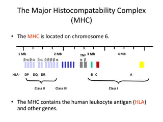
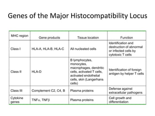
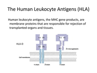
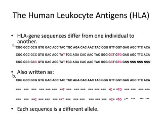
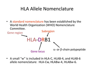
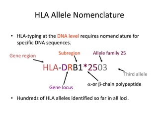
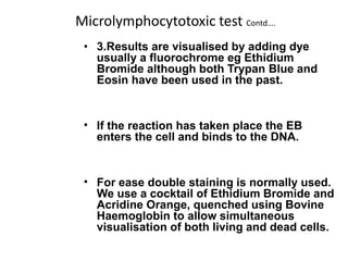
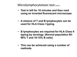
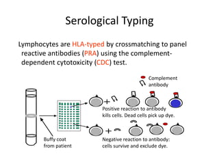
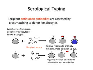
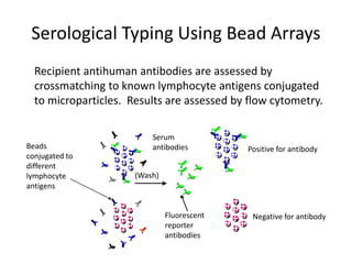
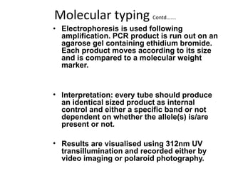
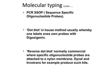
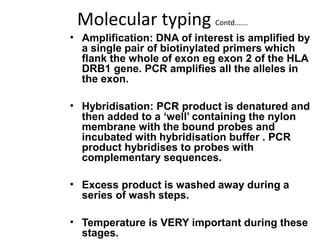
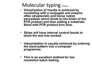
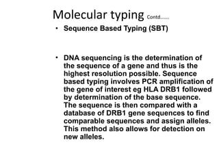
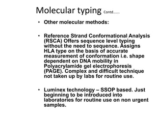
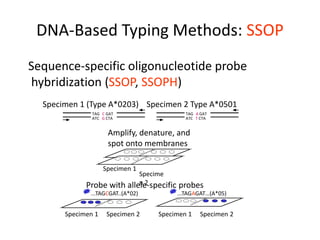
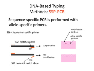
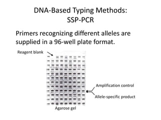
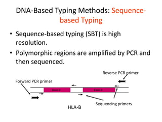
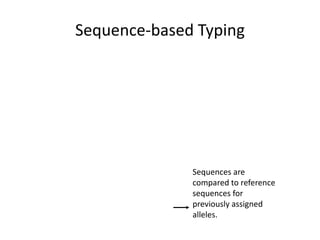
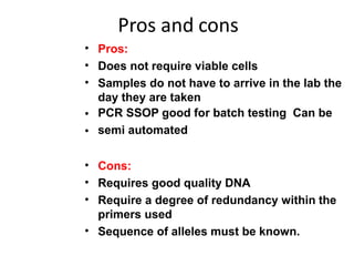
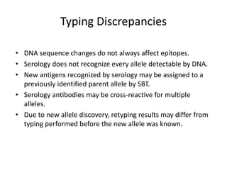
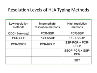
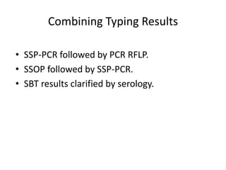
HLA Typing
The Major Histocompatability Complex
(MHC)
• The MHC is located on chromosome 6.
• The MHC contains the human leukocyte antigen (HLA)
and other genes.
1 Mb 2 Mb 3 Mb 4 Mb
HLA- DP DQ DR B C A
Class II Class III Class I
b a b a b a b b b b a a b
TNF
Genes of the Major Histocompatibility Locus
MHC region
Gene products Tissue location Function
Class I HLA-A, HLA-B, HLA-C All nucleated cells
Identification and
destruction of abnormal
or infected cells by
cytotoxic T cells
Class II HLA-D
B lymphocytes,
monocytes,
macrophages, dendritic
cells, activated T cells,
activated endothelial
cells, skin (Langerhans
cells)
Identification of foreign
antigen by helper T cells
Class III Complement C2, C4, B Plasma proteins
Defense against
extracellular pathogens
Cytokine
genes
TNFa, TNFb Plasma proteins
Cell growth and
differentiation
The Human Leukocyte Antigens (HLA)
Human leukocyte antigens, the MHC gene products, are
membrane proteins that are responsible for rejection of
transplanted organs and tissues.
b 2microglobulin
a 1 b1
a 2 b2
a 2 a1
a 3
HLA-D
Cell membrane
a chain b chain a chain
The Human Leukocyte Antigens (HLA)
• HLA-gene sequences differ from one individual to
another.
• Also written as:
• Each sequence is a different allele.
CGG GCC GCG GTG GAC ACC TAC TGC AGA CAC AAC TAC GGG GTT GGT GAG AGC TTC ACA
CGG GCC GCC GTG GAC ACC TAT TGC AGA CAC AAC TAC GGG GCT GTG GAG AGC TTC ACA
CGG GCC GCC GTG GAC ACC TAT TGC AGA CAC AAC TAC GGG GCT GTG GNN NNN NNN NNN
CGG GCC GCG GTG GAC ACC TAC TGC AGA CAC AAC TAC GGG GTT GGT GAG AGC TTC ACA
--- --- --- --- --- --- --T --- --- --- --- --- --- -C - -TG --- --- --- ---
--- --- --C --- --- --- --T --- --- --- --- --- --- -C- -TG -** *** *** ***
a.
b.
HLA Allele Nomenclature
• A standard nomenclature has been established by the
World Health Organization (WHO) Nomenclature
Committee.
• A small “w” is included in HLA-C, HLAB-4, and HLAB-6
allele nomenclature: HLA-Cw, HLABw-4, HLABw-6.
HLA-DRB1
Gene region
Gene locus
Subregion
a- or b-chain polypeptide
HLA Allele Nomenclature
• HLA-typing at the DNA level requires nomenclature for
specific DNA sequences.
• Hundreds of HLA alleles identified so far in all loci.
HLA-DRB1*2503
Gene region
Gene locus
Subregion
a-or b-chain polypeptide
Allele family 25
Third allele
HLA Alleles are Inherited in Blocks as
Haplotypes
HLA-Typing
• Every person (except identical twins) has different
sets of HLA alleles.
• Transplanted organs are allografts, in which the
donor organ and the recipient are genetically
different.
• Compatibility (matching) of the HLA of the donor
and the recipient increases the chance for a
successful engraftment.
• Matching is determined by comparing alleles.
• Resolution is the level of detail with which an allele
is determined.
TYPING METHODS
• SEROLOGY used to be the ‘gold’
standard. Now being superseded by
molecular techniques as they
become more robust and time
efficient.
•
CELLULAR rarely used now. Originally
used for Class II typing.
• MOLECULAR fast becoming the method of
choice.
SEROLOGY
• Complement Dependent Cytotoxicity
(CDC)
• Viable peripheral blood lymphocytes
are obtained by discontinuous density
gradient centrifugation using Ficoll /
Tryosil or Ficoll / Sodium Metrizoate at
a density of 1.077 at 19º - 22ºC.
• Microlymphocytotoxic test: 3 stages
Microlymphocytotoxic test
• 1.Viable lymphocytes are incubated
with HLA specific antibodies. If the
specific antigen is present on the
cell the antibody is bound.
• 2.Rabbit serum as a source of
complement is added, incubate. If
antibody is bound to the HLA antigen
on the cell surface it activates the
complement which damages the cell
membrane making it permeable to
vital stains.
Microlymphocytotoxic test Contd….
• 3.Results are visualised by adding dye
usually a fluorochrome eg Ethidium
Bromide although both Trypan Blue and
Eosin have been used in the past.
• If the reaction has taken place the EB
enters the cell and binds to the DNA.
• For ease double staining is normally used.
We use a cocktail of Ethidium Bromide and
Acridine Orange, quenched using Bovine
Haemoglobin to allow simultaneous
visualisation of both living and dead cells.
Microlymphocytotoxic test Contd….
• Test is left for 10 minutes and then read
using an inverted fluorescent microscope.
• A mixture of T and B lymphocytes can be
used for HLA Class I typing.
• B lymphocytes are required for HLA Class II
typing by serology. (Normal population 85-
90% T and 10-15% B cells)
• This can be achieved using a number of
methods.
Microlymphocytotoxic test
Contd….
• In the past neuraminidase treated sheep red
blood cell rosetting and nylon wool have
been used.
• Immunomagnetic bead separation is the
current method of choice.
• It utilises polystyrene microspheres with a
magnetisable core coated in monoclonal
antibody for a HLA Class II b chain
monomorphic epitope. Positive selection.
Serological Typing
Lymphocytes are HLA-typed by crossmatching to panel
reactive antibodies (PRA) using the complement-
dependent cytotoxicity (CDC) test.
Complement
antibody
Negative reaction to antibody:
cells survive and exclude dye.
Buffy coat
from patient
Positive reaction to antibody
kills cells. Dead cells pick up dye.
Serological Typing
Recipient antihuman antibodies are assessed by
crossmatching to donor lymphocytes.
Recipient serum
Lymphocytes from organ
donor or lymphocytes of
known HLA types
Positive reaction to antibody
kills cells. Dead cells pick up dye.
Negative reaction to antibody:
cells survive and exclude dye.
Serological Typing Using Bead Arrays
Recipient antihuman antibodies are assessed by
crossmatching to known lymphocyte antigens conjugated
to microparticles. Results are assessed by flow cytometry.
Positive for antibody
Serum
antibodies
(Wash)
Negative for antibody
Beads
conjugated to
different
lymphocyte
antigens
Fluorescent
reporter
antibodies
Other Serological Typing
Methods
• Cytotoxic and noncytotoxic methods with flow
cytometry detection.
• Enzyme-linked immunosorbent assay (ELISA) with
solubilized HLA antigens.
• Mixed lymphocyte culture measuring growth of
lymphocytes activated by cross-reactivity.
• Measure of HLA-protein mobility differences in one-
dimensional gel isoelectric focusing or two-
dimensional gel electrophoresis.
Pros and cons
• Pros:
• Easily performed does not require expensive
equipment.
• Takes around three hours to perform
• Low level resolution, with good antisera reliable
results
• Cons:
• Requires large volumes of blood
• Requires viable lymphocytes
• Difficult to find good antisera for rarer antigens in
different populations
Cellular typing
• Not / Rarely used by laboratories
these days.
• Requires panels of homozygous
typing cells.
• Cell culture method therefore takes
a long time. Labour intensive
involves use of radioisotopes.
Molecular typing
• All commonly used molecular methods
require good quality genomic DNA. There
are numerous methods for extraction of
DNA from whole blood.
• There are ‘in house’ methods based on
Miller et al’s Salting Out which are cheap
and easy but labour intensive.
• There are also numerous commercial kits
available such as individual matrix capture
columns, beads and semi automated
systems. This however can increase the
cost per extraction from around 65p to
£3.60p.
Molecular typing Contd…….
• All methods rely on DNA extraction from the
nucleated cells following cell lysis and
protein digestion.
• The application of molecular techniques to
HLA typing began around 1987 when the
Southern Blot technique was used to
identify restriction fragment length
polymorphisms (RFLP’s) associated with
known serological DR/DQ and cellular Dw
defined specificities.
• Around 1992 polymerase chain reaction
(PCR) methods were developed.
Molecular typing Contd…….
• PCR
• Three steps per cycle–
denaturation, annealing and
extension. Amplification is
exponential yielding 2 power n
where n = number of cycles.
• The introduction of the
programmable Thermal Cycler
revolutionised the use of PCR
within the routine laboratory.
Molecular typing Contd…….
• PCT SSP (Sequence Specific Priming)
• Can be used for HLA Class I and II typing
using a panel of primer pairs either for low
to medium resolution whereby primers
amplify groups of alleles or high resolution
whereby primer pairs amplify specific
alleles. Each PCR reaction takes place in a
separate tube therefore the number of
tubes depends on the level of resolution.
Each tube also contains a pair of primers
for part of the human growth hormone gene
as an internal control. These are at a much
lower concentration thus do not compete
with specific primers.
Molecular typing Contd…….
• Electrophoresis is used following
amplification. PCR product is run out on an
agarose gel containing ethidium bromide.
Each product moves according to its size
and is compared to a molecular weight
marker.
• Interpretation: every tube should produce
an identical sized product as internal
control and either a specific band or not
dependent on whether the allele(s) is/are
present or not.
• Results are visualised using 312nm UV
transillumination and recorded either by
video imaging or polaroid photography.
Molecular typing Contd…….
• PCR SSOP ( Sequence Specific
Oligonucleotide Probes).
• ‘Dot blot’ in house method usually whereby
one labels ones own probes with
Digoxigenin.
• ‘Reverse dot blot’ normally commercial
where specific oligonucleotide probes are
attached to a nylon membrane. Dynal and
Innotrans for example produce such kits.
Molecular typing Contd…….
• Amplification: DNA of interest is amplified by
a single pair of biotinylated primers which
flank the whole of exon eg exon 2 of the HLA
DRB1 gene. PCR amplifies all the alleles in
the exon.
• Hybridisation: PCR product is denatured and
then added to a ‘well’ containing the nylon
membrane with the bound probes and
incubated with hybridisation buffer . PCR
product hybridises to probes with
complementary sequences.
• Excess product is washed away during a
series of wash steps.
• Temperature is VERY important during these
stages.
Molecular typing Contd…….
• Visualisation of results is achieved by
incubating with a conjugate and enzyme
often streptavidin and horse radish
peroxidase which binds to the biotin of the
PCR product and then adding a substrate.
Band with PCR product turn blue.
• Strips will have internal control bands to
show the test has worked.
• Interpretation is usually achieved by entering
the band pattern into a computer
programme.
• This is an excellent method for low
resolution batch testing.
Molecular typing Contd…….
• Sequence Based Typing (SBT)
• DNA sequencing is the determination of
the sequence of a gene and thus is the
highest resolution possible. Sequence
based typing involves PCR amplification of
the gene of interest eg HLA DRB1 followed
by determination of the base sequence.
The sequence is then compared with a
database of DRB1 gene sequences to find
comparable sequences and assign alleles.
This method also allows for detection on
new alleles.
Molecular typing Contd…….
• Other molecular methods:
• Reference Strand Conformational Analysis
(RSCA) Offers sequence level typing
without the need to sequence. Assigns
HLA type on the basis of accurate
measurement of conformation i.e. shape
dependent on DNA mobility in
Polyacrylamide gel electrophoresis
(PAGE). Complex and difficult technique
not taken up by labs for routine use.
• Luminex technology – SSOP based. Just
beginning to be introduced into
laboratories for routine use on non urgent
samples.
DNA-Based Typing
Methods
• DNA typing focuses on the most polymorphic
loci in the MHC, HLA-B, and HLA-DRB.
• Whole-blood patient specimens collected in
anticoagulant are used for DNA typing.
• Cell lines of known HLA type are used for
reference samples.
DNA-Based Typing Methods: SSOP
Sequence-specific oligonucleotide probe
hybridization (SSOP, SSOPH)
TAG C GAT
ATC G CTA
TAG A GAT
ATC T CTA
Specimen 1 (Type A*0203) Specimen 2 Type A*0501
Amplify, denature, and
spot onto membranes
Specimen 1
Specime
n 2
Probe with allele-specific probes
...TAGCGAT..(A*02) ...TAGAGAT…(A*05)
Specimen 1 Specimen 2 Specimen 1 Specimen 2
DNA-Based Typing
Methods: SSP-PCR
Sequence-specific PCR is performed with
allele-specific primers.
SSP
Amplification
No
amplification
SSP
Amplification
controls
Allele-specific
product
SSP= Sequence-specific primer
SSP matches allele
SSP does not match allele
DNA-Based Typing Methods:
SSP-PCR
Primers recognizing different alleles are
supplied in a 96-well plate format.
Amplification control
Allele-specific product
Reagent blank
Agarose gel
DNA-Based Typing Methods: Sequence-
based Typing
• Sequence-based typing (SBT) is high
resolution.
• Polymorphic regions are amplified by PCR and
then sequenced.
Exon 2 Exon 3
HLA-B
Forward PCR primer
Sequencing primers
Reverse PCR primer
Sequence-based Typing
Sequences are
compared to reference
sequences for
previously assigned
alleles.
Pros and cons
•
•
•
•
•
Pros:
Does not require viable cells
Samples do not have to arrive in the lab the
day they are taken
PCR SSOP good for batch testing Can be
semi automated
•
•
•
•
Cons:
Requires good quality DNA
Require a degree of redundancy within the
primers used
Sequence of alleles must be known.
Typing Discrepancies
• DNA sequence changes do not always affect epitopes.
• Serology does not recognize every allele detectable by DNA.
• New antigens recognized by serology may be assigned to a
previously identified parent allele by SBT.
• Serology antibodies may be cross-reactive for multiple
alleles.
• Due to new allele discovery, retyping results may differ from
typing performed before the new allele was known.
Resolution Levels of HLA Typing Methods
Low resolution
methods
Intermediate
resolution methods
High resolution
methods
CDC (Serology) PCR-SSP PCR-SSP
PCR-SSP PCR-SSOP PCR-SSOP
PCR-SSOP PCR-RFLP
SSP-PCR + PCR-
RFLP
SSOP-PCR + SSP-
PCR
SBT
Combining Typing Results
• SSP-PCR followed by PCR RFLP.
• SSOP followed by SSP-PCR.
• SBT results clarified by serology.
Summary
• The MHC is a polymorphic locus encoding the HLA genes.
• Antigens encoded by the HLA genes are responsible for
allograft tissue and organ rejection. Identifying and
matching alleles increases the chance of successful organ
and tissue transplant.
• HLA antigens and their corresponding sequence alleles are
determined by serological- and DNA- based methods.
• Serology identifies functional antigen recognition, while
sequence analysis identifies genetic alleles with high
resolution.

HLA typing.ppt

  • 1.
  • 2.
    The Major HistocompatabilityComplex (MHC) • The MHC is located on chromosome 6. • The MHC contains the human leukocyte antigen (HLA) and other genes. 1 Mb 2 Mb 3 Mb 4 Mb HLA- DP DQ DR B C A Class II Class III Class I b a b a b a b b b b a a b TNF
  • 3.
    Genes of theMajor Histocompatibility Locus MHC region Gene products Tissue location Function Class I HLA-A, HLA-B, HLA-C All nucleated cells Identification and destruction of abnormal or infected cells by cytotoxic T cells Class II HLA-D B lymphocytes, monocytes, macrophages, dendritic cells, activated T cells, activated endothelial cells, skin (Langerhans cells) Identification of foreign antigen by helper T cells Class III Complement C2, C4, B Plasma proteins Defense against extracellular pathogens Cytokine genes TNFa, TNFb Plasma proteins Cell growth and differentiation
  • 4.
    The Human LeukocyteAntigens (HLA) Human leukocyte antigens, the MHC gene products, are membrane proteins that are responsible for rejection of transplanted organs and tissues. b 2microglobulin a 1 b1 a 2 b2 a 2 a1 a 3 HLA-D Cell membrane a chain b chain a chain
  • 5.
    The Human LeukocyteAntigens (HLA) • HLA-gene sequences differ from one individual to another. • Also written as: • Each sequence is a different allele. CGG GCC GCG GTG GAC ACC TAC TGC AGA CAC AAC TAC GGG GTT GGT GAG AGC TTC ACA CGG GCC GCC GTG GAC ACC TAT TGC AGA CAC AAC TAC GGG GCT GTG GAG AGC TTC ACA CGG GCC GCC GTG GAC ACC TAT TGC AGA CAC AAC TAC GGG GCT GTG GNN NNN NNN NNN CGG GCC GCG GTG GAC ACC TAC TGC AGA CAC AAC TAC GGG GTT GGT GAG AGC TTC ACA --- --- --- --- --- --- --T --- --- --- --- --- --- -C - -TG --- --- --- --- --- --- --C --- --- --- --T --- --- --- --- --- --- -C- -TG -** *** *** *** a. b.
  • 6.
    HLA Allele Nomenclature •A standard nomenclature has been established by the World Health Organization (WHO) Nomenclature Committee. • A small “w” is included in HLA-C, HLAB-4, and HLAB-6 allele nomenclature: HLA-Cw, HLABw-4, HLABw-6. HLA-DRB1 Gene region Gene locus Subregion a- or b-chain polypeptide
  • 7.
    HLA Allele Nomenclature •HLA-typing at the DNA level requires nomenclature for specific DNA sequences. • Hundreds of HLA alleles identified so far in all loci. HLA-DRB1*2503 Gene region Gene locus Subregion a-or b-chain polypeptide Allele family 25 Third allele
  • 8.
    HLA Alleles areInherited in Blocks as Haplotypes
  • 9.
    HLA-Typing • Every person(except identical twins) has different sets of HLA alleles. • Transplanted organs are allografts, in which the donor organ and the recipient are genetically different. • Compatibility (matching) of the HLA of the donor and the recipient increases the chance for a successful engraftment. • Matching is determined by comparing alleles. • Resolution is the level of detail with which an allele is determined.
  • 10.
    TYPING METHODS • SEROLOGYused to be the ‘gold’ standard. Now being superseded by molecular techniques as they become more robust and time efficient. • CELLULAR rarely used now. Originally used for Class II typing. • MOLECULAR fast becoming the method of choice.
  • 11.
    SEROLOGY • Complement DependentCytotoxicity (CDC) • Viable peripheral blood lymphocytes are obtained by discontinuous density gradient centrifugation using Ficoll / Tryosil or Ficoll / Sodium Metrizoate at a density of 1.077 at 19º - 22ºC. • Microlymphocytotoxic test: 3 stages
  • 12.
    Microlymphocytotoxic test • 1.Viablelymphocytes are incubated with HLA specific antibodies. If the specific antigen is present on the cell the antibody is bound. • 2.Rabbit serum as a source of complement is added, incubate. If antibody is bound to the HLA antigen on the cell surface it activates the complement which damages the cell membrane making it permeable to vital stains.
  • 13.
    Microlymphocytotoxic test Contd…. •3.Results are visualised by adding dye usually a fluorochrome eg Ethidium Bromide although both Trypan Blue and Eosin have been used in the past. • If the reaction has taken place the EB enters the cell and binds to the DNA. • For ease double staining is normally used. We use a cocktail of Ethidium Bromide and Acridine Orange, quenched using Bovine Haemoglobin to allow simultaneous visualisation of both living and dead cells.
  • 14.
    Microlymphocytotoxic test Contd…. •Test is left for 10 minutes and then read using an inverted fluorescent microscope. • A mixture of T and B lymphocytes can be used for HLA Class I typing. • B lymphocytes are required for HLA Class II typing by serology. (Normal population 85- 90% T and 10-15% B cells) • This can be achieved using a number of methods.
  • 15.
    Microlymphocytotoxic test Contd…. • Inthe past neuraminidase treated sheep red blood cell rosetting and nylon wool have been used. • Immunomagnetic bead separation is the current method of choice. • It utilises polystyrene microspheres with a magnetisable core coated in monoclonal antibody for a HLA Class II b chain monomorphic epitope. Positive selection.
  • 16.
    Serological Typing Lymphocytes areHLA-typed by crossmatching to panel reactive antibodies (PRA) using the complement- dependent cytotoxicity (CDC) test. Complement antibody Negative reaction to antibody: cells survive and exclude dye. Buffy coat from patient Positive reaction to antibody kills cells. Dead cells pick up dye.
  • 17.
    Serological Typing Recipient antihumanantibodies are assessed by crossmatching to donor lymphocytes. Recipient serum Lymphocytes from organ donor or lymphocytes of known HLA types Positive reaction to antibody kills cells. Dead cells pick up dye. Negative reaction to antibody: cells survive and exclude dye.
  • 18.
    Serological Typing UsingBead Arrays Recipient antihuman antibodies are assessed by crossmatching to known lymphocyte antigens conjugated to microparticles. Results are assessed by flow cytometry. Positive for antibody Serum antibodies (Wash) Negative for antibody Beads conjugated to different lymphocyte antigens Fluorescent reporter antibodies
  • 19.
    Other Serological Typing Methods •Cytotoxic and noncytotoxic methods with flow cytometry detection. • Enzyme-linked immunosorbent assay (ELISA) with solubilized HLA antigens. • Mixed lymphocyte culture measuring growth of lymphocytes activated by cross-reactivity. • Measure of HLA-protein mobility differences in one- dimensional gel isoelectric focusing or two- dimensional gel electrophoresis.
  • 20.
    Pros and cons •Pros: • Easily performed does not require expensive equipment. • Takes around three hours to perform • Low level resolution, with good antisera reliable results • Cons: • Requires large volumes of blood • Requires viable lymphocytes • Difficult to find good antisera for rarer antigens in different populations
  • 21.
    Cellular typing • Not/ Rarely used by laboratories these days. • Requires panels of homozygous typing cells. • Cell culture method therefore takes a long time. Labour intensive involves use of radioisotopes.
  • 22.
    Molecular typing • Allcommonly used molecular methods require good quality genomic DNA. There are numerous methods for extraction of DNA from whole blood. • There are ‘in house’ methods based on Miller et al’s Salting Out which are cheap and easy but labour intensive. • There are also numerous commercial kits available such as individual matrix capture columns, beads and semi automated systems. This however can increase the cost per extraction from around 65p to £3.60p.
  • 23.
    Molecular typing Contd……. •All methods rely on DNA extraction from the nucleated cells following cell lysis and protein digestion. • The application of molecular techniques to HLA typing began around 1987 when the Southern Blot technique was used to identify restriction fragment length polymorphisms (RFLP’s) associated with known serological DR/DQ and cellular Dw defined specificities. • Around 1992 polymerase chain reaction (PCR) methods were developed.
  • 24.
    Molecular typing Contd……. •PCR • Three steps per cycle– denaturation, annealing and extension. Amplification is exponential yielding 2 power n where n = number of cycles. • The introduction of the programmable Thermal Cycler revolutionised the use of PCR within the routine laboratory.
  • 25.
    Molecular typing Contd……. •PCT SSP (Sequence Specific Priming) • Can be used for HLA Class I and II typing using a panel of primer pairs either for low to medium resolution whereby primers amplify groups of alleles or high resolution whereby primer pairs amplify specific alleles. Each PCR reaction takes place in a separate tube therefore the number of tubes depends on the level of resolution. Each tube also contains a pair of primers for part of the human growth hormone gene as an internal control. These are at a much lower concentration thus do not compete with specific primers.
  • 26.
    Molecular typing Contd……. •Electrophoresis is used following amplification. PCR product is run out on an agarose gel containing ethidium bromide. Each product moves according to its size and is compared to a molecular weight marker. • Interpretation: every tube should produce an identical sized product as internal control and either a specific band or not dependent on whether the allele(s) is/are present or not. • Results are visualised using 312nm UV transillumination and recorded either by video imaging or polaroid photography.
  • 27.
    Molecular typing Contd……. •PCR SSOP ( Sequence Specific Oligonucleotide Probes). • ‘Dot blot’ in house method usually whereby one labels ones own probes with Digoxigenin. • ‘Reverse dot blot’ normally commercial where specific oligonucleotide probes are attached to a nylon membrane. Dynal and Innotrans for example produce such kits.
  • 28.
    Molecular typing Contd……. •Amplification: DNA of interest is amplified by a single pair of biotinylated primers which flank the whole of exon eg exon 2 of the HLA DRB1 gene. PCR amplifies all the alleles in the exon. • Hybridisation: PCR product is denatured and then added to a ‘well’ containing the nylon membrane with the bound probes and incubated with hybridisation buffer . PCR product hybridises to probes with complementary sequences. • Excess product is washed away during a series of wash steps. • Temperature is VERY important during these stages.
  • 29.
    Molecular typing Contd……. •Visualisation of results is achieved by incubating with a conjugate and enzyme often streptavidin and horse radish peroxidase which binds to the biotin of the PCR product and then adding a substrate. Band with PCR product turn blue. • Strips will have internal control bands to show the test has worked. • Interpretation is usually achieved by entering the band pattern into a computer programme. • This is an excellent method for low resolution batch testing.
  • 30.
    Molecular typing Contd……. •Sequence Based Typing (SBT) • DNA sequencing is the determination of the sequence of a gene and thus is the highest resolution possible. Sequence based typing involves PCR amplification of the gene of interest eg HLA DRB1 followed by determination of the base sequence. The sequence is then compared with a database of DRB1 gene sequences to find comparable sequences and assign alleles. This method also allows for detection on new alleles.
  • 31.
    Molecular typing Contd……. •Other molecular methods: • Reference Strand Conformational Analysis (RSCA) Offers sequence level typing without the need to sequence. Assigns HLA type on the basis of accurate measurement of conformation i.e. shape dependent on DNA mobility in Polyacrylamide gel electrophoresis (PAGE). Complex and difficult technique not taken up by labs for routine use. • Luminex technology – SSOP based. Just beginning to be introduced into laboratories for routine use on non urgent samples.
  • 32.
    DNA-Based Typing Methods • DNAtyping focuses on the most polymorphic loci in the MHC, HLA-B, and HLA-DRB. • Whole-blood patient specimens collected in anticoagulant are used for DNA typing. • Cell lines of known HLA type are used for reference samples.
  • 33.
    DNA-Based Typing Methods:SSOP Sequence-specific oligonucleotide probe hybridization (SSOP, SSOPH) TAG C GAT ATC G CTA TAG A GAT ATC T CTA Specimen 1 (Type A*0203) Specimen 2 Type A*0501 Amplify, denature, and spot onto membranes Specimen 1 Specime n 2 Probe with allele-specific probes ...TAGCGAT..(A*02) ...TAGAGAT…(A*05) Specimen 1 Specimen 2 Specimen 1 Specimen 2
  • 34.
    DNA-Based Typing Methods: SSP-PCR Sequence-specificPCR is performed with allele-specific primers. SSP Amplification No amplification SSP Amplification controls Allele-specific product SSP= Sequence-specific primer SSP matches allele SSP does not match allele
  • 35.
    DNA-Based Typing Methods: SSP-PCR Primersrecognizing different alleles are supplied in a 96-well plate format. Amplification control Allele-specific product Reagent blank Agarose gel
  • 36.
    DNA-Based Typing Methods:Sequence- based Typing • Sequence-based typing (SBT) is high resolution. • Polymorphic regions are amplified by PCR and then sequenced. Exon 2 Exon 3 HLA-B Forward PCR primer Sequencing primers Reverse PCR primer
  • 37.
    Sequence-based Typing Sequences are comparedto reference sequences for previously assigned alleles.
  • 38.
    Pros and cons • • • • • Pros: Doesnot require viable cells Samples do not have to arrive in the lab the day they are taken PCR SSOP good for batch testing Can be semi automated • • • • Cons: Requires good quality DNA Require a degree of redundancy within the primers used Sequence of alleles must be known.
  • 39.
    Typing Discrepancies • DNAsequence changes do not always affect epitopes. • Serology does not recognize every allele detectable by DNA. • New antigens recognized by serology may be assigned to a previously identified parent allele by SBT. • Serology antibodies may be cross-reactive for multiple alleles. • Due to new allele discovery, retyping results may differ from typing performed before the new allele was known.
  • 40.
    Resolution Levels ofHLA Typing Methods Low resolution methods Intermediate resolution methods High resolution methods CDC (Serology) PCR-SSP PCR-SSP PCR-SSP PCR-SSOP PCR-SSOP PCR-SSOP PCR-RFLP SSP-PCR + PCR- RFLP SSOP-PCR + SSP- PCR SBT
  • 41.
    Combining Typing Results •SSP-PCR followed by PCR RFLP. • SSOP followed by SSP-PCR. • SBT results clarified by serology.
  • 42.
    Summary • The MHCis a polymorphic locus encoding the HLA genes. • Antigens encoded by the HLA genes are responsible for allograft tissue and organ rejection. Identifying and matching alleles increases the chance of successful organ and tissue transplant. • HLA antigens and their corresponding sequence alleles are determined by serological- and DNA- based methods. • Serology identifies functional antigen recognition, while sequence analysis identifies genetic alleles with high resolution.