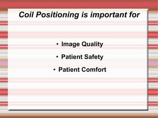
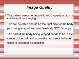
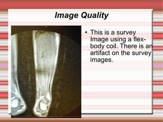
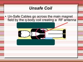
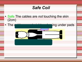
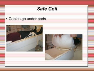
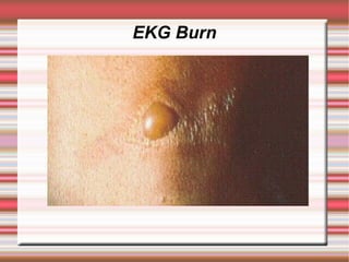
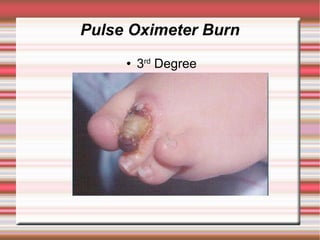
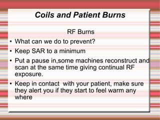
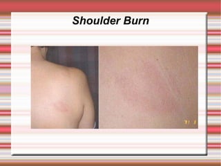
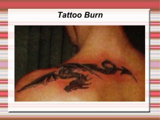
Coil Positioning/Safety


By
Marie Thomas
Coil Positioning is important for


                Image Quality
                Patient Safety
                Patient Comfort
Image Quality
Image Quality
   The patient needs to be positioned properly in or on a
    coil for optimal imaging
   The coil selected should be the right size for the body
    part being imaged (ex. liver flex-body NOT Q-body)
   The part of the body being imaged needs to be in the
    center of the coil, and in turn the coil needs to be as
    close to isocenter as possible.
Image Quality
       
            This Survey image is
           in good position. It
           has good signal
           coverage, the next
           slide shows the
           image received from
           this survey
Image Quality
          Good coil position
           has given this image
           nice even signal over
           the area of interest
Image Quality
          This Survey is not in
           a good position. The
           signal coverage area
           is too high. The
           following is what you
           get.
Image Quality
          An Artifact caused by
           not enough signal,
           poor uneven fat-
           saturation. In order to
           see the top of the
           tibia the window level
           had to be increased
           dramatically, causing
           the fluid to be to
           bright and the
           articular cartilage
           unseen.
Image Quality
          This is a survey
           Image using a flex-
           body coil. There is an
           artifact on the survey
           images.
Image Quality
          The resultant Image
Image Quality
          The coil was
           repositioned and......
Image Quality
          Artifact is gone!
          Revealing some
           pathology in the area
           of the artifact.
Image Quality Conclusion
   An extra minute or two to center the patient
    properly,can make a big difference. A lot of
    coils have a line to show you the center or ends
     of its active area, have a look at yours.
   When you look at your survey, remember to
    look at YOUR SIGNAL COVERAGE as well as
    the anatomy. This will help tell you how good
    your coil positioning is.
Image Quality Conclusion
   On sequences with lots of signal some
    T1's you might “get away” with poorer coil
    position but on Fat- Sat's or low NSA
    sequences you wont.
   If the anatomy your aiming for is not in the
    center, take a minute and reposition it
    might save you ten minutes later on
Coils and Patient Burns
   There are two types of burns
   RF Burns
   Cable Heating Burns
Coils and Patient Burns
                  Cable Heating burns
   What can we do to prevent them?
   NO LOOPS (loops cause conduction causing
    heat causing burns)
   Don't let a cable touch a patients skin/QBC
    directly
   Keep cables aligned with the magnet
   Make sure any gating leads are properly
    attached, no air in between and MR compatible
   Check your cables for frayed parts
Coils and Patient Burns

                 Types of Loops!
   Cable Loops
   Loops between cables and QBC
   Loops between cables and patients
   Loops within patient
Unsafe Coil
   Un-Safe Cables go across the main magnet
    field by the q-body coil creating a RF antenna
Safe Coil
   Safe The cables are not touching the skin
    (2cm)
   The cables are straight and going under pads
Safe Coil
   Cables go under pads
EKG Burn
Pulse Oximeter Burn
        3rd Degree
Coils and Patient Burns

                        RF Burns
   What can we do to prevent?
   Keep SAR to a minimum
   Put a pause in,some machines reconstruct and
    scan at the same time giving continual RF
    exposure.
   Keep in contact with your patient, make sure
    they alert you if they start to feel warm any
    where
Shoulder Burn
Tattoo Burn
Burn--
RF-or Loop burn?
           This patient was
            having his thighs
            scanned due to there
            different sizes.
            Apparently his legs
            were only touching for
            one scan. After the
            exam, he complained
            of an itching, he
            stayed at the facility
            and 3 hours later...
RF-or Loop burn?



    These 3rd degree burns came to the surface?

    What do you think?
Remember
              Burns can happen
           Screen your patient well
   Keep in contact with them during the exam
        Keep your SAR to a minimum
            Check your equipment
       Check your coil/cable positioning

Coil Safe Positioning in MRI

  • 1.
  • 2.
    Coil Positioning isimportant for  Image Quality  Patient Safety  Patient Comfort
  • 3.
  • 4.
    Image Quality  The patient needs to be positioned properly in or on a coil for optimal imaging  The coil selected should be the right size for the body part being imaged (ex. liver flex-body NOT Q-body)  The part of the body being imaged needs to be in the center of the coil, and in turn the coil needs to be as close to isocenter as possible.
  • 5.
    Image Quality  This Survey image is in good position. It has good signal coverage, the next slide shows the image received from this survey
  • 6.
    Image Quality  Good coil position has given this image nice even signal over the area of interest
  • 7.
    Image Quality  This Survey is not in a good position. The signal coverage area is too high. The following is what you get.
  • 8.
    Image Quality  An Artifact caused by not enough signal, poor uneven fat- saturation. In order to see the top of the tibia the window level had to be increased dramatically, causing the fluid to be to bright and the articular cartilage unseen.
  • 9.
    Image Quality  This is a survey Image using a flex- body coil. There is an artifact on the survey images.
  • 10.
    Image Quality  The resultant Image
  • 11.
    Image Quality  The coil was repositioned and......
  • 12.
    Image Quality  Artifact is gone!  Revealing some pathology in the area of the artifact.
  • 13.
    Image Quality Conclusion  An extra minute or two to center the patient properly,can make a big difference. A lot of coils have a line to show you the center or ends of its active area, have a look at yours.  When you look at your survey, remember to look at YOUR SIGNAL COVERAGE as well as the anatomy. This will help tell you how good your coil positioning is.
  • 14.
    Image Quality Conclusion  On sequences with lots of signal some T1's you might “get away” with poorer coil position but on Fat- Sat's or low NSA sequences you wont.  If the anatomy your aiming for is not in the center, take a minute and reposition it might save you ten minutes later on
  • 15.
    Coils and PatientBurns  There are two types of burns  RF Burns  Cable Heating Burns
  • 16.
    Coils and PatientBurns Cable Heating burns  What can we do to prevent them?  NO LOOPS (loops cause conduction causing heat causing burns)  Don't let a cable touch a patients skin/QBC directly  Keep cables aligned with the magnet  Make sure any gating leads are properly attached, no air in between and MR compatible  Check your cables for frayed parts
  • 17.
    Coils and PatientBurns Types of Loops!  Cable Loops  Loops between cables and QBC  Loops between cables and patients  Loops within patient
  • 18.
    Unsafe Coil  Un-Safe Cables go across the main magnet field by the q-body coil creating a RF antenna
  • 19.
    Safe Coil  Safe The cables are not touching the skin (2cm)  The cables are straight and going under pads
  • 20.
    Safe Coil  Cables go under pads
  • 21.
  • 22.
    Pulse Oximeter Burn  3rd Degree
  • 23.
    Coils and PatientBurns RF Burns  What can we do to prevent?  Keep SAR to a minimum  Put a pause in,some machines reconstruct and scan at the same time giving continual RF exposure.  Keep in contact with your patient, make sure they alert you if they start to feel warm any where
  • 24.
  • 25.
  • 26.
  • 27.
    RF-or Loop burn?  This patient was having his thighs scanned due to there different sizes. Apparently his legs were only touching for one scan. After the exam, he complained of an itching, he stayed at the facility and 3 hours later...
  • 28.
    RF-or Loop burn?  These 3rd degree burns came to the surface?  What do you think?
  • 29.
    Remember  Burns can happen  Screen your patient well  Keep in contact with them during the exam  Keep your SAR to a minimum  Check your equipment  Check your coil/cable positioning