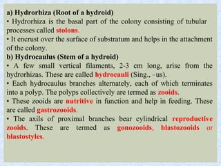
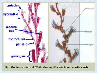
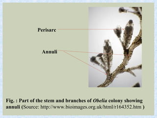
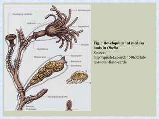
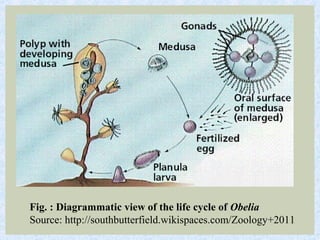
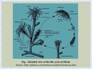
Obelia is a colonial marine cnidarian that exists in both a sessile polyp stage and a free-swimming medusa stage. It has a branching structure made of hydrocaulus and hydrorhiza that support gastrozooids for feeding and gonozooids for asexual reproduction. Gonozooids bud numerous small medusae that detach and transition Obelia to its sexual medusa phase, where it reproduces sexually to complete its life cycle.
It discusses basic information regarding a hemichordate animal called Balanoglossus or Acorn worm, which is also a good connecting link between the non-chordates and chordates.
ORIGIN OF CHORDATES
Animal kingdom is basically divided into two sub kingdoms:
Non-chordata- including animals without notochord.
Chordata- This comprising animals having notochord or chorda dorsalis.
Chordates were evolved sometime 500 million years ago during Cambrian period (invertebrates were also began to evolve in this period) .
Chamberlain (1900) pointed out that all modern chordates possess glomerular kidneys that are designed to remove excess water from body.
It is believed that Chordates have originated from invertebrates.
It is difficult to determine from which invertebrate group the chordates were developed.
Chordate ancestors were soft bodied animals. Hence they were not preserved as Fossils.
However, early fossils of chordates have all been recovered from marine sediments and even modern protochordates are all marine forms.
Also glomerular kidneys are also found in some marine forms such as myxinoids and sharks. That makes the marine origin of chordates more believable.
Chordates evolved from some deuterostome ancestor (echinoderms, hemichordates, pogonophorans etc.) as they have similarities in embryonic development, type of coelom and larval stages.
Many theories infers origin of chordates, hemichordates and echinoderms from a common ancestor.
are worm-like parasites. The clinically relevant groups are separated according to their general external shape and the host organ they inhabit. There are both hermaphroditic and bisexual species.
The definitive classification is based on the external and internal morphology of egg, larval, and adult stages.
Helminth is a general term meaning worm. The helminths are invertebrates characterized by elongated, flat or round bodies.
In flatworms or platyhelminths (platy from the Greek root meaning “flat”) include flukes and tapeworms.
Roundworms are nematodes (nemato from the Greek root meaning “thread”).
Sponges,are pore bearing,multicellular,diploblastic animals that belong to phylum Porifera
Body of all sponges is perforated by large number of pores called ostia through which water enters Inside body and flows through a system of criss-crossing canals known as canal system
Three main types of canal systems in the order of increasing complexity are Asconoid, Syconoid and Leuconoid type.
The invertebrates, or invertebrates, are animals that do not contain bony structures, such as the cranium and vertebrae. The simplest of all the invertebrates are the Parazoans, which include only the phylum Porifera: the sponges.
Parazoans (“beside animals”) do not display tissue-level organization, although they do have specialized cells that perform specific functions. Sponge larvae are able to swim; however, adults are non-motile and spend their life attached to a substratum.
Since water is vital to sponges for excretion, feeding, and gas exchange, their body structure facilitates the movement of water through the sponge. Structures such as canals, chambers, and cavities enable water to move through the sponge to nearly all body cells.
Chordata is the last phylum of kingdom Animalia.
Which is further subdivided into subphylums, divisions and classes.
The Slides shows the classification of the phylum along with the basis on which it is classified.
(includes examples along with pictures for easy understanding and memorizing)
Cnidaria is a phylum containing over 9,000 species found only in aquatic and mostly marine environments. All cnidarians have radial symmetrical. There are two major body forms among the Cnidaria - the polyp and the medusa. Sea anemones and corals have the polyp form, while jellyfish are typical medusae.
purpose of delivering a speech varies depending on the occasion or the audience it is delivered to. A wedding speech, for example, is usually delivered to express a congratulatory remark to the newly wed couple or to propose a toast to the audience. In order to ensure a proper delivery of words and ideas, speakers may practice their speaking through speech exercises like tongue twisters.
In this presentation, Phylum Cnidaria, Coelenterata is described. After watching this you will learn the characteristics of Phylum Cnidaria, Coelenterata, The Body Wall and Nematocysts, Alternation of Generations, Maintenance of Functions, Reproduction, Class Hydrozoa, Class Staurozoa, Class Scyphozoa, Class Cubozoa, Class Anthozoa, locomotion, Digestion, Nutrition,Exchanges with the Environment, Nervous and Sensory Functions, Reproduction and Development, cnidocytes, alternation of generations, polyps, medusa, dactylozooids gastrozooid, gonozoid, obelia, aurelia Jelly fish, coral reef, Hydra, Gonionemus, siphonophora, example and taxonomy of Phylum Cnidaria. It is part of BS Zoology Course Animal diversity.
Corals are marine invertebrates in class Anthozoa of phylum Cnidaria typically living in compact colonies of many identical individual "polyps".
Corals are gastrovascular marine organisms. Each one of these animals is known as a coral
"polyp". Coral Polyps are tiny, primitive marine organisms.
A single polyp has a tube-shaped body with a mouth which is surrounded by tentacles.
The polyp of hard corals produces a stony skeleton of calcium carbonate which form the base. Often the skeleton forms a cup-like structure in which the polyp lives. Coral polyps in colonies make up the cora reefs.
Development of Chordata: From Embryogenesis to Morphogenesis"mishisajjad566
This topic explores the developmental processes that shape the Chordata phylum, including embryogenesis, morphogenesis, and organogenesis. It covers the formation of the notochord, nerve cord, and post-anal tail, as well as the development of chordate characteristics such as gill slits and pharyngeal pouches.
This topic delves into the developmental biology of Enteropneusta, a subphylum of Chordata that includes acorn worms. It examines the embryonic development of Enteropneusta, including gastrulation, neurulation, and organogenesis, and discusses the unique features of their developmental processes, such as the formation of the proboscis and the development of their distinctive body shape.
Note: Enteropneusta is a subphylum of Chordata that includes acorn worms and other related species. They are marine animals that belong to the phylum Hemichordata
A chart showing the fate of each part of an early embryo, in a particular blastula stage is called fate maps. It is done because the correct interpretation of gastrulation is impossible without the knowledge of the position which are the presumptive germinal layers (Ectoderm, Mesoderm and Endoderm) occupy in blastula.
Fate mapping is a method used in developmental biology to study the embryonic origin of various adult tissues and structures. The "fate" of each cell or group of cells is mapped onto the embryo, showing which parts of the embryo will develop into which tissue. When carried out at single-cell resolution, this process is called cell lineage tracing. It is also used to trace the development of tumors.
DNA sequencing is the process of determining the sequence of nucleotides (A, T, G, and C) in the DNA. It includes method or technology that is used to determine the order of the four bases: adenine, thymine, guanine and cytosine.
The chain-termination method developed by Frederick Sanger and coworkers in 1977. This method used fewer toxic chemicals and lower amounts of radioactivity than the Maxam and Gilbert method. Because of its comparative ease, the Sanger method was soon automated and was the method used in the first generation of DNA sequencers.
published a DNA sequencing method in 1977 based on chemical modification of DNA and subsequent cleavage at specific bases. Also known as chemical sequencing, this method allowed purified samples of double-stranded DNA to be used without further cloning.
Maxam-Gilbert sequencing requires radioactive labeling at one 5' end of the DNA and purification of the DNA fragment to be sequenced. Chemical treatment then generates breaks at a small proportion of one or two of the four nucleotide bases in each of four reactions (G, A+G, C, C+T). The concentration of the modifying chemicals is controlled to introduce on average one modification per DNA molecule. Thus a series of labeled fragments is generated, from the radiolabeled end to the first "cut" site in each molecule. The fragments in the four reactions are electrophoresed side by side in denaturing acrylamide gels for size separation. To visualize the fragments, the gel is exposed to X-ray film for autoradiography, yielding a series of dark bands each corresponding to a radiolabeled DNA fragment, from which the sequence may be inferred.
Cloning is the process of producing genetically identical individuals of an organism either naturally or artificially.
It is the process of taking genetic information from one living thing and creating identical copies of it. The copied material is called a clone.
Nature has been doing it for millions of years. For example, identical twins have almost identical DNA, and asexual reproduction in some plants and organisms can produce genetically identical offspring.
Cloning in biotechnology refers to the process of creating clones of organisms or copies of cells or DNA fragments (molecular cloning).
Bacteriophage- types, structure and morphology of t4 phage, morphogenesisDr. Dinesh C. Sharma
Escherichia virus T4 is a species of bacteriophages that infect Escherichia coli bacteria. It is a member of virus subfamily Tevenvirinae (not to be confused with T-even bacteriophages, which is an alternate name of the species). T4 is capable of undergoing only a lytic lifecycle and not the lysogenic lifecycle.
Each cell of a multicellular organism contain the same genetic material, but the expression of the gene is different in different type of cell group. On the basis of expression requirement they are grouped in to
Structural Gene- Mostly expressed once in a life
Vital Gene- Involved in of vital biochemical processes such as respiration and need to be expressed all the time
Functional Gene- Genes are not expressed all the time. They are switched on an off at need
The regulation of Gene required in case of functional gene and its explained by Francois Jacob, Jacques Monod and Andre Lwoff (Nobal Prize in 1961)
From studies and predictions such as Dreyer and Bennett's, it shows that the light chains and heavy chains are encoded by separate multigene families on different chromosomes. They are referred to as gene segments and are separated by non-coding regions. The rearrangement and organization of these gene segments during the maturation of B cells produce functional proteins. The entire process of rearrangement and organization of these gene segments is the vital source where our body immune system gets its capabilities to recognize and respond to variety of antigens.
The cells of the B line synthesize immunoglobulins. They are either produced at a membrane (on the surface of the B-lymphocytes) or are secreted (by the plasmocytes)
Theory of preformation,
Epigenetic theory,
Theory of pengenesis,
Recapitulation theory,
Germplasm theory,
Mosaic theory,
Regulated theory,
Gradient theory
Theory of organizers.
Sericulture is the cultivation of silkworms to produce silk. Bombyx mori (the caterpillar of the domesticated silk moth) is the most widely used species of silkworms.
Introduction:
RNA interference (RNAi) or Post-Transcriptional Gene Silencing (PTGS) is an important biological process for modulating eukaryotic gene expression.
It is highly conserved process of posttranscriptional gene silencing by which double stranded RNA (dsRNA) causes sequence-specific degradation of mRNA sequences.
dsRNA-induced gene silencing (RNAi) is reported in a wide range of eukaryotes ranging from worms, insects, mammals and plants.
This process mediates resistance to both endogenous parasitic and exogenous pathogenic nucleic acids, and regulates the expression of protein-coding genes.
What are small ncRNAs?
micro RNA (miRNA)
short interfering RNA (siRNA)
Properties of small non-coding RNA:
Involved in silencing mRNA transcripts.
Called “small” because they are usually only about 21-24 nucleotides long.
Synthesized by first cutting up longer precursor sequences (like the 61nt one that Lee discovered).
Silence an mRNA by base pairing with some sequence on the mRNA.
Discovery of siRNA?
The first small RNA:
In 1993 Rosalind Lee (Victor Ambros lab) was studying a non- coding gene in C. elegans, lin-4, that was involved in silencing of another gene, lin-14, at the appropriate time in the
development of the worm C. elegans.
Two small transcripts of lin-4 (22nt and 61nt) were found to be complementary to a sequence in the 3' UTR of lin-14.
Because lin-4 encoded no protein, she deduced that it must be these transcripts that are causing the silencing by RNA-RNA interactions.
Types of RNAi ( non coding RNA)
MiRNA
Length (23-25 nt)
Trans acting
Binds with target MRNA in mismatch
Translation inhibition
Si RNA
Length 21 nt.
Cis acting
Bind with target Mrna in perfect complementary sequence
Piwi-RNA
Length ; 25 to 36 nt.
Expressed in Germ Cells
Regulates trnasposomes activity
MECHANISM OF RNAI:
First the double-stranded RNA teams up with a protein complex named Dicer, which cuts the long RNA into short pieces.
Then another protein complex called RISC (RNA-induced silencing complex) discards one of the two RNA strands.
The RISC-docked, single-stranded RNA then pairs with the homologous mRNA and destroys it.
THE RISC COMPLEX:
RISC is large(>500kD) RNA multi- protein Binding complex which triggers MRNA degradation in response to MRNA
Unwinding of double stranded Si RNA by ATP independent Helicase
Active component of RISC is Ago proteins( ENDONUCLEASE) which cleave target MRNA.
DICER: endonuclease (RNase Family III)
Argonaute: Central Component of the RNA-Induced Silencing Complex (RISC)
One strand of the dsRNA produced by Dicer is retained in the RISC complex in association with Argonaute
ARGONAUTE PROTEIN :
1.PAZ(PIWI/Argonaute/ Zwille)- Recognition of target MRNA
2.PIWI (p-element induced wimpy Testis)- breaks Phosphodiester bond of mRNA.)RNAse H activity.
MiRNA:
The Double-stranded RNAs are naturally produced in eukaryotic cells during development, and they have a key role in regulating gene expression .
The increased availability of biomedical data, particularly in the public domain, offers the opportunity to better understand human health and to develop effective therapeutics for a wide range of unmet medical needs. However, data scientists remain stymied by the fact that data remain hard to find and to productively reuse because data and their metadata i) are wholly inaccessible, ii) are in non-standard or incompatible representations, iii) do not conform to community standards, and iv) have unclear or highly restricted terms and conditions that preclude legitimate reuse. These limitations require a rethink on data can be made machine and AI-ready - the key motivation behind the FAIR Guiding Principles. Concurrently, while recent efforts have explored the use of deep learning to fuse disparate data into predictive models for a wide range of biomedical applications, these models often fail even when the correct answer is already known, and fail to explain individual predictions in terms that data scientists can appreciate. These limitations suggest that new methods to produce practical artificial intelligence are still needed.
In this talk, I will discuss our work in (1) building an integrative knowledge infrastructure to prepare FAIR and "AI-ready" data and services along with (2) neurosymbolic AI methods to improve the quality of predictions and to generate plausible explanations. Attention is given to standards, platforms, and methods to wrangle knowledge into simple, but effective semantic and latent representations, and to make these available into standards-compliant and discoverable interfaces that can be used in model building, validation, and explanation. Our work, and those of others in the field, creates a baseline for building trustworthy and easy to deploy AI models in biomedicine.
Bio
Dr. Michel Dumontier is the Distinguished Professor of Data Science at Maastricht University, founder and executive director of the Institute of Data Science, and co-founder of the FAIR (Findable, Accessible, Interoperable and Reusable) data principles. His research explores socio-technological approaches for responsible discovery science, which includes collaborative multi-modal knowledge graphs, privacy-preserving distributed data mining, and AI methods for drug discovery and personalized medicine. His work is supported through the Dutch National Research Agenda, the Netherlands Organisation for Scientific Research, Horizon Europe, the European Open Science Cloud, the US National Institutes of Health, and a Marie-Curie Innovative Training Network. He is the editor-in-chief for the journal Data Science and is internationally recognized for his contributions in bioinformatics, biomedical informatics, and semantic technologies including ontologies and linked data.
Professional air quality monitoring systems provide immediate, on-site data for analysis, compliance, and decision-making.
Monitor common gases, weather parameters, particulates.
Multi-source connectivity as the driver of solar wind variability in the heli...Sérgio Sacani
The ambient solar wind that flls the heliosphere originates from multiple
sources in the solar corona and is highly structured. It is often described
as high-speed, relatively homogeneous, plasma streams from coronal
holes and slow-speed, highly variable, streams whose source regions are
under debate. A key goal of ESA/NASA’s Solar Orbiter mission is to identify
solar wind sources and understand what drives the complexity seen in the
heliosphere. By combining magnetic feld modelling and spectroscopic
techniques with high-resolution observations and measurements, we show
that the solar wind variability detected in situ by Solar Orbiter in March
2022 is driven by spatio-temporal changes in the magnetic connectivity to
multiple sources in the solar atmosphere. The magnetic feld footpoints
connected to the spacecraft moved from the boundaries of a coronal hole
to one active region (12961) and then across to another region (12957). This
is refected in the in situ measurements, which show the transition from fast
to highly Alfvénic then to slow solar wind that is disrupted by the arrival of
a coronal mass ejection. Our results describe solar wind variability at 0.5 au
but are applicable to near-Earth observatories.
Observation of Io’s Resurfacing via Plume Deposition Using Ground-based Adapt...Sérgio Sacani
Since volcanic activity was first discovered on Io from Voyager images in 1979, changes
on Io’s surface have been monitored from both spacecraft and ground-based telescopes.
Here, we present the highest spatial resolution images of Io ever obtained from a groundbased telescope. These images, acquired by the SHARK-VIS instrument on the Large
Binocular Telescope, show evidence of a major resurfacing event on Io’s trailing hemisphere. When compared to the most recent spacecraft images, the SHARK-VIS images
show that a plume deposit from a powerful eruption at Pillan Patera has covered part
of the long-lived Pele plume deposit. Although this type of resurfacing event may be common on Io, few have been detected due to the rarity of spacecraft visits and the previously low spatial resolution available from Earth-based telescopes. The SHARK-VIS instrument ushers in a new era of high resolution imaging of Io’s surface using adaptive
optics at visible wavelengths.
THE IMPORTANCE OF MARTIAN ATMOSPHERE SAMPLE RETURN.Sérgio Sacani
The return of a sample of near-surface atmosphere from Mars would facilitate answers to several first-order science questions surrounding the formation and evolution of the planet. One of the important aspects of terrestrial planet formation in general is the role that primary atmospheres played in influencing the chemistry and structure of the planets and their antecedents. Studies of the martian atmosphere can be used to investigate the role of a primary atmosphere in its history. Atmosphere samples would also inform our understanding of the near-surface chemistry of the planet, and ultimately the prospects for life. High-precision isotopic analyses of constituent gases are needed to address these questions, requiring that the analyses are made on returned samples rather than in situ.
1. The word Obelia is probably derived from the Greek word –
‘obeliās’, which means a loaf baked on a spit; obel (ós) - a spit +
-ias noun suffix.
2. Table of Contents
• Introduction
• Habit and Habitat
• Morphology -• Hydrorhiza • Hydrocaulus
• Living Tissue of Obelia - Coenosarc -• Epidermis • Gastrodermis • Protective
Covering - Perisarc
• Morphology of a Gastrozooid
• Morphology of a Gonozooid
• Morphology of a Medusa
• Locomotion in Obelia
• Nutrition in Obelia
• Respiration in Obelia
• Excretion and Osmoregulation in Obelia
• Sense Organ - Statocyst
• Reproduction in Obelia -• Asexual Reproduction • Sexual Reproduction •
Metagenesis • Polymorphism
• Summary
• Exercise/Practice
• Glossary
• References/Bibliography/Further Reading
3. INTRODUCTION
Obelia is a sedentary colonial marine cnidarian which
grows upright in a branching tree-like form and has
several specialized feeding and reproductive polyps. It is
commonly called sea-fur and exists in both asexual,
sessile, polypoid stage and sexual, free-swimming
medusoid phase.
The common species of Obelia are:
a) Obelia geniculata (Knotted thread hydroid)
b) Obelia longissima (Sessile hydroid)
c) Obelia dichotoma (Sea thread hydroid)
d) Obelia bidentata (Double toothed hydroid)
4. HABIT AND HABITAT
Obelia is cosmopolitan in distribution, only exception
being the high-arctic and Antarctic seas. They grow in
shallow water, in intertidal rock pools and are usually
found up to 80-100 meters of depth from the water's
surface. The medusa stage of Obelia species is commonly
found in coastal and offshore plankton around the world.
The colonies of Obelia are often found as a delicate fur-like
growth on the rocks, stones, mollusc shells, sea weeds,
wooden pilings and wharves. Obelia geniculata normally
grows on kelp fronds, especially on Laminaria hyperborea
in conditions of moderate wave exposure.
5. FIG. : OBELIA SP. GROWING ON (A) KELP STIPES
OF LAMINARIA HYPERBOREA; (B) ROCKY BOTTOM
Source: Smale, D.A., Burrows, M. T., Moore, P., O'Connor, N. and Hawkins, S. J.
(2013) Threats and knowledge gaps for ecosystem services provided by kelp forests:
a northeast Atlantic perspective. Ecology and Evolution, 3: 4016–4038.
6. MORPHOLOGY
Obelia is a very small marine
hydroid. It looks like a small
branching tree exhibiting
whitish or brown colour. The
height of Obelia varies from 2
cm or more.
The body of Obelia consists
of two kinds of filaments,
horizontal hydrorhiza and
vertical hydrocaulus.
Fig. : Colony of Obelia geniculata
7. a) Hydrorhiza (Root of a hydroid)
• Hydrorhiza is the basal part of the colony consisting of tubular
processes called stolons.
• It encrust over the surface of substratum and helps in the attachment
of the colony.
b) Hydrocaulus (Stem of a hydroid)
• A few small vertical filaments, 2-3 cm long, arise from the
hydrorhizas. These are called hydrocauli (Sing., –us).
• Each hydrocaulus branches alternately, each of which terminates
into a polyp. The polyps collectively are termed as zooids.
• These zooids are nutritive in function and help in feeding. These
are called gastrozooids.
• The axils of proximal branches bear cylindrical reproductive
zooids. These are termed as gonozooids, blastozooids or
blastostyles.
8. Fig. : Outline structure of Obelia showing alternate branches with zooids
9. LIVING TISSUE OF OBELIA -
COENOSARC
Whole colony of Obelia; hydrorhiza, hydrocaulus and
zooids; contain living tissue, called coenenchyme or
coenosarc.
• The coenosarc is diploblastic comprising of two layers;
outer epidermis and inner, gastrodermis. A middle non-
cellular layer of mesoglea is present in between epidermis
and gastrodermis.
• A narrow canal, called coenosarcal canal runs through
whole colony of Obelia which is continuous with the
gastrovascular cavity of the zooids. The continuity of the
canal system helps to transport the digested food throughout
the colony.
10. EPIDERMIS
• The epidermis is thin and made up of
typical cells of Cnidaria. These include;
epithelio-muscular cells, mucus-secreting
cells, interstitial cells, nerve cells and
nematoblasts.
• The nematocysts are basitrichous
isorhizas. These consist of an oval capsule, a
long thread bearing spines and open at the tip.
11. GASTRODERMIS
It forms the lining of
gastrovascular cavity and
consists of endothelio-muscular
cells, nutritive cells, gland cells
and nerve cells.
12. PROTECTIVE COVERING – PERISARC
Entire colony of Obelia is surrounded by a protective covering, called
perisarc. It is non-cellular, tough, transparent, yellowish-brown and
cuticular in nature and is called perisarc or periderm.
• It makes the vertical part of the colony firm and rigid. Perisarc is
secreted by the epidermis and is separated from the coenosarc by a
thin fluid-filled space.
• However, the coenosarc and perisarc are in contact making the
colony more rigid.
• At some points, the perisarc is arranged in flexible rings called
annuli. These allow the swaying movements due to the force of water
currents.
• The perisarc of hydranth is termed as hydrotheca and that of
gonozooid is called gonotheca.
13. Fig. : Part of the stem and branches of Obelia colony showing
annuli (Source: http://www.bioimages.org.uk/html/r164352.htm )
Annuli
Perisarc
14. MORPHOLOGY OF A GASTROZOOID
Gastrozooid of Obelia is a feeding polyp. Its function is to feed the
whole colony.
• Gastrozooid is a tubular and diplobastic zooid with a central
gastrovascular cavity continuous with the coenosarcal canal.
• The polyp is attached to the hydrocaulus by a hollow stalk while its
distal end is produced into a conical elevation called manubrium or
hypostome.
• The apical portion of the manubrium bears a terminal mouth
encircled by numerous long, solid tentacles, often 24, loaded with
nematoblasts.
• The perisarc of gastrozooid, called hydrotheca, is transparent and
cup-shaped invaginated as a platform or shelf at the base of the
gastrozooids for polyp to rest.
15. • The gastrozooid and
hydrotheca collectively form
hydranth.
• In case of any emergent
situation, the polyp can
withdraw itself into the
hydrotheca and the tentacles
fold over the manubrium
covering the mouth. The
presence of shelf prevents the
polyp to retract into the
hydrocaulus.
• The annuli of the perisarc
present around the stalk of
polyp allow the swaying
movements due to the force of
water current.
Fig. : Structure of a gastrozooid and gonozooid
(Source: http://quizlet.com/21150632/lab-test-total-
flash-cards/ )
16. MORPHOLOGY OF A GONOZOOID
The gonozooids, also called blastozooids or blastostyles are cylindrical rod-like
reproductive bodies present in the axils of hydrocaulus and stalk of gastrozooids.
• Gonozooids are less in number than gastrozooids as these are present only in the
proximal part of the colony.
It has a reduced gastrovascular cavity and is devoid of mouth and tentacles. It,
thus, can not feed and receives food digested by the gastrozooids and transported
through the gastrovascular cavity.
• Like other parts of the colony, gonozooids are also enclosed in a perisarc, called
gonotheca. It is constricted distally and constricted by annuli proximally. The
apical part of the gonotheca has an opening called gonopore.
• Gonozooid produces numerous small medusae or gonophores by the asexual
process of budding.
• Mature medusae detach from the gonozooids and escape into the surrounding
water through the gonopore.
• The gonozooids, gonophores and gonotheca collectively form gonangium.
17. MORPHOLOGY OF A MEDUSA
Medusa of Obelia is radially symmetrical, umbrella-like zooid which
measures approximately 6-7 mm in diameter.
• The outer surface of medusa is convex and known as ex-umbrellar
surface, while the inner concave surface is called sub-umbrellar surface.
• A short manubrium containing a quadrangular mouth at its distal end
hangs from the centre of the sub-umbrellar surface.
• The medusa is craspedote type as its edge is produced inwards into an
insignificant rudimentary velum.
• The margins of the medusa bear initially 16 short, contractile tentacles;
which gradually increase in number.
• The mouth open into a short gullet which leads to a wide expanded
stomach from which arise four narrow, radial canals which mark the four
principal per-radii. The radial canals extend till the margin of the umbrella
and open into a circular canal running parallel to the margin.
18. The radius bisecting two per-radii is called
inter-radius (four in number) and that
bisecting per-radius and adjacent inter-radius
is termed as ad-radius (eight numbers). The
tentacles present at the end of these radii are
named accordingly, such as per-radial
tentacles; inter-radial tentacles and so on.
• Whole system of canals is lined by inner
layer of gastrodermis and both the ex-
umbrellar and sub-umbrellar surfaces are
covered by epidermis.
19. Nervous system consists of two diffused
nerve nets which are concentrated
around the margins of the umbrella and
form two circular nerve rings.
• Eight receptor organs, called
statocysts, are present at the bases of
ad-radial tentacles. These are the organs
of balance, muscular co-ordination and
equilibrium.
• Medusa possesses four gonads on the
sub-umbrellar surface. These are per-
radial in position and each of these is
present in the middle of each radial
canal.
• These are dioecious, male and female
medusae being separate individuals.
Fig. : Oral view of a medusa
Source:
http://quizlet.com/6754939/
zoology-mid-term-flash-cards/
20. Fig.: Lateral view of a medusa
Source: http://quizlet.com/21150632/lab-test-total-flash-cards/
21. LOCOMOTION IN OBELIA
Movement in Polyps
The polypoid colony of Obelia is sessile and
attached to the substratum. It does not move from
place to place. However, polyps exhibit certain
movements under the force of water currents due to
the presence of annuli in the perisarc. The polyps
can also undergo
contraction and extension because of the presence of
longitudinal and circular muscles in their body wall.
22. Locomotion in Medusa a) Hydro propulsion: Medusae
are free swimming forms. They generally swim in the water
by jet propulsion method. The contraction and expansion
of bell muscles alternatively closes and opens the bell
which forces water out of the sub-umbrellar cavity
downwards and propels the body in upward direction. The
contraction of the epidermal muscle tails of the sub-
umbrellar surface helps in the closure of the bell cavity
while the opening of the bell is brought about by elastic
mesoglea and contraction of the muscle tails in the middle
of upper surface. This kind of jet propulsion method is
called hydro propulsion. b) Passive drifting: Medusae
also drift and float passively in sea water under the force of
strong water currents and wind. Thick mesoglea of
medusae provides them buoyancy and helps in floating.
23. NUTRITION IN OBELIA
Nutrition in Polyps The gastrozooids are the nutritive zooids of the
Obelia colony. They are primarily carnivorous and feed upon small
crustaceans, tadpoles, worms, insect larvae, etc. The gastrozooids
capture the food with the help of nematocysts present on the tentacles.
The food is pushed into the gastrovascular cavity through the mouth
where the proteolytic enzymes secreted by the gastrodermal gland
cells partially digested the food. The semi-digested food is engulfed
by the food vacuoles of the nutritive cells for complete digestion.
Thus, digestion is both extracellular and intracellular. The digested
products of the food are distributed throughout the body by cell-to-
cell diffusion helped by beating of flagella of gastrodermal cells; the
gastrovascular cavity thus serving for both digestion and
transportation of food. The undigested food material is egested
through the mouth of the gastrozooids.
24. Nutrition in Medusa The process of feeding
in medusa is similar to that in polyps.
Medusa is strictly carnivorous and captures
food with the help of tentacles beset with
nematocysts. As in polyps, the food is
digested both extracellularly and
intracellularly but exclusively in stomach.
The digested food is distributed to whole
body through the network of radial and
circular canals present in medusa.
25. RESPIRATION IN OBELIA
Obelia does not have any respiratory organs and the gas
exchange takes place by diffusion through the general body
surface. Oxygen diffuses directly from the surrounding
water into the epidermal cells and carbon dioxide is
diffused out.
The diffusion of gases can also take place during
circulation of water in the gastrovascular cavity of polyp or
medusa as there is a continuous influx of water. Here,
exchange of gases takes place between water and the
gastrodermal cells from where oxygen diffuses to each cells
of Obelia.
26. EXCRETION AND OSMOREGULATION IN
OBELIA
Obelia does not have special excretory or
osmoregulatory organs. It excretes
nitrogenous waste in the form of ammonia
that diffuses through the body wall. Excess
water is thrown out of the gastrovascular
cavity through the mouth. Thus, mouth
being the single opening functions as a
contractile vacuole also.
27. SENSE ORGANS – STATOCYST
Polyps of Obelia are sessile zooids and they do
not require any sense organs. However,
medusae are free-swimming zooids and while
swimming, their body may tilt and lose balance.
Thus, they possess balancing organs, statocysts
with the help of which they can regain their
position. Structure A statocyst is a fluid-filled
sac lined by sensory epithelial cells. The basal
part of the cells is connected to the nerve cells
while the inner ends bear sensory processes.
The cavity of statocyst contains a round particle
of calcium carbonate, called statolith or otolith.
The particle is movable and is secreted by a
large cell, lithocyte.
Fig. : Structure of a
Statocyst
Source:
http://upload.wikimedia.
org/wikipedia/commons/
7/7e/Statocyst.jpg
28. Function The statocysts help in balance
and equilibrium of medusa. While
swimming, if the medusa tilts, the movable
particle of statolith rolls over the tilted side
and presses against the sensory processes.
The stimulated cells transmit the nerve
impulse to the nerve ring which is
connected to the muscle tails. The nerve
impulse causes the rapid contraction of the
muscle tails of the stimulated side
regaining the original position of medusa.
29. REPRODUCTION IN OBELIA
The life cycle of Obelia includes both polyp and medusa stages.
Polyp is an asexual form and reproduces by asexual means while
medusa is a sexual zooid and reproduces sexually.
a) Asexual Reproduction
The polyps reproduce asexually by the process of budding. The
hydrocaulus gives rise to a number of gastrozooids and as the colony
matures, blastostyles bud from the axils of proximal gastrozooids and
hydrocaulus.
Each blastostyle produces a large number of medusa buds in spring
and summer. These buds gradually develop and mature. When fully
formed, they detach from the blastostyles and escape into the water
through the gonopore.
30. Fig. : Development of medusa
buds in Obelia
Source:
http://quizlet.com/21150632/lab-
test-total-flash-cards/
31. B) SEXUAL REPRODUCTION
The sexual reproduction in Obelia takes place in the medusa
stage; the male and female medusa being separate. The
medusae produce ova and sperms and release them into the
water where fertilization takes place. Sperms may also enter
the female medusa along with the water current and
fertilization may take place inside the body of female
medusa. Development of Fertilized Egg
• The fertilized egg undergoes complete and equal cleavage
resulting in the formation of solid ball of cells, called morula.
• It develops a central cavity, blastocoel surrounded by
loosely arranged blastomeres. This hollow blastula is termed
as coeloblastula.
32. • Gradually, the new cells cut off from the blastomeres and start
migrating in the blastocoel from one end of the coeloblastula.
Slowly, entire blastocoel is filled with the cells and hollow blastula
converts into solid gastrula, called stereogastrula by delamination.
• The outer surface of the embryo becomes ciliated forming a
ciliated larva, planula larva. It is double layered ovoid larva,
consisting of outer ciliated ectoderm and inner solid mass of
endodermal cells. It actively swims in the water and helps in the
dispersal of species.
• After a short period of time, the larva settles down and attaches
itself to the substratum by one of its ends.
• The attached end forms a basal disc while a mouth surrounded by
tentacles is formed at the distal end. This sessile stage is termed as
hydrula stage as it resembles a hydra.
• Gradually, hydrula undergoes asexual reproduction repeatedly and
converts into an adult Obelia colony.
33. Fig. : Diagrammatic view of the life cycle of Obelia
Source: http://southbutterfield.wikispaces.com/Zoology+2011
34. METAGENESIS
The life cycle of Obelia represents a remarkable
example of alternation of generation where the
asexual and sessile phase of Obelia reproduces
asexually by budding and gives rise to sexual and
free-swimming medusa. The medusa reproduces
sexually and forms new polyps.
Thus, a diploid asexual hydroid phase alternates
with another diploid sexual medusoid phase. This
phenomenon of alternation between two diploid
phases is termed as metagenesis.
35. Fig. : Detailed view of the life cycle of Obelia
Source: http://palaeos.com/metazoa/cnidaria/hydrozoa.htm
36. POLYMORPHISM
Thus, the life cycle of Obelia includes three distinct types of zooids;
a) Nutritive polyps – hydranths
b) Asexual reproductive polyps – blastostyles
c) Sexual reproductive polyps – medusa
This phenomenon, where Obelia is represented by structurally and
functionally different individuals, is called polymorphism. Initially
the colony of Obelia is represented by only two forms, gastrozooids
and blastozooids and is called dimorphic. Later, when gonophores
develop on the blastozooids by the process of budding, the colony is
considered trimorphic represented by three kinds of zooids.
37. Fig. : Mature colony of Obelia
Source: http://www.bioimages.org.uk/html/r164352.htm