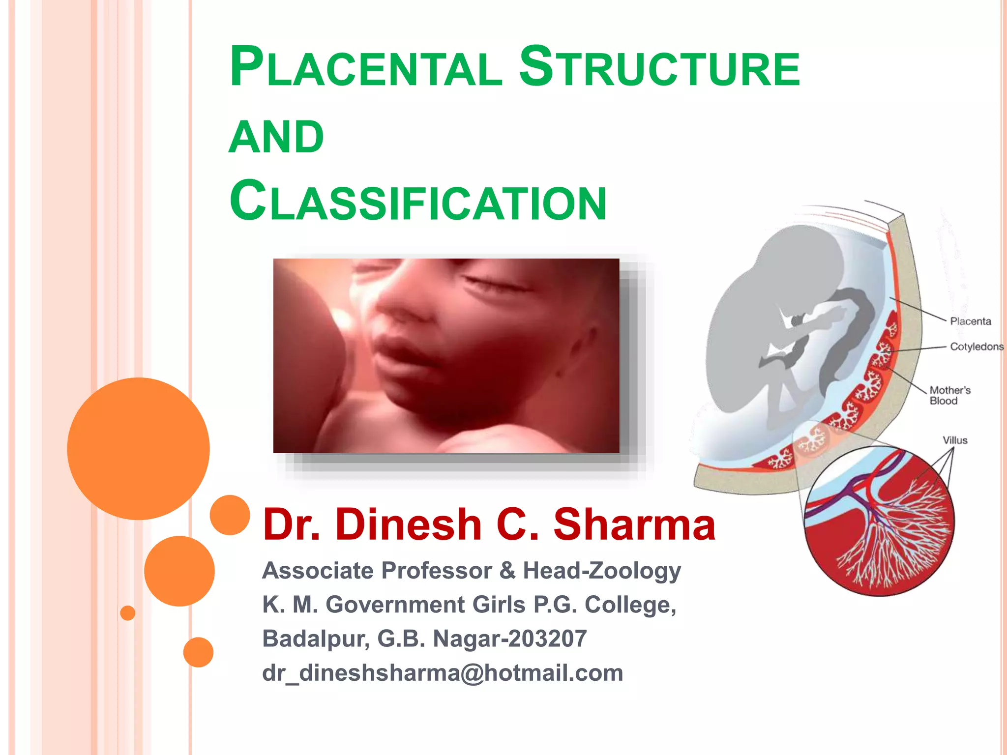
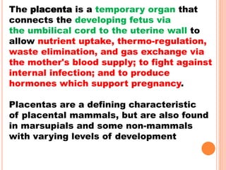
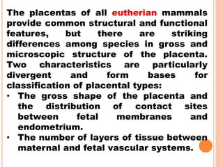
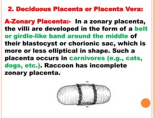
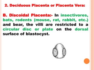
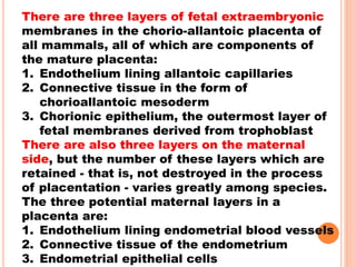
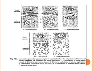
1. The document discusses the structure and classification of placentas. It describes the different types of placentas found across mammals based on their gross shape, layers between maternal and fetal blood, and histological structure.
2. The major placenta types discussed are epithelio-chorial (found in marsupials and some ungulates), syndesmo-chorial (ruminant ungulates), endothelio-chorial (carnivores), haemochorial (primates and bats), and haemo-endothelial (rodents). Each type involves a different number of layers separating maternal and fetal blood and tissues.
3. Placentas are also classified
In all viviparous animals, embryonic development takes place inside the uterus of the mother, because the eggs are microlecithal and the amount of stored yolk is not sufficient for the developing embryo. Such embryos get attached to the uterine wall to draw essential substances from the maternal circulation through the placenta.
The term implantation is used to describe the attachment of the developing embryo to the endometrium.
After fertilization, the embryo reaches the uterus in the blastocyst stage. Then attached to the wall of the uterus. Though the implantation may occur at any period between the sixth to the tenth day after the fertilization generally it occurs on the seventh day after fertilization.
presentation on oogenesis of fertilisation process full details about it u will never find it anywhere else have full details about the ovum formation polar bodies and everything . so explore here
Vittelogenesis is a word developed from Latin vitellus-yolk, and genero-produce
Vitellogenesis (also known as yolk deposition) is the process of yolk formation via nutrients being deposited in the oocyte, or female germ cell involved in reproduction of lecithotrophic organisms. In insects, it starts when the fat body stimulates the release of juvenile hormones and produces vitellogenin protein.
Yolks is the most usual form of food storage in the egg.
Yolks appear in the oocyte in the secondary period of their growth called vittelogenesis.
Thus,the formation and deposition of yolks is known as vittelogenesis
Characteristic
Yolks is a complex variable assembled component.
The principle component are protein,phospholipid and fats in different combination.
Depending upon these component yolks is distinguished into protein yolks and fatty acid
For eg- the avian contain 48.19% water , 16.6 % protein, 32.6% phospholipids and fats and 1% carbohydrates.
The Puerperium & Lactation Early Embryogenesis & Materna.docxarnoldmeredith47041
The Puerperium & Lactation
Early Embryogenesis &
Maternal Recognition of Pregnancy
Ovulation & Fertilization
Cyclicity
Regulation of
Reproduction
Tract Function
Puberty
Prenatal
Development
Spermatogenesis
Regulat ion of
Reproduction
Tract Function
Puberty
Prenatal
Development
Take Home Message
Gestation is the period of time that a female is pregnant. During gestation, the
placenta forms a major organ of pregnancy that provides an inteJface for metabolic
exchange between the dam and the f etus. Placentas are described mmplwlogically
according to the distribution of villi on the chorionic smface ami the degree of separa..:.
tion between matemal and fetal blood. The placenta is also an endocrine organ that
secretes hormones responsible for: 1) maintenance of pregnancy; 2) stimulation of the
matemal mammmy gland and 3) ensures fetal growth. Parturition is brought about
by secretion of fetal corticoitls and requires removal of the progesterone block. Par-
turition consists of three stages. They are: 1) initiation of myometrial contractions;
2) expulsion ofthe f etus and 3) e.:\:pulsion oftlzefetalmembranes.
The word gestation literally means "the act
of carry ing or being carried". Thus, gestation means
the action or process of carrying or being carried' in
the uterus between conception and birth . · Gest ation
and pregnancy are synonymous and thus, gestation
length means the length of pregnancy. Attachment
of the conceph1s to form an intimate, but temporary,
relationship with the uterus is an evolutionary step that
provides significant advantage to the conceph1s . The
phenomenon of intrauterine development ensures that
the developing conceptus will receive adequate nutri-
tion and protection during its development. In contrast,
lower fon11S of animals lay eggs (oviparous). The
survival of potential offspring of oviparous animals
is jeopardized because the female cannot completely
protect the eggs from environmental and predatmy dan-
ger. Thus, from an evolutionary perspective, eutherian
mammals (mammals with a placenta), are "equipped"
with an in-utero protection mechanism that is highly
successful after the placenta is formed.
The final prepartum steps of
reproduction are:
• formation of a placenta
• acquisition of endocrine function of
the placenta
• initiation of parturition
The term implantation is often used to mean
attachment of the placental membranes to the endo-
metrium in most animals. Achmlly, true implantation
is a phenomenon in humans in which the conceptus
"buries" itself into the uterine endometrium. The con-
ceptus temporarily disappears beneath the surface. In
most other species, the conceph1s does not truly implant,
but rather attaches to the endometrial surface and never
disappears from the luminal compartment.
The placenta is an organ of metabolic inter-
change between the conceph1s and the dam. It is also
an endocrine organ. The placenta is co.
A chart showing the fate of each part of an early embryo, in a particular blastula stage is called fate maps. It is done because the correct interpretation of gastrulation is impossible without the knowledge of the position which are the presumptive germinal layers (Ectoderm, Mesoderm and Endoderm) occupy in blastula.
Fate mapping is a method used in developmental biology to study the embryonic origin of various adult tissues and structures. The "fate" of each cell or group of cells is mapped onto the embryo, showing which parts of the embryo will develop into which tissue. When carried out at single-cell resolution, this process is called cell lineage tracing. It is also used to trace the development of tumors.
DNA sequencing is the process of determining the sequence of nucleotides (A, T, G, and C) in the DNA. It includes method or technology that is used to determine the order of the four bases: adenine, thymine, guanine and cytosine.
The chain-termination method developed by Frederick Sanger and coworkers in 1977. This method used fewer toxic chemicals and lower amounts of radioactivity than the Maxam and Gilbert method. Because of its comparative ease, the Sanger method was soon automated and was the method used in the first generation of DNA sequencers.
published a DNA sequencing method in 1977 based on chemical modification of DNA and subsequent cleavage at specific bases. Also known as chemical sequencing, this method allowed purified samples of double-stranded DNA to be used without further cloning.
Maxam-Gilbert sequencing requires radioactive labeling at one 5' end of the DNA and purification of the DNA fragment to be sequenced. Chemical treatment then generates breaks at a small proportion of one or two of the four nucleotide bases in each of four reactions (G, A+G, C, C+T). The concentration of the modifying chemicals is controlled to introduce on average one modification per DNA molecule. Thus a series of labeled fragments is generated, from the radiolabeled end to the first "cut" site in each molecule. The fragments in the four reactions are electrophoresed side by side in denaturing acrylamide gels for size separation. To visualize the fragments, the gel is exposed to X-ray film for autoradiography, yielding a series of dark bands each corresponding to a radiolabeled DNA fragment, from which the sequence may be inferred.
Cloning is the process of producing genetically identical individuals of an organism either naturally or artificially.
It is the process of taking genetic information from one living thing and creating identical copies of it. The copied material is called a clone.
Nature has been doing it for millions of years. For example, identical twins have almost identical DNA, and asexual reproduction in some plants and organisms can produce genetically identical offspring.
Cloning in biotechnology refers to the process of creating clones of organisms or copies of cells or DNA fragments (molecular cloning).
Bacteriophage- types, structure and morphology of t4 phage, morphogenesisDr. Dinesh C. Sharma
Escherichia virus T4 is a species of bacteriophages that infect Escherichia coli bacteria. It is a member of virus subfamily Tevenvirinae (not to be confused with T-even bacteriophages, which is an alternate name of the species). T4 is capable of undergoing only a lytic lifecycle and not the lysogenic lifecycle.
Each cell of a multicellular organism contain the same genetic material, but the expression of the gene is different in different type of cell group. On the basis of expression requirement they are grouped in to
Structural Gene- Mostly expressed once in a life
Vital Gene- Involved in of vital biochemical processes such as respiration and need to be expressed all the time
Functional Gene- Genes are not expressed all the time. They are switched on an off at need
The regulation of Gene required in case of functional gene and its explained by Francois Jacob, Jacques Monod and Andre Lwoff (Nobal Prize in 1961)
From studies and predictions such as Dreyer and Bennett's, it shows that the light chains and heavy chains are encoded by separate multigene families on different chromosomes. They are referred to as gene segments and are separated by non-coding regions. The rearrangement and organization of these gene segments during the maturation of B cells produce functional proteins. The entire process of rearrangement and organization of these gene segments is the vital source where our body immune system gets its capabilities to recognize and respond to variety of antigens.
The cells of the B line synthesize immunoglobulins. They are either produced at a membrane (on the surface of the B-lymphocytes) or are secreted (by the plasmocytes)
Theory of preformation,
Epigenetic theory,
Theory of pengenesis,
Recapitulation theory,
Germplasm theory,
Mosaic theory,
Regulated theory,
Gradient theory
Theory of organizers.
Sericulture is the cultivation of silkworms to produce silk. Bombyx mori (the caterpillar of the domesticated silk moth) is the most widely used species of silkworms.
Francesca Gottschalk - How can education support child empowerment.pptxEduSkills OECD
Francesca Gottschalk from the OECD’s Centre for Educational Research and Innovation presents at the Ask an Expert Webinar: How can education support child empowerment?
Read| The latest issue of The Challenger is here! We are thrilled to announce that our school paper has qualified for the NATIONAL SCHOOLS PRESS CONFERENCE (NSPC) 2024. Thank you for your unwavering support and trust. Dive into the stories that made us stand out!
Introduction to AI for Nonprofits with Tapp NetworkTechSoup
Dive into the world of AI! Experts Jon Hill and Tareq Monaur will guide you through AI's role in enhancing nonprofit websites and basic marketing strategies, making it easy to understand and apply.
Macroeconomics- Movie Location
This will be used as part of your Personal Professional Portfolio once graded.
Objective:
Prepare a presentation or a paper using research, basic comparative analysis, data organization and application of economic information. You will make an informed assessment of an economic climate outside of the United States to accomplish an entertainment industry objective.
2024.06.01 Introducing a competency framework for languag learning materials ...Sandy Millin
http://sandymillin.wordpress.com/iateflwebinar2024
Published classroom materials form the basis of syllabuses, drive teacher professional development, and have a potentially huge influence on learners, teachers and education systems. All teachers also create their own materials, whether a few sentences on a blackboard, a highly-structured fully-realised online course, or anything in between. Despite this, the knowledge and skills needed to create effective language learning materials are rarely part of teacher training, and are mostly learnt by trial and error.
Knowledge and skills frameworks, generally called competency frameworks, for ELT teachers, trainers and managers have existed for a few years now. However, until I created one for my MA dissertation, there wasn’t one drawing together what we need to know and do to be able to effectively produce language learning materials.
This webinar will introduce you to my framework, highlighting the key competencies I identified from my research. It will also show how anybody involved in language teaching (any language, not just English!), teacher training, managing schools or developing language learning materials can benefit from using the framework.
The French Revolution, which began in 1789, was a period of radical social and political upheaval in France. It marked the decline of absolute monarchies, the rise of secular and democratic republics, and the eventual rise of Napoleon Bonaparte. This revolutionary period is crucial in understanding the transition from feudalism to modernity in Europe.
For more information, visit-www.vavaclasses.com
How to Make a Field invisible in Odoo 17Celine George
It is possible to hide or invisible some fields in odoo. Commonly using “invisible” attribute in the field definition to invisible the fields. This slide will show how to make a field invisible in odoo 17.
2. The placenta is a temporary organ that
connects the developing fetus via
the umbilical cord to the uterine wall to
allow nutrient uptake, thermo-regulation,
waste elimination, and gas exchange via
the mother's blood supply; to fight against
internal infection; and to produce
hormones which support pregnancy.
Placentas are a defining characteristic
of placental mammals, but are also found
in marsupials and some non-mammals
with varying levels of development
3. The placentas of all eutherian mammals
provide common structural and functional
features, but there are striking
differences among species in gross and
microscopic structure of the placenta.
Two characteristics are particularly
divergent and form bases for
classification of placental types:
• The gross shape of the placenta and
the distribution of contact sites
between fetal membranes and
endometrium.
• The number of layers of tissue between
maternal and fetal vascular systems.
4. On the basis of mode of Impanation
Placenta
Superficial
Carnivore,
Dog,
Monkey
Eccentric
Squirrel, Rat
Interstitial
Hedgehog, Ape,
Human, Bat,
Guinea pig
5. 1. Superficial Implantation:-Growth of the
chorionic sac brings it into contact with
the lining of the main uterine cavity. This
type of implantation is called central
implantation, e.g., ungulates, carnivores,
monkey.
2. Eccentric Implantation:-The chorionic
sac lies for a time in a fold or pocket
which looses off from the main cavity,
e.g., beaver, rat, squirrel.
3. Interstitial Implantation:-The chorionic
sac penetrates into the substance of the
uterine lining, e.g., hedgehog, guinea pig,
some bats, ape and man.
8. 1. Non-Deciduous Placenta or Semiplacenta:
In most mammals, the implantation is superficial, i.e,
the blastocyst lies in the cavity of the uterus in
contact with the uterine wall. The contact may be
made more intimate by the surface of the blastocyst
by forming finger-like outgrowths which penetrate
into depressions in the wall of the uterus. Such
outgrowths are initially formed by the trophoblast
(i.e., the epithelial layer covering the blastocyst), but
later on the connective tissue and blood vessels
invade the outgrowths.
These outgrowths are called chorionic villi, the blood
vessels of chorionic villi are the branches of allantoic
blood vessels in case of chorio-allantoic placenta. (In
chorio-vitelline placenta, vitelline blood vessels give
their branches to chorionic villi).
9. At the time of birth, when parturition (the
separation of the foetus and its membranes from
the mother’s body) occurs, the chorionic villi are
simply drawn out from the depressions in the
wall of the uterus and, thus, maternal and foetal
tissues are separated without further damage to
the uterine wall and no bleeding occurs.
This type of placenta is called non-deciduate or
non- deciduous placenta and is found in pigs,
cattle and some other mammals. Further, the
chorionic villi of a non-deciduate placenta,
because lie in apposition with the endometrium,
but, do not fuse with it, so such a placenta is
also called semiplacenta.
10. 2. Deciduous Placenta or Placenta Vera:
In other mammals, however, the degree of intimacy
between maternal and foetal tissues becomes further
increased. The wall of the uterus becomes eroded to
various degrees through the action of the trophoblast and
the embryonic tissues penetrate into the uterine wall,
establishing a more intimate contact and facilitating the
passage of substances from the mother to the foetus and
from the foetus to the mother.
Here because the chorionic villi fuse with the eroded
uterine mucosa, such placenta is called placenta vera
(true placenta). At the end of pregnancy the uterine wall is
no longer intact and when the foetus with its membranes
including the chorion is removed, more or less extensive
haemorrhage from the uterine wall ensues (i.e., at birth,
when such placenta is discharged, the uterine lining also
tears away with some bleeding). Such a type of placenta
found in higher eutherian mammals and is called
deciduate or deciduous placenta.
11. The maternal tissues which are
expelled at birth in the case of
deciduate placenta are called
deciduae. The haemorrhage at
parturition is normally stopped by
the same mechanism as serves for
the expulsion of the newborn, the
contraction of the muscular wall of
the uterus constricts the blood
vessels and, thus, slows down the
flow of blood, until clotting of the
blood stops the haemorrhage
altogether.
12. 3. Contra-Deciduate Placenta:-
In Perameles and Talpa (mole),
somewhat modified type of
deciduate placenta occurs,
which is called contra-
deciduate placenta. In such
case, not only there is a loss of
maternal tissue but also of the
foetal portion of the placenta,
both of which absorbed in situ
by maternal leucocytes.
13. 1. Non-Deciduous Placenta or Semiplacenta:
A. Diffuse Placenta:- In some mammals
(e.g., ungulates, pig, sow, mare, horse,
lemur, etc.) the chorionic villi remain
scattered all over the surface of the
chorion and their placentae are
correspondingly expensive.
14. 1. Non-Deciduous Placenta or Semiplacenta:
B. Cotyledonary Placenta:- In a
cotyledonary placenta, the villi are found
in groups or patches, while the rest of
the chorion surface is smooth. The
rosettes or patches of villi are called
cotyledons, and the placenta of this type
is found in ruminants (cud-chewing),
ungulates such as cattle, sheep and
deer.
15. 1. Non-Deciduous Placenta or Semiplacenta:
C. Intermediate Placenta:-
It is a rare type, it shows free villi
on cotyledons. Villi are arranged in
cotyledons as well as scattered.
Hence it is called intermediate type
placenta
In these three types of placenta
during parturition the foetus will
not damage uterus.
Camel and giraffe
16. 2. Deciduous Placenta or Placenta Vera:
A-Zonary Placenta:- In a zonary placenta,
the villi are developed in the form of a belt
or girdle-like band around the middle of
their blastocyst or chorionic sac, which is
more or less elliptical in shape. Such a
placenta occurs in carnivores (e.g., cats,
dogs, etc.). Raccoon has incomplete
zonary placenta.
17. 2. Deciduous Placenta or Placenta Vera:
B. Discoidal Placenta:- In insectivores,
bats, rodents (mouse, rat, rabbit, etc.)
and bear, the villi are restricted to a
circular disc or plate on the dorsal
surface of blastocyst.
18. 2. Deciduous Placenta or Placenta Vera:
C. Metadiscoidal Placenta:- In primates also
discoidal placenta is found but of special type,
i.e., chorionic villi are at first scattered but later
on become restricted to one or two discs.
a-Monodiscoidal Thus, in man the placenta has
a single disc-shaped villous area and is called
monodiscoidal placenta.
b- Bidiscoidal- In the monkeys, the placenta
consists of two disc-shaped villous areas and
such a placenta is called bidiscoidal placenta.
19. C. Classification of Placenta
According to Histology:
On histological basis, following
types of mammalian placentae
have been recognised:
20. In placenta formation six tissue or
membranes participated :
(i) The endothelium of the maternal blood vessels;
(ii) Endometrial connective tissue (mesenchyme);
(iii) Uterine epithelium;
(iv) The ectoderm of the chorion or chorionic epithelium;
(v) Chorionic connective tissue (foetal mesenchyme)
(vi) The endothelium of foetal blood vessels.
22. There are three layers of fetal extraembryonic
membranes in the chorio-allantoic placenta of
all mammals, all of which are components of
the mature placenta:
1. Endothelium lining allantoic capillaries
2. Connective tissue in the form of
chorioallantoic mesoderm
3. Chorionic epithelium, the outermost layer of
fetal membranes derived from trophoblast
There are also three layers on the maternal
side, but the number of these layers which are
retained - that is, not destroyed in the process
of placentation - varies greatly among species.
The three potential maternal layers in a
placenta are:
1. Endothelium lining endometrial blood vessels
2. Connective tissue of the endometrium
3. Endometrial epithelial cells
26. 1. Epithelio-Chorial Placenta:
The epithelio-chorial type placenta is most primitive type
and it is found in marsupials, ungulates (pig, horse, cow,
cattle, etc.) and lemurs.
In this case, placenta is formed of six tissue or
membranes:
I. The endothelium of the maternal blood vessels;
II. Endometrial connective tissue (mesenchyme);
III. Uterine epithelium;
IV. The ectoderm of the chorion or chorionic epithelium;
V. Chorionic connective tissue (foetal mesenchyme) and
VI. The endothelium of foetal blood vessels.
Because, the immediate contact of the two halves of the
placenta involves chorionic epithelium and uterine
epithelium, this type of placenta is called epithelio-
chorial placenta. The villi of an epithelio-chorial
placenta, push in the wall of uterus and lie in pocket-like
depressions of the uterine wall.
27. The foetal chorion is in contact with epithelium of the uterus
hence it is called epithelio-chorial placenta. In between foetal,
maternal parts six layers are present. If all the six layers are
present the placenta is called epithelio-chorial placenta.
28. 2. Syndesmo-Chorial Placenta:
In the ruminant ungulates (cattle, sheep), the
foetal and maternal components are fused so
intimately as to result in a destruction of the
uterine epithelium, thus, bringing the chorion
into contact with the connective tissue of the
uterine mucosa. Only five barriers or tissues,
therefore, lie between the two (viz., foetal and
uterine) blood streams. This type of placenta is
called syndesmo-chorial placenta. The allanto-
chorianic vith will pierce into the uterus of the
mother, the chorion will come in contact with
syndesmose of mother’s uterus. Hence iti s
called syndesmose chorial.
29. 3. Endothelio-Chorial Placenta:- In
carnivores (dogs, cats, bears, etc.),
the uterine mucosa is also reduced
and the chorionic epithelium comes
in contact with endoethelial walls of
the maternal (uterine) blood
vessels. In such a case, therefore,
there lies only four barriers between
the foetal and maternal blood
streams. This type of placenta is
called endothelio-chorial placenta.
30. The chorion of the
foetus will come in
contact with the
endothelim of
mother ‘s uterus,
hence it is called
endothelio-chorial
placenta.
31. 4. Haemo-Chorial Placenta:- In the
haemo-chorial placenta of primates,
insectivores (moles, shrews), and
chiropterans (bats), a reduction of the
barriers to three occurs, i.e., the
endothelial walls of maternal (uterine)
blood vessels also disappear and the
chorionic epithelium is bathed directly in
maternal blood sinuses. Actually, the
chorionic villi are surrounded by spaces
(sinuses) devoid of endothelial lining,
into which maternal blood enters
through the uterine arteries flows out
through the uterine veins.
32. The placental
connections are
more intimate. The
chorion of foetus
will float in the
blood pools of
mother’s uterus.
Hence it is called
haemochorial
placenta
33. 5. Haemo-Endothelial Placenta:- In
haemo-endothelial placenta of higher
rodents (rat, guinea pig, rabbit), the
number of barriers between the
maternal and foetal blood streams is
further reduced to two. In them, the
chorionic villi lose their epithelial and
connective tissue layers to such a
degree that, in most places, the bare
endothelial lining of their blood vessels
alone separates the foetal blood from
the maternal blood sinuses.