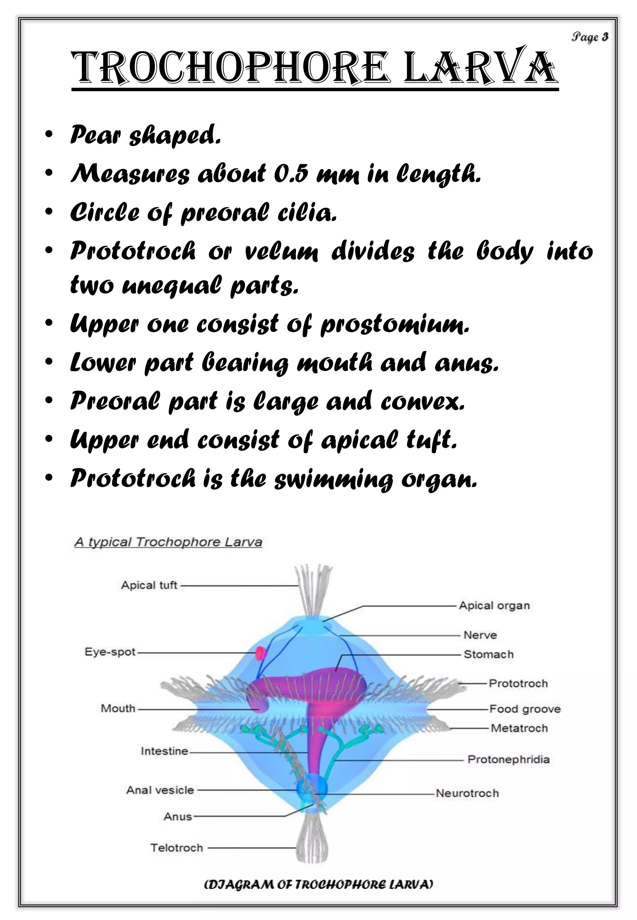
A PROJECT ON
“THE LARVAL FORMS OF
MOLLUSCA”
BY-
Sambit Bhai Patel
CONTENTS
1. INTRODUCTION TO MOLLUSCS _ _ _ _ _ _ _ _ _ _ _ _ _ _ _ page 1
2. TYPES OF LARVAL FORMS IN MOLLUSCS _ _ _ _ _ _ _ _ _ _ _ page 2
3. TROCHOPHORE LARVA _ _ _ _ _ _ _ _ _ _ _ _ _ _ _ _ _ _ page 3
4. VALIGER LARVA _ _ _ _ _ _ _ _ _ _ _ _ _ _ _ _ _ _ _ _ _ _ page 5
5. GLOCHIDIUM LARVA _ _ _ _ _ _ _ _ _ _ _ _ _ _ _ _ _ _ _ _ page 7
6. CONCLUSION _ _ _ _ _ _ _ _ _ _ _ _ _ _ _ _ _ _ _ _ _ _ _ _ page 9
7. SOURCE _ _ _ _ _ _ _ _ _ _ _ _ _ _ _ _ _ _ _ _ _ _ _ _ _ _ _page 10
introduction
• Mollusca = mollis (L.) = soft bodies.
• 1st used by Aristotle to the cuttle fish .
• Second largest phylum of animal
kingdom.
• Animal having soft bodies with in shell
and ventral muscular foot.
• Found in all possible habitat except in the
air.
• Most of them are marine.
• Dioecious or monoecious.
• Fertilization either internal or external.
• Development is either direct or indirect.
• Direct development -- no larval stage ,
indirect development– with larval stage.
Types of larva
• 3 types of larva are found in Mollusca.
TROCHOPHORE VELIGER GLOCHIDIUM
Trochophore larva
• Pear shaped.
• Measures about 0.5 mm in length.
• Circle of preoral cilia.
• Prototroch or velum divides the body into
two unequal parts.
• Upper one consist of prostomium.
• Lower part bearing mouth and anus.
• Preoral part is large and convex.
• Upper end consist of apical tuft.
• Prototroch is the swimming organ.
• Comprises mouth.
• Oesophagus.
• Stomach.
• Intestine (mesenteric).
• Sides of the mouth two ciliated elevations
present each consisting of a single cell.
• Lower end bearing a bunch of cilia called
“telotroch”.
• Generally planktonic and feeds on tiny
suspended particle, living or dead.
• Body divided in 3 regions -- pretrochal
-- pygidium
--growth zone.
• Pretrochal region consist of apical plate,
prototroch , the area surround the mouth.
• Pygidium consist of telotroch and the area
surrounding the anus.
• Growth zone lies between the mouth and
telotroch.
• The trochophore larva later develops into a
veliger larva
Veliger larva
• Preoral ciliate area.
• Velum begin to protrude on both sides as a
bilobed flap.
• Very delicate.
• Anterior end of the larva provided with eyes
and Tentacles.
• Larva has a shell.
• Velum is developed from the prototroch of
the trochophore.
Page 5
• Larval heart and kidney present which is
situated at the anterior end of the body
immediately behind the velum.
• Statocyst and gill-rudiments present.
• Long cilia of the velum function
Locomotion.
• suspension feeding.
• Alimentary canal is complete.
• Anus is shifted to anterior side.
• Foot usually bearing an operculum.
• During development of veliger larva
torsion occur.
Glochidium larva
• Glochidium larva enclosed by two valves.
• Each edge of which bears a hook.
• Shell valves cover a larval mantle.
• Bears four groups of sensory bristles.
• Rudimentary foot is present.
• Attached a long adhesive thread called
“Byssal thread”.
• Neither mouth nor anus.
• Measures from 0.1 mm to 0.5mm .
• Highly modified for a parasitic existence on
fish.
• They clamp on the body and other part of
the body.
• The larval mantle contains phagocytic cells
that feed on the tissue of host and obtain
nutrition for development.
• This period lasts for about 10-30 days.
• In the mean time the parasite is surrounded
by the overgrowth of skin of fish forming a
cyst.
• Some of the larger freshwater molluscans
may produce as many as 30,00,000
glochidia.
CONCLUSION
• Free-swimming larva are usually formed
when the adult is fixed.
• Internal parasites generally have a stage
which may be called larval stage in which
they are transferred either by active or
passive migration to a new host.
source
• Google
• Modern textbook of
zoology invertebrate book
by R.L. Kotpal.
• Biology of Mollusca book
by D.R. Khanna &P.R. Yadav.
• Wikipedia.
• www.biologyjunction.com/
mollusca
• ecoursesonline.iasri.res.in/m
od/resource/view.php?id=85576
• Invertebrate zoology book
by barnes,fox,ruppert.
Larval form of mollusca

Larval form of mollusca

  • 1.
    A PROJECT ON “THELARVAL FORMS OF MOLLUSCA” BY- Sambit Bhai Patel
  • 2.
    CONTENTS 1. INTRODUCTION TOMOLLUSCS _ _ _ _ _ _ _ _ _ _ _ _ _ _ _ page 1 2. TYPES OF LARVAL FORMS IN MOLLUSCS _ _ _ _ _ _ _ _ _ _ _ page 2 3. TROCHOPHORE LARVA _ _ _ _ _ _ _ _ _ _ _ _ _ _ _ _ _ _ page 3 4. VALIGER LARVA _ _ _ _ _ _ _ _ _ _ _ _ _ _ _ _ _ _ _ _ _ _ page 5 5. GLOCHIDIUM LARVA _ _ _ _ _ _ _ _ _ _ _ _ _ _ _ _ _ _ _ _ page 7 6. CONCLUSION _ _ _ _ _ _ _ _ _ _ _ _ _ _ _ _ _ _ _ _ _ _ _ _ page 9 7. SOURCE _ _ _ _ _ _ _ _ _ _ _ _ _ _ _ _ _ _ _ _ _ _ _ _ _ _ _page 10
  • 3.
    introduction • Mollusca =mollis (L.) = soft bodies. • 1st used by Aristotle to the cuttle fish . • Second largest phylum of animal kingdom. • Animal having soft bodies with in shell and ventral muscular foot. • Found in all possible habitat except in the air. • Most of them are marine. • Dioecious or monoecious. • Fertilization either internal or external. • Development is either direct or indirect. • Direct development -- no larval stage , indirect development– with larval stage.
  • 4.
    Types of larva •3 types of larva are found in Mollusca. TROCHOPHORE VELIGER GLOCHIDIUM
  • 5.
    Trochophore larva • Pearshaped. • Measures about 0.5 mm in length. • Circle of preoral cilia. • Prototroch or velum divides the body into two unequal parts. • Upper one consist of prostomium. • Lower part bearing mouth and anus. • Preoral part is large and convex. • Upper end consist of apical tuft. • Prototroch is the swimming organ.
  • 6.
    • Comprises mouth. •Oesophagus. • Stomach. • Intestine (mesenteric). • Sides of the mouth two ciliated elevations present each consisting of a single cell. • Lower end bearing a bunch of cilia called “telotroch”. • Generally planktonic and feeds on tiny suspended particle, living or dead. • Body divided in 3 regions -- pretrochal -- pygidium --growth zone. • Pretrochal region consist of apical plate, prototroch , the area surround the mouth. • Pygidium consist of telotroch and the area surrounding the anus. • Growth zone lies between the mouth and telotroch. • The trochophore larva later develops into a veliger larva
  • 7.
    Veliger larva • Preoralciliate area. • Velum begin to protrude on both sides as a bilobed flap. • Very delicate. • Anterior end of the larva provided with eyes and Tentacles. • Larva has a shell. • Velum is developed from the prototroch of the trochophore. Page 5
  • 8.
    • Larval heartand kidney present which is situated at the anterior end of the body immediately behind the velum. • Statocyst and gill-rudiments present. • Long cilia of the velum function Locomotion. • suspension feeding. • Alimentary canal is complete. • Anus is shifted to anterior side. • Foot usually bearing an operculum. • During development of veliger larva torsion occur.
  • 9.
    Glochidium larva • Glochidiumlarva enclosed by two valves. • Each edge of which bears a hook. • Shell valves cover a larval mantle. • Bears four groups of sensory bristles. • Rudimentary foot is present. • Attached a long adhesive thread called “Byssal thread”. • Neither mouth nor anus. • Measures from 0.1 mm to 0.5mm .
  • 10.
    • Highly modifiedfor a parasitic existence on fish. • They clamp on the body and other part of the body. • The larval mantle contains phagocytic cells that feed on the tissue of host and obtain nutrition for development. • This period lasts for about 10-30 days. • In the mean time the parasite is surrounded by the overgrowth of skin of fish forming a cyst. • Some of the larger freshwater molluscans may produce as many as 30,00,000 glochidia.
  • 11.
    CONCLUSION • Free-swimming larvaare usually formed when the adult is fixed. • Internal parasites generally have a stage which may be called larval stage in which they are transferred either by active or passive migration to a new host.
  • 12.
    source • Google • Moderntextbook of zoology invertebrate book by R.L. Kotpal. • Biology of Mollusca book by D.R. Khanna &P.R. Yadav. • Wikipedia. • www.biologyjunction.com/ mollusca • ecoursesonline.iasri.res.in/m od/resource/view.php?id=85576 • Invertebrate zoology book by barnes,fox,ruppert.