Coagulants and anticoagulants
•Download as DOCX, PDF•
34 likes•18,515 views

This document discusses coagulants and anticoagulants used in medicinal chemistry. It provides background on blood coagulation pathways and factors. Coagulants discussed include vitamin K, fibrinogen, antihaemophilic factor, desmopressin, and others. Vitamin K is important for the synthesis of clotting factors and its deficiency can cause bleeding. Anticoagulants lower blood coagulability and include heparins, low molecular weight heparins, danaparoid, lepirudin, ancrod, and oral coumarin derivatives like warfarin.

More Related Content
What's hot
What's hot (20)
Viewers also liked
Viewers also liked (20)
Similar to Coagulants and anticoagulants
Similar to Coagulants and anticoagulants (20)
Recently uploaded
Recently uploaded (20)
Coagulants and anticoagulants
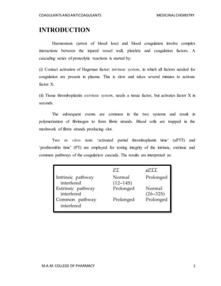
- 1. COAGULANTSANDANTICOAGULANTS MEDICINALCHEMISTRY M.A.M. COLLEGE OF PHARMACY 1 INTRODUCTION Haemostasis (arrest of blood loss) and blood coagulation involve complex interactions between the injured vessel wall, platelets and coagulation factors. A cascading series of proteolytic reactions is started by: (i) Contact activation of Hageman factor: intrinsic system, in which all factors needed for coagulation are present in plasma. This is slow and takes several minutes to activate factor X. (ii) Tissue thromboplastin: extrinsic system, needs a tissue factor, but activates factor X in seconds. The subsequent events are common in the two systems and result in polymerization of fibrinogen to form fibrin strands. Blood cells are trapped in the meshwork of fibrin strands producing clot. Two in vitro tests ‘activated partial thromboplastin time’ (aPTT) and ‘prothrombin time’ (PT) are employed for testing integrity of the intrinsic, extrinsic and common pathways of the coagulation cascade. The results are interpreted as:
- 2. COAGULANTSANDANTICOAGULANTS MEDICINALCHEMISTRY M.A.M. COLLEGE OF PHARMACY 2 Most clotting factors are proteins present in plasma in the inactive (zymogen) form. By partial proteolysis they themselves become an active protease and activate the next factor. In addition to its critical role in cleaving and polymerizing fibrinogen, thrombin activates many upstream factors (especially f. XI, VIII and V) of the intrinsic and common pathways—amplifying its own generation and continuation of clot formation. It is also a potent activator of platelets. On the other hand, factors like antithrombin, protein C, protein S, antithromboplastin and the fibrinolysin system tend to oppose coagulation and lyse formed clot. Thus, a check and balance system operates to maintain blood in a fluid state while in circulation and allows rapid hemostasis following injury. Roman Numerical Nomenclature of Blood-Clotting Factors and Some Common Synonyms
- 3. COAGULANTSANDANTICOAGULANTS MEDICINALCHEMISTRY M.A.M. COLLEGE OF PHARMACY 3 The coagulation cascade. The vit. K dependent factors have been encircled,* Inactivated by heparin a—activated form; Pl.Ph.—Platelet phospholipid; HMW—High molecular weight. COAGULANTS These are substances which promote coagulation, and are indicated in haemorrhagic states. Fresh whole blood or plasma provide all the factors needed for coagulation and are the best therapy for deficiency of any clotting factor; also they act immediately. Other drugs used to restore haemostasis are: 1. Vitamin K K1 (from plants, : Phytonadione fat-soluble) (Phylloquinone) K3 (synthetic) *Fat-soluble : Menadione, Acetomenaphthone *Water-soluble : Menadione sod. Bisulfite : Menadione sod.diphosphate
- 4. COAGULANTSANDANTICOAGULANTS MEDICINALCHEMISTRY M.A.M. COLLEGE OF PHARMACY 4 2. Miscellaneous Fibrinogen (human), Antihaemophilic factor Desmopressin, Adrenochrome monosemicarbazone Rutin, Ethamsylate VITAMIN K It is a fat-soluble dietary principle required for the synthesis of clotting factors. Dam (1929) produced bleeding disorder in chicken by feeding deficient diet. This was later found to be due to decreased concentration of prothrombin in blood and that it could be cured by a fat soluble fraction of hog liver. This factor was called Koagulations vitamin (vit K) and soon its structure was worked out. A similar vitamin was isolated in 1939 from alfalfa grass and labelled vit K1, while that from sardine (sea fish) meal was labelled K2. Synthetic compounds have been produced and labelled K3. CHEMISTRY Chemically, all vitamin Ks are 2-methyl 1,4-naphthoquinone derivatives containing variable aliphatic side chains at C3. Phylloquinone invariably contains a phytyl side chain. Menaquinones are a series of compounds that have a longer side chain with more unsaturation. This side chain may be composed of 1 to 13 prenyl (isoprenyl) units.
- 5. COAGULANTSANDANTICOAGULANTS MEDICINALCHEMISTRY M.A.M. COLLEGE OF PHARMACY 5 Many other closely related compounds possess vitamin K activity. Of particular historical note are the water-soluble menadione (2-methyl-1,4-naphthoquinone; vitamin K3), which is no longer marketed because of toxicities, and menadiol (2- methylnaphthalene-1,4-diol; vitamin K4). Menadione contains the naphthoquinone ring without any attached prenyls and can be thought of as MK-0. Menadiol is menadione with the two keto groups reduced to hydroxyls. Both compounds are bioactivated to the active form through alkylation. Phylloquinone naturally occurs as a trans-isomer and has an R,R,E configuration. The synthetic, commercially available form is a mixture of cis- and transisomers, with no more than 20% cis. Fig: 1 Fig: 2 Fig: 1-Phylloquinone (2–Methyl–3–phytyl–1,4–naphthoquinone). Fig: 2-Menaquinone general structure. Menaquinone-4 (n=4) and menaquinone-6 (n=6) are the most common forms. Mechanism of action The accepted mechanism for vitamin K is to function as a cofactor in the posttranslational synthesis of -carboxyglutamic acid (Gla) from glutamic acid (Glu) residues. The discovery of Gla in clarified the mechanism of vitamin K and led to the identification of additional vitamin K–dependent proteins. All vitamin K–dependent proteins contain propeptide possessing Glu residues. Vitamin K participates in the carboxylation of several specific Glu residues to form Gla residues, which function as Ca+2-binding sites on the vitamin K–dependent proteins . Further, the Gla residues are
- 6. COAGULANTSANDANTICOAGULANTS MEDICINALCHEMISTRY M.A.M. COLLEGE OF PHARMACY 6 found in essentially the same region, near the amino terminus, and this region is called the Gla domain. Of the 14 known vitamin K–dependent proteins, four function as procoagulants, three function as anticoagulants, two are involved in bone and extracellular matrix homeostasis, one in cell proliferation, and four represent transmembrane proteins whose function has not been identified. All of these proteins have considerable structural homology. All contain 10 to 12 Gla residues, which are important for their proper activity. For example, prothrombin has 10 Gla residues; loss of just two residues decreases its coagulation activity by 80%. Figure: Calcium binding to Gla residues on vitamin K–dependent proteins. This synthesis of Gla residues from Glu residues requires reduced vitamin K (a hydroquinone, KH2), carbon dioxide, and molecular oxygen. Although the reaction does not require ATP, it uses the energy from the oxidation of KH2 to execute the carboxylation of glutamic acid. The enzyme, -glutamyl carboxylase (GGCX; EC 6.4.-.-), must create a carbanion by extracting a proton from the glutamate -carbon. This requires a base with a pKa of 26 to 28. The anion of the hydroquinone, however, has a pKa of only about 9. A proposed mechanism for this carboxylation creates such a base from vitamin K. Vitamin K is reduced to its hydroquinone form (vitamin KH2). Molecular oxygen is incorporated into the conjugate acid form of vitamin KH2 to form a peroxy anion, which subsequently forms a dioxetane intermediate. The peroxy bond is cleaved by the adjacent enolate anion to produce an intermediate sufficiently basic to deprotonate the -carbon. Extraction of the proton allows the carboxylase to carboxylate the Glu residue. The
- 7. COAGULANTSANDANTICOAGULANTS MEDICINALCHEMISTRY M.A.M. COLLEGE OF PHARMACY 7 vitamin K intermediate is converted to vitamin K oxide, which must be reduced back to vitamin K. Vitamin K oxide is recycled back to vitamin K by vitamin K–epoxide reductase (VKOR; EC 1.1.4.1) and NAD(P)H dehydrogenase (quinone) (EC 1.6.5.2), also known as vitamin K reductase. Both of these enzymes are dithiol dependent and are inhibited by the 4-hydroxycoumarin anticoagulants such as warfarin. Figure: The vitamin K cycle showing its role in -carboxylation of glutamate by GGCX. (R, phytyl side chain.) Utilization Fat-soluble forms of vit K are absorbed from the intestine via lymph and require bile salts for absorption, while water-soluble forms are absorbed directly into portal blood. An active transport process in the jejunum has been demonstrated for K1, while K2 and K3 are absorbed by simple diffusion. Vitamin K is only temporarily concentrated in liver, but there are no significant stores in the body. It is metabolized in liver by side chain cleavage and glucuronide conjugation; excreted in bile and urine.
- 8. COAGULANTSANDANTICOAGULANTS MEDICINALCHEMISTRY M.A.M. COLLEGE OF PHARMACY 8 Deficiency Deficiency of vit K occurs due to liver disease, obstructive jaundice, malabsorption, long-term antimicrobial therapy which alters intestinal flora. However, deficient diet is rarely responsible. The most important manifestation is bleeding tendency due to lowering of the levels of prothrombin and other clotting factors in blood. Haematuria is usually first to occur; other common sites of bleeding are g.i.t., nose and under the skin—ecchymoses. Use The only use of vit K is in prophylaxis and treatment of bleeding due to deficiency of clotting factors in the following situations: (a) Dietary deficiency: of vit K is very rare in adults. However, when it occurs 5–10 mg/day oral or parenteral vit K rapidly corrects the defects. (b) Prolonged antimicrobial therapy: treat in the same way as dietary deficiency of vit K. (c) Obstructive jaundice or malabsorption syndromes: (sprue, regional ileitis, stea- torrhoea, etc.): vit K 10 mg i.m./day, or orally along with bile salts. (d) Liver disease (cirrhosis, viral hepatitis): associated bleeding responds poorly to vit K. Because of hepatocellular damage, synthesis of clotting factors is inadequate despite the presence of vit K. However, vit K may be of some use if its absorption had been affected due to lack of bile salts. (e) Newborns: All newborns have low levels of prothrombin and other clotting factors. Further decrease occurs in the next few days. The cause is both lower capacity to synthesize clotting factors as well as deficiency of vit K. The defect is exaggerated in the premature infant. Vit K 1 mg i.m. soon after birth has been recommended routinely. Some prefer administering 5–10 mg i.m. to the mother 4–12 hours before delivery. Haemorrhagic disease of the newborn can be effectively prevented/treated by such medication. Menadione (K3) should not be used for this purpose .
- 9. COAGULANTSANDANTICOAGULANTS MEDICINALCHEMISTRY M.A.M. COLLEGE OF PHARMACY 9 (f) Overdose of oral anticoagulants: This is the most important indication of vit K. Phytonadione (K1) is the preparation of choice, because it acts most rapidly; dose depends on the severity of hypoprothrombinaemia (measured INR) and bleeding. Unnecessary high dose is to be avoided because it will render the patient unresponsive to oral anticoagulants for several days. Severe: 10 mg i.m. followed by 5 mg 4 hourly; bleeding generally stops in 6–12 hours, but normal levels of coagulation factors are restored only after 24 hr. This dose of vit K will block anticoagulant action for 7–10 days. Moderate: 10 mg i.m. followed by 5 mg once or twice according to response. Mild: Just omit a few doses of the anticoagulant. (g) Prolonged high dose salicylate therapy causes hypoprothrombinemia; vit K should be given prophylactically. If bleeding occurs—treat as for oral anticoagulants. Toxicity Rapid i.v. injection of emulsified vit K produces flushing, breathlessness, a sense of constriction in the chest, fall in BP; few deaths are on record. It is probably due to emulsion form of the preparation. Menadione and its water-soluble derivatives can cause haemolysis in a dose-dependent manner. Patients with G-6-PD deficiency and neonates are especially susceptible. In the newborn menadione or its salts can precipitate kernicterus: (a) by inducing haemolysis and increasing bilirubin load. (b) by competitively inhibiting glucuronidation of bilirubin. Glucuronide conjugation is, as such, inadequate in neonates. Because of poor efficacy and higher toxicity, there is little justification to use menadione and its water soluble salts for any indication.
- 10. COAGULANTSANDANTICOAGULANTS MEDICINALCHEMISTRY M.A.M. COLLEGE OF PHARMACY 10 FIBRINOGEN The fibrinogen fraction of human plasma is employed to control bleeding in haemophilia, antihaemophilic globulin (AHG) deficiency and acute afibrinogenemic states; 0.5 g is infused i.v. ANTIHAEMOPHILIC FACTOR It is concentrated human AHG prepared from pooled human plasma. It is indicated (along with human fibrinogen) in haemophilia and AHG deficiency. It is highly effective in controlling bleeding episodes, but action is short-lasting (1 to 2 days). Dose: 5–10 U/kg by i.v. infusion, repeated 6–12 hourly. DESMOPRESSIN It releases factor VIII and von Willebrand’s factor from vascular endothelium and checks bleeding in haemophilia and von Willebrand’s disease. ADRENOCHROME MONOSEMICARBAZONE It is believed to reduce capillary fragility, control oozing from raw surfaces and prevent microvessel bleeding, e.g. epistaxis, haematuria, retinal haemorrhage, secondary haemorrhage from wounds, etc. Its efficacy is uncertain. Dose: 1–5 mg oral, i.m. RUTIN It is a plant glycoside claimed to reduce capillary bleeding. It has been used in a dose of 60 mg oral BD–TDS along with vit C which is believed to facilitate its action. Its efficacy is uncertain. ETHAMSYLATE It reduces capillary bleeding when platelets are adequate; probably exerts antihyaluronidase action—improves capillary wall stability, but does not stabilize fibrin
- 11. COAGULANTSANDANTICOAGULANTS MEDICINALCHEMISTRY M.A.M. COLLEGE OF PHARMACY 11 (not an antifibrinolytic). Ethamsylate has been used in the prevention and treatment of capillary bleeding in menorrhagia, after abortion, PPH, epistaxis, malena, hematuria and after tooth extraction, but efficacy is unsubstantiated. Side effects are nausea, rash, headache, and fall in BP (only after i.v. injection). Dose: 250–500 mg TDS oral/i.v.; ANTICOAGULANTS These are drugs used to reduce the coagulability of blood. They may be classified into: I. Used in vivo A. Parenteral anticoagulants-Heparin, Low molecular weight heparin. Heparinoids- Heparan sulfate, Danaparoid, Lepirudin, Ancrod. B. Oral anticoagulants (i) Coumarin derivatives: Bishydroxycoumarin (dicumarol), Warfarin sodium, Acenocoumarol (Nicoumalone), Ethylbiscoumacetate (ii) Indandione derivative: Phenindione. II. Used in vitro A. Heparin B. Calcium complexing agents: Sodium citrate: 1.65 g for 350 ml of blood; used to keep blood in the fluid state for transfusion; Sodium oxalate: 10 mg for 1 ml blood Sodium edetate: 2 mg for 1 ml blood HEPARIN McLean, a medical student, discovered in 1916 that liver contains a powerful anticoagulant. Howell and Holt (1918) named it ‘heparin’ because it was obtained from liver. However, it could be used clinically only in 1937 when sufficient degree of purification was achieved.
- 12. COAGULANTSANDANTICOAGULANTS MEDICINALCHEMISTRY M.A.M. COLLEGE OF PHARMACY 12 Chemistry and occurrence Heparin is a non-uniform mixture of straight chain mucopolysaccharides with MW 10,000 to 20,000. It contains polymers of two sulfated disaccharide units: D- glucosamine-Liduronic acid D-glucosamine-D-glucuronic acid. It carries strong electronegative charges and is the strongest organic acid present in the body. It occurs in mast cells as a much bigger molecule(MW ~75,000) loosely bound to the granular protein. Thus, heparin is present in all tissues containing mast cells; richest sources are lung, liver and intestinal mucosa. Commercially it is produced from ox lung and pig intestinal mucosa. A lot of heparin is found especially in the liver and lungs. Lysosomes of mast cells contain proteases and glycosidases that evidently destroy heparin-proteoglucan that is contained in them, forming various sulfated oligosaccharides, of which heparin is one; it is present in extracellular fluid, and cleansed samples are used in clinics. Heparin is active only upon parenteral introduction. It is frequently used intravenously. Commercial heparin is essentially a mixture of a number of compounds with various chain lengths and of molecular masses between 5000 and 30,000. Monosaccharides that form heparin are modified by either N-acetyl, or N- or O-sulfate groups, and are joined by glucoside bonds, thus forming polymers like 24.1.6 with different chain lengths. The main monosaccharides that form heparin are 6-sulfate-2- desoxy-2-sulfamino-α-D-glucose (24.1.1), 2-sulfate α-L-iduronic acid (24.1.2), 2- acetamido-2-desoxy α-D-glucose (23.1.3), β-D-glucoronic acid (24.1.4), and α-L- iduronic acid (24.1.5). These sugars are present in commercial heparin in descending order: (24.1.1) > (24.1.2) > (24.1.3) > (24.1.4) > (24.1.5). Because of the presence of sulfonate and carboxyl groups in the molecules, heparin is a strongly acidic compound that is partially neutralized in the body by substituting acidic hydrogen atoms in sulfate groups with sodium ions.
- 13. COAGULANTSANDANTICOAGULANTS MEDICINALCHEMISTRY M.A.M. COLLEGE OF PHARMACY 13 Mechanism of action 1. Anticoagulant Heparin is a powerful and instantaneously acting anticoagulant, effective both in vivo and in vitro. It acts indirectly by activating plasma antithrombin III (AT III, a serine proteinase inhibitor) and may be other similar cofactors. The heparin-AT III complex then binds to clotting factors of the intrinsic and common pathways (Xa, IIa, IXa, XIa, XIIa and XIIIa) and inactivates them but not factor VIIa operative in the extrinsic pathway. At low concentrations of heparin, factor Xa mediated conversion of prothrombin to thrombin is selectively affected. The anticoagulant action is exerted mainly by inhibition of factor Xa as well as thrombin (IIa) mediated conversion of fibrinogen to fibrin. Low concentrations of heparin prolong aPTT without significantly prolonging PT. High concentrations prolong both. Thus, low concentrations interfere selectively with the intrinsic pathway, affecting amplification and continuation of clotting, while high concentrations affect the common pathway as well. Antithrombin III is itself a substrate for the protease clotting factors; binds with the protease to form a stable complex (suicide inhibitor). However, in the absence of heparin, the two interact very slowly. Heparin enhances the action of AT III in two ways: (a) Long heparin molecule provides a scaffolding for the clotting factors (mainly Xa and IIa) as well as AT III to get bound and interact with each other.
- 14. COAGULANTSANDANTICOAGULANTS MEDICINALCHEMISTRY M.A.M. COLLEGE OF PHARMACY 14 (b) Heparin induces conformational change in AT III to expose its interactive sites. Recently, it has been shown that a specific pentasaccharide sequence, which is present in only some of the heparin molecules, binds to AT III with high affinity to induce the conformational change needed for rapid interaction with clotting factors. Inhibition of IIa requires both the mechanisms, but Xa inhibition can occur by mechanism ‘b’ alone. This probably explains why low molecular weight heparin, which is insufficient to provide a long scaffolding, selectively inhibits factor Xa. Higher doses of heparin given for some time cause reduction in AT-III levels, probably a compensatory phenomenon. Sudden stoppage of conventional-dose therapy may result in rebound increase in coagulability for few days. Figure: Heparin-antithrombin does not inhibit fibrin bound thrombin. 2. Antiplatelet Heparin in higher doses inhibits platelet aggregation and prolongs bleeding time. 3. Lipaemia clearing Injection of heparin clears turbid post-prandial lipaemic by releasing a lipoprotein lipase from the vessel wall and tissues, which hydrolyses triglycerides of chylomicra and very low density lipoproteins to free fatty acids; these then pass into tissues and the plasma looks clear. This action requires lower concentration of heparin than that needed for anticoagulation. Facilitation of fatty acid transport may be the physiological function of heparin; but since, it is not found in circulating blood and its storage form in tissues is much less active, this seems only conjectural.
- 15. COAGULANTSANDANTICOAGULANTS MEDICINALCHEMISTRY M.A.M. COLLEGE OF PHARMACY 15 ADVERSE EFFECTS 1. Bleeding due to overdose is the most serious complication of heparin therapy. Haematuria is generally the first sign. With proper monitoring, serious bleeding is reported in 1–3% patients. 2. Thrombocytopenia is another common problem. Generally it is mild and transient; occurs due to aggregation of platelets. Occasionally serious thromboembolic events result. In some patients antibodies are formed to the heparin-platelet complex and marked depletion of platelets occurs—heparin should be discontinued. Even LMW heparins are not safe in such patients. 3. Transient and reversible alopecia is infrequent. Serum transaminase levels may rise. 4. Osteoporosis may develop on long-term use of relatively high doses. 5. Hypersensitivity reactions are rare—urticaria, rigor, fever and anaphylaxis. Patients with allergic diathesis are more liable. Contraindications 1. Bleeding disorders, heparin induced thrombocytopenia. 2. Severe hypertension, (risk of cerebral haemorrhage), threatened abortion, piles, g.i. ulcers (risk of aggravated bleeding). 3. Subacute bacterial endocarditis (risk of embolism), large malignancies (risk of bleeding in the central necrosed area of the tumour), tuberculosis (risk of hemoptysis). 4. Ocular and neurosurgery, lumbar puncture. 5. Chronic alcoholics, cirrhosis, renal failure. 6. Aspirin and other antiplatelet drugs should be used very cautiously during heparin therapy.
- 16. COAGULANTSANDANTICOAGULANTS MEDICINALCHEMISTRY M.A.M. COLLEGE OF PHARMACY 16 Low molecular weight (LMW) heparins Heparin has been fractionated into LMW forms (MW 3000–7000) by different techniques. LMW heparins have a different anticoagulant profile; selectively inhibit factor Xa with little effect on IIa. They act only by inducing conformational change in AT III and not by bringing together AT III and thrombin. As a result, LMW heparins have smaller effect on aPTT and whole blood clotting time than unfractionated heparin (UFH) relative to antifactor Xa activity. Also, they appear to have lesser antiplatelet action—less interference with haemostasis. Thrombocytopenia is less frequent. A lower incidence of haemorrhagic complications compared to UFH has been reported in some studies, but not in others. However, major bleeding may be less frequent. The more important advantages of LMW heparins are pharmacokinetic: • Better subcutaneous bioavailability (70–90%) compared to UFH (20–30%): Variability in response is minimized. • Longer and more consistent mono exponential t½: once daily s.c. administration. • Since aPTT/clotting times are not prolonged, laboratory monitoring is not needed; dose is calculated on body weight basis. Most studies have found LMW heparins to be equally efficacious to UFH. Indications of LMW heparins are: 1. Prophylaxis of deep vein thrombosis and pulmonary embolism in high-risk patients undergoing surgery; stroke or other immobilized patients. 2. Treatment of established deep vein thrombosis. 3. Unstable angina. 4. To maintain patency of cannulae and shunts in dialysis patients, and in extracorporeal circulation. A number of LMW heparins have been marketed. They differ in composition, pharmacokinetics and dosage. A number of LMW heparins have been marketed. They differ in composition, pharmacokinetics and dosage.
- 17. COAGULANTSANDANTICOAGULANTS MEDICINALCHEMISTRY M.A.M. COLLEGE OF PHARMACY 17 Enoxaparin, Reviparin, Nadroparin, Dalteparin: for treatment of deep vein thrombosis Pamparin: for unstable angina and prophylaxis of DVT; Ardeparin Fondaparinux: Fondaparinux is a prototype of a novel class of anticoagulants with significant advantages compared to their structurally related heparin. Based on the active site of the heparins, fondaparinux is a synthetic, highly sulfonated pentasaccharide. The immediate advantage of fondaparinux is that as a synthetic drug, its composition will not change, which results in improved pharmacokinetics and a more selective anticoagulant action. Mechanism of action The development of fondaparinux, a synthetically derived pentasaccharide that binds specifically to and activates antithrombin III, is a further refinement on the mechanism of action of heparin. Fondaparinux and a related analogue, idraparinux, are specific, indirect inhibitors of activated factor Xa via their activation of antithrombin. Fondaparinux has strategically located sulfonates that bind to antithrombin. Fondaparinux is structurally related to the antithrombotic binding site of heparin. Unlike heparin or LMWHs, however, these inhibitors have no effect on thrombin, because they lack the longer saccharide chains required for binding to thrombin. The highly sulfated heparins
- 18. COAGULANTSANDANTICOAGULANTS MEDICINALCHEMISTRY M.A.M. COLLEGE OF PHARMACY 18 exhibit nonselective binding to a number of additional proteins, resulting in decreased bioavailability and significant variation in activity. Therapeutic application Fondaparinux is the first selective factor Xa inhibitor that is approved for the prophylaxis of DVT, which may occur in patients undergoing hip fracture surgery or hip or knee replacement surgery. The most common side effect is major and minor bleeding, and the patient must be carefully monitored. The drug is not to be used when spinal anesthesia or spinal puncture is employed because of the potential for developing a blood clot in the spine. Fondaparinux has not been reported to cause thrombocytopenia, a condition seen with heparin (56,59,60). It is 100% bioavailable, with little or no protein binding. HEPARINOIDS Heparan sulfate It is a heparin-like natural substance found on cell surface and intercellular matrix in many tissues. It is a less potent anticoagulant than heparin, but may have a more favourable profile of action. Danaparoid It is a preparation containing mainly heparin sulfate, obtained from pig gut mucosa, which is used in cases with heparin induced thrombocytopenia. Lepirudin This recombinant preparation of hirudin (a polypeptide anticoagulant secreted by salivary glands of leech) acts by inhibiting thrombin directly. It is indicated in patients with heparin induced thrombocytopenia.
- 19. COAGULANTSANDANTICOAGULANTS MEDICINALCHEMISTRY M.A.M. COLLEGE OF PHARMACY 19 Ancrod It is an enzyme obtained from Malayan pit viper venom. It degrades fibrinogen into an unstable form of fibrin which is taken up by RE cells. Thus, fibrinogen gets depleted and an apparent heparin like effect results. It is given only by slow infusion: 2 U/kg over 6 hours for deep vein thrombosis in patients who develop thrombocytopenia or hypersensitivity reactions to heparin and require immediate anticoagulation. Bivalirudin Heparin antagonist Protamine Sulfate, USP. Protamine sulfate has an anticoagulant effect, but if used in the proper amount, it counteracts the action of heparin and is used as an antidote for the latter in cases of over dosage. It is administered intravenously in a dose that depends on the circumstances.
- 20. COAGULANTSANDANTICOAGULANTS MEDICINALCHEMISTRY M.A.M. COLLEGE OF PHARMACY 20 ORAL ANTICOAGULANTS Action and mechanism Warfarin and its congeners act as anticoagulants only in vivo, not in vitro. This is so because they act indirectly by interfering with the synthesis of vit K dependent clotting factors in liver. They apparently behave as competitive antagonists of vit K and reduce the plasma levels of functional clotting factors in a dose-dependent manner. In fact, they interfere with regeneration of the active hydroquinone form of vit K which carries out the final step of γ carboxylating glutamate residues of prothrombin and factors VII, IX and X. This carboxylation is essential for the ability of the clotting factors to bind Ca2+ and to get bound to phospholipid surfaces, necessary for coagulation sequence to proceed. Warfarin: Warfarin, 3-(α-acetonylbenzyl)-4-hydroxycoumarin, is synthesized via Michael reaction by attaching 4-hydroxycoumarin to benzalacetone in the presence of pyridine . Warfarin is used as an anticoagulant for preventing and treating deep venous thrombosis
- 21. COAGULANTSANDANTICOAGULANTS MEDICINALCHEMISTRY M.A.M. COLLEGE OF PHARMACY 21 and pulmonary embolism. Synonyms of this drug are cumadin, panwarfin, sofrain, warnerin, and others. Dicoumarol: Dicoumarol, 3,3′-methylene-bis(4-hydroxycoumarin), is synthesized from 4- hydroxycoumarine, which is in turn synthesized from salicylic acid methyl ester by cyclization to a chromone derivative using sodium or sodium methoxide; or from o- oxyacetophenone by reacting it with diethylcarbonate in the presence of sodium ethoxide. Condensation of the resulting 4-hydroxycoumarin with formaldehyde as a phenol component gives dicoumarol . This drug is used for preventing and treating thrombosis, thrombophlebitis, thromboemolium, and for preventing thrombo-formation in post-operational periods. Synonyms of this drug are bishydroxycoumarin, dicumol, cromolyn, and others.
- 22. COAGULANTSANDANTICOAGULANTS MEDICINALCHEMISTRY M.A.M. COLLEGE OF PHARMACY 22 Ethyl biscoumacetate: Ethyl biscoumacetate, the ethyl ester of bis-(4-hydroxy-3-coumarinyl)-acetic acid, is synthesized analogously from 4-hydroxycoumarine, but using ethylglyoxylate, its semiacetal or glyoxylic acid instead of formaldehyde. This drug is used for the same indications as dicoumarin. Synonyms of this drug are neodicoumarin, ethyldicourmarol, tremexan, dicumacyl, and others. Acenocoumarin: Acenocoumarin, 3-(α-acetonyl-p-nitrobenzyl)-4-hydroxycoumarin , is synthesized by a scheme completely analogous to making warfarin, but using p-nitrobenzalacetone. It is used for the same indications for preventing and treating thrombosis and pulmonary embolism. A synonym of this drug is sintrom. Phenprocoumon: Phenprocoumon, 3-(α-ethylbenzyl)-4-hydroxycoumarin, is synthesized by acylating sodium salts of diethyl ester (1-phenylpropyl)butyric acid with acetylsalicylic acid chloride, which forms the compound, which upon reaction with sodium ethoxide cyclizes to 3-(α-ethylbenzyl)-2-carboethoxy-4-hydroxycoumarin. Alkaline hydrolysis of this product and further decarboxylation gives phenprocoumon. Phenprocoumon is used for the same indications as all of the forementioned drugs. Synonyms of this drug are marcoumar and liquamar.
- 23. COAGULANTSANDANTICOAGULANTS MEDICINALCHEMISTRY M.A.M. COLLEGE OF PHARMACY 23 Phenindione: Phenindione, 3-phenylindan-1,3-dion, is synthesized in two ways. The first consists of condensating benzaldehyde with phthalide in the presence of sodium ethoxide. Evidently, the resulting phenylmethylenphthalide rearranges under the reaction conditions to give the desired phenindione. The second method consists of condensation of phenylacetic acid with phthalic anhydride, forming phenylmethylenphthalide, which rearranges further in the presence of sodium ethoxide to phenindione. Like coumarin derivatives, phenindione, a compound of the indandione class, acts by altering biosynthesis of coagulant proteins in the liver. It is used for preventing and treating thrombosis, thrombophlebitis, and thromboembolism. However, because of a
- 24. COAGULANTSANDANTICOAGULANTS MEDICINALCHEMISTRY M.A.M. COLLEGE OF PHARMACY 24 number of side effects such as polyurea, polydipsia, tachycardia, and others, it is rarely used in practical medicine. Synonyms of this drug are pindone, bindan, gevulin, indan, phenyline, and rectadione. Anisindione: Anisindione, 3-(p-methoxyphenyl)indan-1,3-dion, differs from phenidione only in the presence of a p-methoxy group in the phenyl ring, and it is synthesized in the same manner as phenindione, but by using p-methoxybenzaldehyde or p-methoxyphenylacetic acid. It is used for the same indications as phenindione. Synonyms of this drug are unidone and miradon. Adverse effects Bleeding as a result of extension of the desired pharmacological action is the most important problem: ecchymosis, epistaxis, hematuria, bleeding in the g.i.t. Intracranial or other internal haemorrhages may be fatal. This is more likely if therapy is not properly monitored or interacting drugs/contraindications are present. Contraindications All contraindications to heparin apply to these drugs as well. Factors which enhance the effect of oral anticoagulants should also be taken into consideration. Oral anticoagulants should not be used during pregnancy. Warfarin given in early pregnancy increases birth defects, especially skeletal abnormalities: foetal warfarin syndrome—
- 25. COAGULANTSANDANTICOAGULANTS MEDICINALCHEMISTRY M.A.M. COLLEGE OF PHARMACY 25 hypoplasia of nose, eye socket, hand bones, and growth retardation. Given later in pregnancy, it can cause CNS defects, foetal haemorrhage, foetal death and accentuates neonatal hypoprothrombinemia.