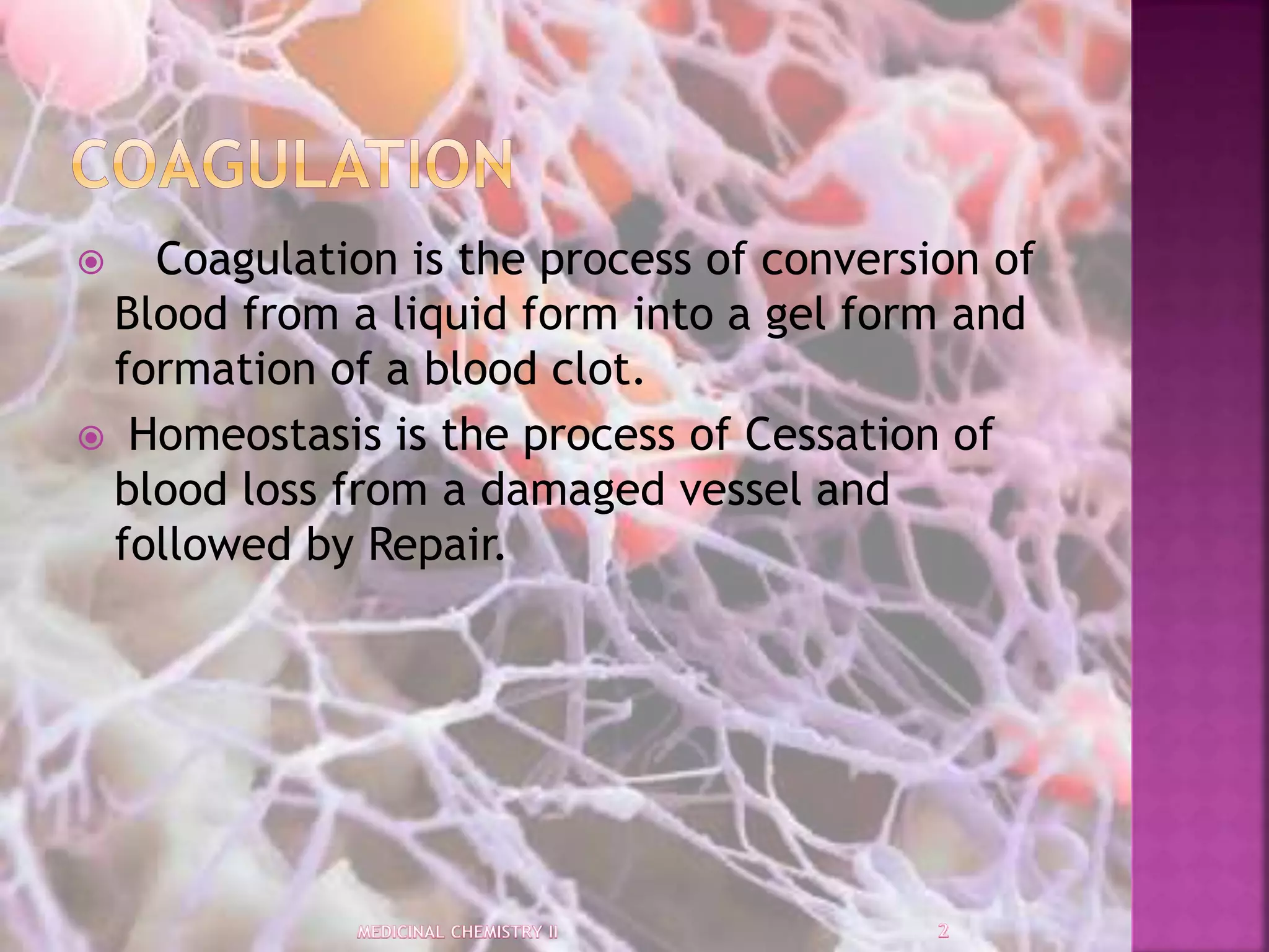
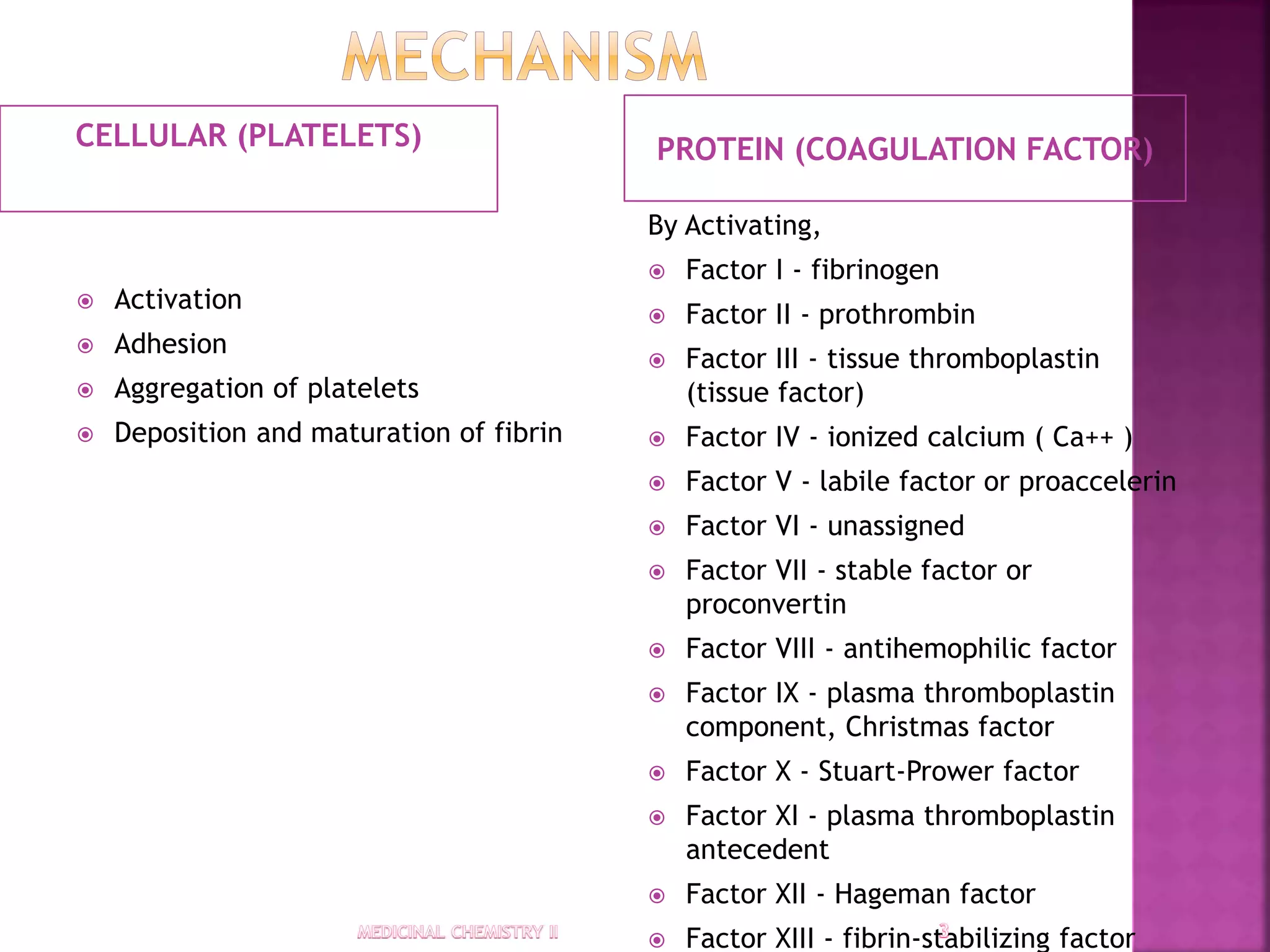
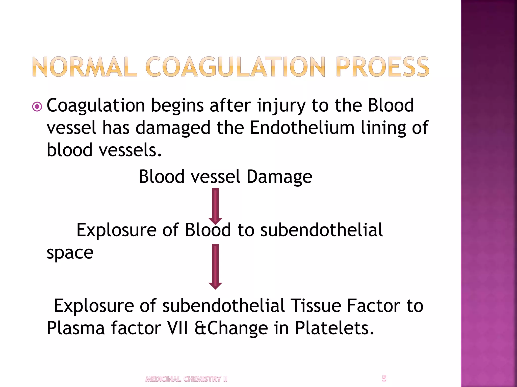
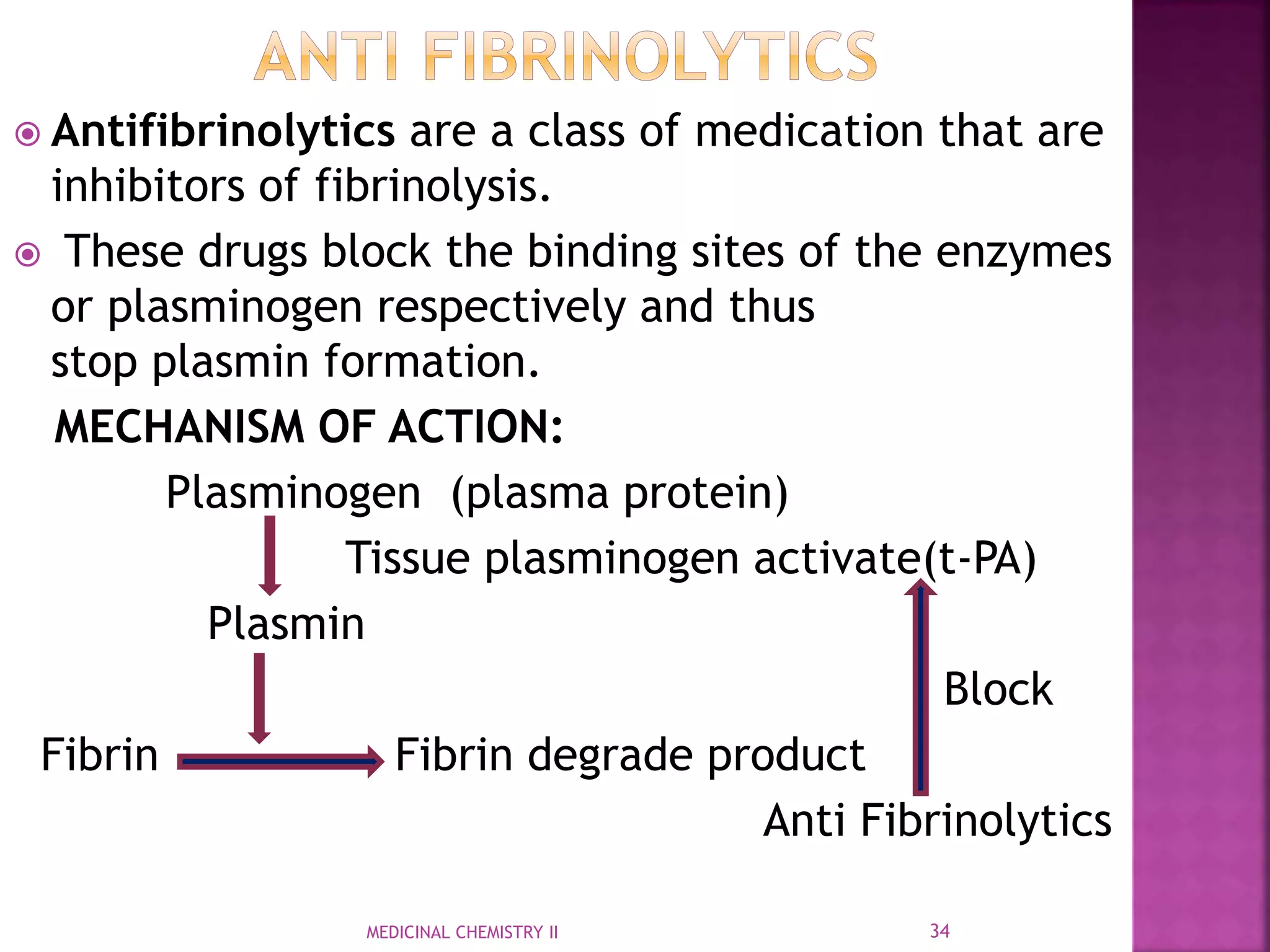
Presented By,
ASWIN K
B.Pharmacy, Final Year,
KMCH College of Pharmacy.
 Coagulation is the process of conversion of
Blood from a liquid form into a gel form and
formation of a blood clot.
 Homeostasis is the process of Cessation of
blood loss from a damaged vessel and
followed by Repair.
CELLULAR (PLATELETS) PROTEIN (COAGULATION FACTOR)
 Activation
 Adhesion
 Aggregation of platelets
 Deposition and maturation of fibrin
By Activating,
 Factor I - fibrinogen
 Factor II - prothrombin
 Factor III - tissue thromboplastin
(tissue factor)
 Factor IV - ionized calcium ( Ca++ )
 Factor V - labile factor or proaccelerin
 Factor VI - unassigned
 Factor VII - stable factor or
proconvertin
 Factor VIII - antihemophilic factor
 Factor IX - plasma thromboplastin
component, Christmas factor
 Factor X - Stuart-Prower factor
 Factor XI - plasma thromboplastin
antecedent
 Factor XII - Hageman factor
 Factor XIII - fibrin-stabilizing factor
 Bleeding (hemorrhage or bruising)
 Obstructive clotting (thrombosis).
 Coagulation begins after injury to the Blood
vessel has damaged the Endothelium lining of
blood vessels.
Blood vessel Damage
Explosure of Blood to subendothelial
space
Explosure of subendothelial Tissue Factor to
Plasma factor VII &Change in Platelets.
 PRIMARY HEMOSTASIS:
Platelets forms a plug at the site of
injury is called primary hemostasis.
 SECONDARY HEMOSTASIS:
Additional coagulation factor/clotting
factors beyond Factor VII forms a Fibrin
strands to strengthen the platelet plug is
called secondary hemostasis.
Endothelium damage
Circulating platelets +underlying collagen
(via gp VI receptor)
Signal cascade &Activation of platelet Integrins
Tight binding of platelet to the extracellular matrix
Process of adhere platelet to the site of injury
 Activated platelets release ADP , Serotonin, PAF,
Platelet factor 4, Thromboxane A2.
 Activated platelets also activate other platelets.
 Activated platelets changes its shape from
Spherical to Satellite.
 Fibrinogen Cross-links between glycoprotein &
aggregation of adjacent platelets.
Activated platelet Release granular substances
Activate Gq-linked receptor
Increases the coc.of Calcium in the platelet’s cytosol
Activate protein kinase C
act
Phospholipase A2
Modify Integrin membrane glycoprotein &
Increases affinity to bind Fibrinogen
1.Calcium & phospholipids:
Require for tenase & prothrombinase
complexes to function.
2.Vitamin K:
Gamma-glutamyl carboxylase
add
-COOH group to glutamic acid residues
(F,II,VII,IX,X Protein S,C,Z)
Vitamin K
Vitamin K epoxide reductase
Active form
Maturation of Clotting Factors
Protein C:
Thrombin Act on Protein C
Activated Protein C
APC Protein + S Phospholipid (Co factor)
Degrade Fva &FVIIIa
Serine protease inhibitor (serpin)
degrade
FIXa,Fxa,FXIIa,FXIIa
 Limit the action of TF
 Inhibit excessive TF-mediated activation of
FVII &FV
Plasminogen (plasma protein)
Tissue plasminogen activate(t-PA)
Plasmin
Fibrin Fibrin degrade product
Endothelium
release
PG I2
Activate
Gs protein – linked receptor
Adenyl cyclase
cAMP
Cytosolic Calcium level decreases
 It is the process of Desorption &
Reorganisation of Blood clot.
 Anticoagulants also called as blood thinners.
 These are chemical substances that prevent
or reduce coagulation of blood & prolonging
the clotting time.
Heparin also known as unfractionated heparin
(UFH).
HISTORY::
1. Heparin was discovered by Jay
McLean and William Henry Howell in 1916.
2. In 1918, Howell coined the term 'heparin' for
this type of fat-soluble anticoagulant.
3. In the early 1920s, Howell isolated a water-
soluble polysaccharide anticoagulant, which he
also termed 'heparin',
CHEMISTRY:
1. Native heparin is a polymer with
a molecular weight ranging from 3 to
30 kDa and commercial heparin
preparations is in the range of 12 to 15 kDa.
2. [Heparin is a member of
the glycosaminoglycan family
of carbohydrates and consists of a variably
sulfated repeating disaccharide unit
MOA:
Binds with antithrombin III
Inactivate thrombin,factor Xa and protease
ANTODOTE:
1.Protamine sulfate
(1 mg per 100 units of heparin that had been
given over the past four hours).
21MEDICINAL CHEMISTRY II
 Heparin (M.Wt- 5000 -40,000Da) is a
naturally occurring polysaccharide that
inhibits coagulation.
 LMWHs are defined as heparin salts having
an average molecular weight of less than
8000 Da
ANTIDOTE:
1.Protamine
AntiThrombin
(Serine protease inhibitor)
Degrade Activate
FIXa,Fxa,FXIIa,FXIIa
LMWHs
DRUGS:
1. Bemiparin
2. Nadroparin
3. Reviparin
4. Enoxaparin
5. Parnaparin
6. Certoparin
Vitamin
Vitamin K epoxide reductase
Active form
Block
Maturation of Clotting Factors
Vitamin K antagonists
Dicumarol
Phenindione
Coumarin
Warfarin
Ethyl Bicoumacetate
Fibrinogen
Thrombin
Fibrin (clot) Inhibit
DTI
Bivalent:
Bivalirudin
:
Hirudin
Bivalirudin
Lepirudin
Desirudin
Univalent:
Argatroban
Inogatran
Melagatran (and its
prodrug ximelagatran)
Dabigatran
Argatroban
ximelagatran
 These are a class of anticoagulant drugs which act
directly upon Factor X in the coagulation cascade,
without using antithrombin as a mediator.
 These compounds as substitutes for the currently
administered vitamin K antagonists or low
molecular weight heparin.
 ADVANTAGES:
1.Rapid onset and offset of action.
Rivaroxaban
Apixaban Edoxaban
DRUGS:
1.Rivaroxaban
2.Apixaban
3.Edoxaban
4.Darexaban
5.Eribaxaban
Fondaparinux
Idraparinux sodium
DRUGS:
1.Fondaparinux
2.Idraparinux sodium
3.Idrabiotaparinum
 These are agents which decrease platelet
aggregation and inhibit thrombus formation
 They are effective in the arterial
circulation, where anticoagulants have little
effect.
 Irreversible cyclooxygenase inhibitors
 Aspirin
 Triflusal
 Adenosine diphosphate (ADP) receptor inhibitors
 Cangrelor
 Clopidogrel
 Prasugrel
 Ticlopidine
 Phosphodiesterase inhibitors
 Cilostazol
 Protease-activated receptor-1 (PAR-1) antagonists
 Vorapaxar
 Glycoprotein IIB/IIIA inhibitors (intravenous use only)
 Abciximab
 Eptifibatide
 Tirofiban
 Adenosine reuptake inhibitors
 Dipyridamole
 Thromboxane inhibitors
 Thromboxane synthase inhibitors
 Thromboxane receptor antagonists
Aspirin
Trifusal
Ticlopidine
Clopidogrel
Tirofiban
 Antifibrinolytics are a class of medication that are
inhibitors of fibrinolysis.
 These drugs block the binding sites of the enzymes
or plasminogen respectively and thus
stop plasmin formation.
MECHANISM OF ACTION:
Plasminogen (plasma protein)
Tissue plasminogen activate(t-PA)
Plasmin
Block
Fibrin Fibrin degrade product
Anti Fibrinolytics
MEDICINAL CHEMISTRY II 34
6-aminocaproic acid
Tranexamic acid
DRUGS:
1.Aminocaproic acid
2.Tranexamic acid (Cyclokapron)
3.Ethamsylate
4.Aprotinin
 Strokes – where a blood clot restricts the flow of
blood to your brain, causing brain cells to die and
possibly resulting in permanent brain damage or
death.
 Transient ischemic attacks (TIAs) – also called "mini-
strokes", these have similar symptoms to a stroke,
but the effects usually last less than 24 hours.
 Heart attacks– where a blood clot blocks a blood
vessel supplying your heart, starving it of oxygen and
causing chest pain and sometimes death.
 Deep vein thrombosis (DVT) – where a blood clot
forms in one of the deep veins in your body, usually
your legs, causing pain and swelling.
 Pulmonary embolism – where a blood clot blocks one
of the blood vessels around the lungs, stopping the
supply of blood to the lungs.
MEDICINAL CHEMISTRY II 37

Anticoagulant-medicinal chemistry

  • 1.
    Presented By, ASWIN K B.Pharmacy,Final Year, KMCH College of Pharmacy.
  • 2.
     Coagulation isthe process of conversion of Blood from a liquid form into a gel form and formation of a blood clot.  Homeostasis is the process of Cessation of blood loss from a damaged vessel and followed by Repair.
  • 3.
    CELLULAR (PLATELETS) PROTEIN(COAGULATION FACTOR)  Activation  Adhesion  Aggregation of platelets  Deposition and maturation of fibrin By Activating,  Factor I - fibrinogen  Factor II - prothrombin  Factor III - tissue thromboplastin (tissue factor)  Factor IV - ionized calcium ( Ca++ )  Factor V - labile factor or proaccelerin  Factor VI - unassigned  Factor VII - stable factor or proconvertin  Factor VIII - antihemophilic factor  Factor IX - plasma thromboplastin component, Christmas factor  Factor X - Stuart-Prower factor  Factor XI - plasma thromboplastin antecedent  Factor XII - Hageman factor  Factor XIII - fibrin-stabilizing factor
  • 4.
     Bleeding (hemorrhageor bruising)  Obstructive clotting (thrombosis).
  • 5.
     Coagulation beginsafter injury to the Blood vessel has damaged the Endothelium lining of blood vessels. Blood vessel Damage Explosure of Blood to subendothelial space Explosure of subendothelial Tissue Factor to Plasma factor VII &Change in Platelets.
  • 6.
     PRIMARY HEMOSTASIS: Plateletsforms a plug at the site of injury is called primary hemostasis.  SECONDARY HEMOSTASIS: Additional coagulation factor/clotting factors beyond Factor VII forms a Fibrin strands to strengthen the platelet plug is called secondary hemostasis.
  • 7.
    Endothelium damage Circulating platelets+underlying collagen (via gp VI receptor) Signal cascade &Activation of platelet Integrins Tight binding of platelet to the extracellular matrix Process of adhere platelet to the site of injury
  • 8.
     Activated plateletsrelease ADP , Serotonin, PAF, Platelet factor 4, Thromboxane A2.  Activated platelets also activate other platelets.  Activated platelets changes its shape from Spherical to Satellite.  Fibrinogen Cross-links between glycoprotein & aggregation of adjacent platelets.
  • 9.
    Activated platelet Releasegranular substances Activate Gq-linked receptor Increases the coc.of Calcium in the platelet’s cytosol Activate protein kinase C act Phospholipase A2 Modify Integrin membrane glycoprotein & Increases affinity to bind Fibrinogen
  • 11.
    1.Calcium & phospholipids: Requirefor tenase & prothrombinase complexes to function. 2.Vitamin K: Gamma-glutamyl carboxylase add -COOH group to glutamic acid residues (F,II,VII,IX,X Protein S,C,Z)
  • 12.
    Vitamin K Vitamin Kepoxide reductase Active form Maturation of Clotting Factors
  • 13.
    Protein C: Thrombin Acton Protein C Activated Protein C APC Protein + S Phospholipid (Co factor) Degrade Fva &FVIIIa
  • 14.
    Serine protease inhibitor(serpin) degrade FIXa,Fxa,FXIIa,FXIIa
  • 15.
     Limit theaction of TF  Inhibit excessive TF-mediated activation of FVII &FV
  • 16.
    Plasminogen (plasma protein) Tissueplasminogen activate(t-PA) Plasmin Fibrin Fibrin degrade product
  • 17.
    Endothelium release PG I2 Activate Gs protein– linked receptor Adenyl cyclase cAMP Cytosolic Calcium level decreases
  • 18.
     It isthe process of Desorption & Reorganisation of Blood clot.
  • 19.
     Anticoagulants alsocalled as blood thinners.  These are chemical substances that prevent or reduce coagulation of blood & prolonging the clotting time.
  • 21.
    Heparin also knownas unfractionated heparin (UFH). HISTORY:: 1. Heparin was discovered by Jay McLean and William Henry Howell in 1916. 2. In 1918, Howell coined the term 'heparin' for this type of fat-soluble anticoagulant. 3. In the early 1920s, Howell isolated a water- soluble polysaccharide anticoagulant, which he also termed 'heparin', CHEMISTRY: 1. Native heparin is a polymer with a molecular weight ranging from 3 to 30 kDa and commercial heparin preparations is in the range of 12 to 15 kDa. 2. [Heparin is a member of the glycosaminoglycan family of carbohydrates and consists of a variably sulfated repeating disaccharide unit MOA: Binds with antithrombin III Inactivate thrombin,factor Xa and protease ANTODOTE: 1.Protamine sulfate (1 mg per 100 units of heparin that had been given over the past four hours). 21MEDICINAL CHEMISTRY II
  • 22.
     Heparin (M.Wt-5000 -40,000Da) is a naturally occurring polysaccharide that inhibits coagulation.  LMWHs are defined as heparin salts having an average molecular weight of less than 8000 Da ANTIDOTE: 1.Protamine
  • 23.
    AntiThrombin (Serine protease inhibitor) DegradeActivate FIXa,Fxa,FXIIa,FXIIa LMWHs DRUGS: 1. Bemiparin 2. Nadroparin 3. Reviparin 4. Enoxaparin 5. Parnaparin 6. Certoparin
  • 24.
    Vitamin Vitamin K epoxidereductase Active form Block Maturation of Clotting Factors Vitamin K antagonists
  • 25.
  • 26.
  • 27.
  • 28.
     These area class of anticoagulant drugs which act directly upon Factor X in the coagulation cascade, without using antithrombin as a mediator.  These compounds as substitutes for the currently administered vitamin K antagonists or low molecular weight heparin.  ADVANTAGES: 1.Rapid onset and offset of action.
  • 29.
  • 30.
  • 31.
     These areagents which decrease platelet aggregation and inhibit thrombus formation  They are effective in the arterial circulation, where anticoagulants have little effect.
  • 32.
     Irreversible cyclooxygenaseinhibitors  Aspirin  Triflusal  Adenosine diphosphate (ADP) receptor inhibitors  Cangrelor  Clopidogrel  Prasugrel  Ticlopidine  Phosphodiesterase inhibitors  Cilostazol  Protease-activated receptor-1 (PAR-1) antagonists  Vorapaxar  Glycoprotein IIB/IIIA inhibitors (intravenous use only)  Abciximab  Eptifibatide  Tirofiban  Adenosine reuptake inhibitors  Dipyridamole  Thromboxane inhibitors  Thromboxane synthase inhibitors  Thromboxane receptor antagonists
  • 33.
  • 34.
     Antifibrinolytics area class of medication that are inhibitors of fibrinolysis.  These drugs block the binding sites of the enzymes or plasminogen respectively and thus stop plasmin formation. MECHANISM OF ACTION: Plasminogen (plasma protein) Tissue plasminogen activate(t-PA) Plasmin Block Fibrin Fibrin degrade product Anti Fibrinolytics MEDICINAL CHEMISTRY II 34
  • 35.
    6-aminocaproic acid Tranexamic acid DRUGS: 1.Aminocaproicacid 2.Tranexamic acid (Cyclokapron) 3.Ethamsylate 4.Aprotinin
  • 36.
     Strokes –where a blood clot restricts the flow of blood to your brain, causing brain cells to die and possibly resulting in permanent brain damage or death.  Transient ischemic attacks (TIAs) – also called "mini- strokes", these have similar symptoms to a stroke, but the effects usually last less than 24 hours.  Heart attacks– where a blood clot blocks a blood vessel supplying your heart, starving it of oxygen and causing chest pain and sometimes death.  Deep vein thrombosis (DVT) – where a blood clot forms in one of the deep veins in your body, usually your legs, causing pain and swelling.  Pulmonary embolism – where a blood clot blocks one of the blood vessels around the lungs, stopping the supply of blood to the lungs.
  • 37.