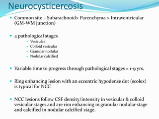
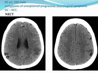
SlideShare a Scribd company logo
Dr. Mac
For Radiology
Mycobacterium Tuberculosis
Pulmonary tuberculosis
Extra-pulmonary tuberculosis
 TB Lymphadenitis
 Pleural TB
 Skeletal TB (Bones & Joints)
 CNS TB – 1% of all Tuberculosis
 Abdominal TB
 Genitourinary TB
 Pericardial TB
CNS Tuberculosis
Leptomeningitis
Pachymeningitis
Cerebritis
Tuberculoma
TB Abscess
Intracranial tuberculoma
 CNS TB almost always secondary to hematogenous
spread
 Tuberculoma can occur either in association with
meningitis or separately
 Tuberculoma
 are well defined focal masses that result from
Mycobacterium Tuberculosis with central caseous necrotic
core
 Etiology / Pathophysiology
 Hematogenous spread (gray-white matter junction lesions)
 Extension of meningitis into parenchyma
Clinical feature
 Symptoms and signs are non-specific
 Include
 Headache
 Fever
 Vomiting
 Seizures
 Neurological deficits e.g. altered sensorium, hemiparesis
Location
 Parenchyma (Supra/Infratentorial)
 Dural
 Subarachnoid space
 Brainstem infrequent, 2.5-8 % of intracranial
tuberculomas.
 Size
 1mm-6cm
Pathology
 3 stages
 Area of cerebritis – focal non-caseating granuloma – caseation
occurs later
 Histologically it consists of a central core of caseating
necrosis with a surrounding wall of a florid granulomatous
reaction containing Langerhans giant cells, epithelioid,
histiocytes and lymphocytes
 Unlike tuberculous abscesses, organisms are uncommon or
absent
 Usually, tuberculomas have a solid granulomatous core and
some may undergo liquefaction
CSF Analysis
 Increased protein
 Increased lymphocyte count
 Low glucose
 May be negative for organisms
Imaging modalities
 CT Scan
 MRI
 MRS
CT Scan
 NECT
 Non-caseating granuloma –isodense or mildly
hyperdense, surrounded by perilesional low-
density edema.
 Caseating granuloma – isodense or slightly
hypodense, edema remains.
 CECT
 Non-caseating granuloma – dense nodular
enhancement
 Caseating granuloma – ring enhancement.
Target sign may be found in some cases, central
area of enhancement surrounded by area of low
attenuation then peripheral rim enhancement.
MRI
 T1WI
 Non-caseating – Iso/Hypointense
 Caseating granuloma
 Solid centre – hypo/isointense
 Necrotic centre – hypointense
 Rim – may be hyperintense
 T2WI
 Non-caseating – hyperintense
 Caseating granuloma
 Solid centre – hypointense
 Necrotic centre – hyperintense
 Rim - hypointense
Continue….
 FLAIR
 Same as T2WI
 DWI
 Usually shows no restriction
 May show hyperintense centre of tuberculoma
 Post-contrast
 Non-caseating granuloma – Nodular homogenous enhancement
 Caseating granuloma – Peripheral rim enhancement
 MRS
 Prominent lipid and lactate peaks
 Reduced NAA
 No amino acid
Case of 32 yrs. Old male with complaints of headache, vomiting,
seizures and blurred vision
T1WI
T2WI
FLAIR
Post-contrast
Case of Tuberculoma + Meningitis
31 yrs. Old with complaints fever & seizures for 15 days
FLAIR
POST-CONTRAST
Tuberculomas with both nodular and ring
enhancing lesions in same patient
6 yrs. Old girl
Complaints of headache & deterioration of consciousness
Dx- CNS military tuberculomas
T2
Axial Post-contrast
Coronal Post-contrast
Sagittal Post-contrast
Natural history & prognosis of CNS Tuberculoma
 Long term morbidity
 Mental retardation
 Paralysis
 Seizures
 Rigidity
 Mortality high in immunocompromised patients
 Complications
 Hydrocephalus
 Stroke
 Cranial neuropathies
 Tuberculomas may take months to years to resolve
Tuberculoma Tubercular
Abscess
Central core Caseating necrosis
surrounded by
granulomatous
reaction.
No DWI restriction
Pus.
So DWI restriction.
Peripheral rim Comparatively thin Comparatively thicker
Mycobacterium
Tuberculosis
May or may not
present
Must be present
Differentials for ring-enhancing lesions in CNS
 Common
 Some primary brain tumor (e.g. anaplastic astrocytoma)
 Metastatic brain tumor
 Abscess
 Granuloma
 Resolving hematoma
 Subacute infarct
 Less common
 Thrombosed vascular malformation
 Demyelinating disease (e.g. multiple sclerosis)
 Uncommon
 Thrombosed aneurysm
 CNS lymphoma in AIDS
Differentials for CNS Tuberculomas
 Abscess
 Metastases
 Neurocysticercosis
 Meningioma (Dural tuberculomas)
Neurocysticercosis
 Common site – Subarachnoid> Parenchyma > Intraventricular
(GM-WM junction)
 4 pathological stages
 Vesicular
 Colloid vesicular
 Granular nodular
 Nodular calcified
 Variable time to progress through pathological stages = 1-9 yrs.
 Ring enhancing lesion with an eccentric hypodense dot (scolex)
is typical for NCC
 NCC lesions follow CSF density/intensity in vesicular & colloid
vesicular stages and are rim enhancing in granular nodular stage
and calcified in nodular calcified stage.
65 yrs. Old male
complaints of unexplained progressive neurological symptoms
Dx – NCC.
NECT
T1
FLAIR
Post-contrast
Vesicular & colloid vesicular stage
Brain Parenchymal metastases
 Patient may have known primary site
 Common location – GM-WM junction
 Metastases usually displace rather than infiltrate tissue
 Disproportionate edema and mass effect
 On DWI, most metastases do not restrict, but often
have a high nuclear to cytoplasm ratio and sometimes
show restricted diffusion
 Post-contrast
 Pattern varies = intense/punctate/nodular/ring enhancement.
 MRS
 Elevated choline
 Lipid/lactate peak often present
 80-85% lack Creatine peak
 Reduced NAA peak
Cerebral abscess
 Has 4 pathological stages
 Early cerebritis
 Late cerebritis
 Early capsule
 Late capsule
 T2WI
 Hypointense rim is typical (due to collagen)
 SWI
 Dual rim sign is typical
 Hyperintense inside & hypointense outside
 DWI
 Shows restriction
 MRS
 Central necrotic area may show peaks of amino acids,
lactate, acetate & succinate.
T1
T1 T2
FLA
IR
T1C+ T1C+
25 yrs. male with headache
Case of Cerebral Abscess
THANK YOU

More Related Content

What's hot

Imaging cns tb
Imaging   cns tbImaging   cns tb
Imaging cns tb
NeurologyKota
 
Imaging hiv
Imaging   hivImaging   hiv
Imaging hiv
NeurologyKota
 
Presentation1.pptx, radiological imaging of sarcoidosis.
Presentation1.pptx, radiological imaging of sarcoidosis.Presentation1.pptx, radiological imaging of sarcoidosis.
Presentation1.pptx, radiological imaging of sarcoidosis.
Abdellah Nazeer
 
Hypoxic ischemic encephalopathy modified
Hypoxic ischemic encephalopathy  modifiedHypoxic ischemic encephalopathy  modified
Hypoxic ischemic encephalopathy modified
Anish Choudhary
 
LUNG MASSES
LUNG MASSESLUNG MASSES
LUNG MASSES
Ameen Rageh
 
Imaging in pediatric brain tumors
Imaging in pediatric brain tumorsImaging in pediatric brain tumors
Imaging in pediatric brain tumors
Dr.Suhas Basavaiah
 
Imaging in multiple ring enhancing brain lesions
Imaging in multiple ring enhancing brain lesionsImaging in multiple ring enhancing brain lesions
Imaging in multiple ring enhancing brain lesions
Sumiya Arshad
 
Presentation1.pptx, radiological imaging of cerebral venous thrombosis.
Presentation1.pptx, radiological imaging of cerebral venous thrombosis.Presentation1.pptx, radiological imaging of cerebral venous thrombosis.
Presentation1.pptx, radiological imaging of cerebral venous thrombosis.
Abdellah Nazeer
 
Diagnostic Imaging of Pineal Region Masses
Diagnostic Imaging of Pineal Region MassesDiagnostic Imaging of Pineal Region Masses
Diagnostic Imaging of Pineal Region Masses
Mohamed M.A. Zaitoun
 
IMAGING OF INTRAVENTRICULAR TUMORS
IMAGING OF INTRAVENTRICULAR TUMORS IMAGING OF INTRAVENTRICULAR TUMORS
IMAGING OF INTRAVENTRICULAR TUMORS
Ameen Rageh
 
Presentation1.pptx, radiological imaging of intra cranial calcification.
Presentation1.pptx, radiological imaging of intra cranial calcification.Presentation1.pptx, radiological imaging of intra cranial calcification.
Presentation1.pptx, radiological imaging of intra cranial calcification.
Abdellah Nazeer
 
Presentation1.pptx sellar and para sellar masses
Presentation1.pptx sellar and para sellar massesPresentation1.pptx sellar and para sellar masses
Presentation1.pptx sellar and para sellar massesAbdellah Nazeer
 
Cavitatoy lung lesions
Cavitatoy lung lesionsCavitatoy lung lesions
Cavitatoy lung lesions
Sumiya Arshad
 
Pediatric brain tumors Dr. Muhammad Bin Zulfiqar
Pediatric brain tumors Dr. Muhammad Bin Zulfiqar Pediatric brain tumors Dr. Muhammad Bin Zulfiqar
Pediatric brain tumors Dr. Muhammad Bin Zulfiqar
Dr. Muhammad Bin Zulfiqar
 
Pediatric chest
Pediatric chestPediatric chest
Pediatric chest
Anish Choudhary
 
CNS infections in HIV
CNS infections in HIVCNS infections in HIV
CNS infections in HIV
Madhu Reddy
 
MEDULLOBLASTOMA
MEDULLOBLASTOMAMEDULLOBLASTOMA
MEDULLOBLASTOMA
suresh Bishokarma
 
Presentation2.pptx. posterior fossa tumour
Presentation2.pptx. posterior fossa tumourPresentation2.pptx. posterior fossa tumour
Presentation2.pptx. posterior fossa tumourAbdellah Nazeer
 
Radioanatomy of mediastinum and approach to mediastinal masses
Radioanatomy of mediastinum and approach to mediastinal massesRadioanatomy of mediastinum and approach to mediastinal masses
Radioanatomy of mediastinum and approach to mediastinal masses
AkankshaMalviya3
 

What's hot (20)

Imaging cns tb
Imaging   cns tbImaging   cns tb
Imaging cns tb
 
Imaging hiv
Imaging   hivImaging   hiv
Imaging hiv
 
Presentation1.pptx, radiological imaging of sarcoidosis.
Presentation1.pptx, radiological imaging of sarcoidosis.Presentation1.pptx, radiological imaging of sarcoidosis.
Presentation1.pptx, radiological imaging of sarcoidosis.
 
Hypoxic ischemic encephalopathy modified
Hypoxic ischemic encephalopathy  modifiedHypoxic ischemic encephalopathy  modified
Hypoxic ischemic encephalopathy modified
 
LUNG MASSES
LUNG MASSESLUNG MASSES
LUNG MASSES
 
Imaging in pediatric brain tumors
Imaging in pediatric brain tumorsImaging in pediatric brain tumors
Imaging in pediatric brain tumors
 
Imaging in multiple ring enhancing brain lesions
Imaging in multiple ring enhancing brain lesionsImaging in multiple ring enhancing brain lesions
Imaging in multiple ring enhancing brain lesions
 
Presentation1.pptx, radiological imaging of cerebral venous thrombosis.
Presentation1.pptx, radiological imaging of cerebral venous thrombosis.Presentation1.pptx, radiological imaging of cerebral venous thrombosis.
Presentation1.pptx, radiological imaging of cerebral venous thrombosis.
 
Diagnostic Imaging of Pineal Region Masses
Diagnostic Imaging of Pineal Region MassesDiagnostic Imaging of Pineal Region Masses
Diagnostic Imaging of Pineal Region Masses
 
IMAGING OF INTRAVENTRICULAR TUMORS
IMAGING OF INTRAVENTRICULAR TUMORS IMAGING OF INTRAVENTRICULAR TUMORS
IMAGING OF INTRAVENTRICULAR TUMORS
 
Presentation1.pptx, radiological imaging of intra cranial calcification.
Presentation1.pptx, radiological imaging of intra cranial calcification.Presentation1.pptx, radiological imaging of intra cranial calcification.
Presentation1.pptx, radiological imaging of intra cranial calcification.
 
Presentation1.pptx sellar and para sellar masses
Presentation1.pptx sellar and para sellar massesPresentation1.pptx sellar and para sellar masses
Presentation1.pptx sellar and para sellar masses
 
IMAGING: NEUROCYSTICERCOSIS
IMAGING: NEUROCYSTICERCOSISIMAGING: NEUROCYSTICERCOSIS
IMAGING: NEUROCYSTICERCOSIS
 
Cavitatoy lung lesions
Cavitatoy lung lesionsCavitatoy lung lesions
Cavitatoy lung lesions
 
Pediatric brain tumors Dr. Muhammad Bin Zulfiqar
Pediatric brain tumors Dr. Muhammad Bin Zulfiqar Pediatric brain tumors Dr. Muhammad Bin Zulfiqar
Pediatric brain tumors Dr. Muhammad Bin Zulfiqar
 
Pediatric chest
Pediatric chestPediatric chest
Pediatric chest
 
CNS infections in HIV
CNS infections in HIVCNS infections in HIV
CNS infections in HIV
 
MEDULLOBLASTOMA
MEDULLOBLASTOMAMEDULLOBLASTOMA
MEDULLOBLASTOMA
 
Presentation2.pptx. posterior fossa tumour
Presentation2.pptx. posterior fossa tumourPresentation2.pptx. posterior fossa tumour
Presentation2.pptx. posterior fossa tumour
 
Radioanatomy of mediastinum and approach to mediastinal masses
Radioanatomy of mediastinum and approach to mediastinal massesRadioanatomy of mediastinum and approach to mediastinal masses
Radioanatomy of mediastinum and approach to mediastinal masses
 

Similar to Cns tuberculoma

imaging in neurology - demyelinating diseases
 imaging in neurology - demyelinating diseases imaging in neurology - demyelinating diseases
imaging in neurology - demyelinating diseases
NeurologyKota
 
Central Nervous System (CNS) TBacterialM
Central Nervous System (CNS) TBacterialMCentral Nervous System (CNS) TBacterialM
Central Nervous System (CNS) TBacterialM
Dr. Ajit Surya Singh
 
Tuberous sclerosis
Tuberous sclerosisTuberous sclerosis
Tuberous sclerosis
amol lahoti
 
Multiple sclerosis
Multiple sclerosisMultiple sclerosis
Multiple sclerosis
suriyaprakash nagarajan
 
Brain abscess
Brain abscessBrain abscess
Brain abscess
macshrestha
 
Tb meningitis
Tb meningitisTb meningitis
Tb meningitis
ajayyadav753
 
Imaging of intracranial infections including COVID 19 pk2 ppt, pdf
Imaging of intracranial infections including COVID 19 pk2 ppt, pdfImaging of intracranial infections including COVID 19 pk2 ppt, pdf
Imaging of intracranial infections including COVID 19 pk2 ppt, pdf
Dr pradeep Kumar
 
Complications of csom Dr.sithanandha Kumar,29.02.2016
Complications of csom  Dr.sithanandha Kumar,29.02.2016Complications of csom  Dr.sithanandha Kumar,29.02.2016
Complications of csom Dr.sithanandha Kumar,29.02.2016
ophthalmgmcri
 
Complications of csom dr.sithanandha kumar,29.02.2016
Complications of csom  dr.sithanandha kumar,29.02.2016Complications of csom  dr.sithanandha kumar,29.02.2016
Complications of csom dr.sithanandha kumar,29.02.2016
ophthalmgmcri
 
Tuberculosis of the Central Nervous system
Tuberculosis of the Central Nervous systemTuberculosis of the Central Nervous system
Tuberculosis of the Central Nervous system
Pramod Krishnan
 
Imaging of spinal cord acute myelopathies
Imaging of spinal cord acute myelopathiesImaging of spinal cord acute myelopathies
Imaging of spinal cord acute myelopathies
Navni Garg
 
Imaging of cns viral infection
Imaging of cns viral infectionImaging of cns viral infection
Imaging of cns viral infection
NeurologyKota
 
Imaging of cns viral infection
Imaging of cns viral infectionImaging of cns viral infection
Imaging of cns viral infection
sks200166
 
imagingofintracranialinfectionsincludingcovid19pk2-200605190339 (1).pptx
imagingofintracranialinfectionsincludingcovid19pk2-200605190339 (1).pptximagingofintracranialinfectionsincludingcovid19pk2-200605190339 (1).pptx
imagingofintracranialinfectionsincludingcovid19pk2-200605190339 (1).pptx
Rose635887
 
Non Traumatic Spinal cord injuries
Non Traumatic Spinal cord injuries   Non Traumatic Spinal cord injuries
Non Traumatic Spinal cord injuries
Dr. Muzahid
 
CNS infections- bacterial and viral-1.pptx
CNS  infections- bacterial and viral-1.pptxCNS  infections- bacterial and viral-1.pptx
CNS infections- bacterial and viral-1.pptx
yashwanthnaik8
 
Brain abscess
Brain   abscessBrain   abscess
Brain abscess
puspa raj koirala
 
Paraparesis.pptx
Paraparesis.pptxParaparesis.pptx
Paraparesis.pptx
NatanA7
 
Tuberculosis of central Nervous System- CNSTB
Tuberculosis of central Nervous System- CNSTBTuberculosis of central Nervous System- CNSTB
Tuberculosis of central Nervous System- CNSTB
YatinBhole
 
ADEM.pdf
ADEM.pdfADEM.pdf
ADEM.pdf
ssusere8f40d
 

Similar to Cns tuberculoma (20)

imaging in neurology - demyelinating diseases
 imaging in neurology - demyelinating diseases imaging in neurology - demyelinating diseases
imaging in neurology - demyelinating diseases
 
Central Nervous System (CNS) TBacterialM
Central Nervous System (CNS) TBacterialMCentral Nervous System (CNS) TBacterialM
Central Nervous System (CNS) TBacterialM
 
Tuberous sclerosis
Tuberous sclerosisTuberous sclerosis
Tuberous sclerosis
 
Multiple sclerosis
Multiple sclerosisMultiple sclerosis
Multiple sclerosis
 
Brain abscess
Brain abscessBrain abscess
Brain abscess
 
Tb meningitis
Tb meningitisTb meningitis
Tb meningitis
 
Imaging of intracranial infections including COVID 19 pk2 ppt, pdf
Imaging of intracranial infections including COVID 19 pk2 ppt, pdfImaging of intracranial infections including COVID 19 pk2 ppt, pdf
Imaging of intracranial infections including COVID 19 pk2 ppt, pdf
 
Complications of csom Dr.sithanandha Kumar,29.02.2016
Complications of csom  Dr.sithanandha Kumar,29.02.2016Complications of csom  Dr.sithanandha Kumar,29.02.2016
Complications of csom Dr.sithanandha Kumar,29.02.2016
 
Complications of csom dr.sithanandha kumar,29.02.2016
Complications of csom  dr.sithanandha kumar,29.02.2016Complications of csom  dr.sithanandha kumar,29.02.2016
Complications of csom dr.sithanandha kumar,29.02.2016
 
Tuberculosis of the Central Nervous system
Tuberculosis of the Central Nervous systemTuberculosis of the Central Nervous system
Tuberculosis of the Central Nervous system
 
Imaging of spinal cord acute myelopathies
Imaging of spinal cord acute myelopathiesImaging of spinal cord acute myelopathies
Imaging of spinal cord acute myelopathies
 
Imaging of cns viral infection
Imaging of cns viral infectionImaging of cns viral infection
Imaging of cns viral infection
 
Imaging of cns viral infection
Imaging of cns viral infectionImaging of cns viral infection
Imaging of cns viral infection
 
imagingofintracranialinfectionsincludingcovid19pk2-200605190339 (1).pptx
imagingofintracranialinfectionsincludingcovid19pk2-200605190339 (1).pptximagingofintracranialinfectionsincludingcovid19pk2-200605190339 (1).pptx
imagingofintracranialinfectionsincludingcovid19pk2-200605190339 (1).pptx
 
Non Traumatic Spinal cord injuries
Non Traumatic Spinal cord injuries   Non Traumatic Spinal cord injuries
Non Traumatic Spinal cord injuries
 
CNS infections- bacterial and viral-1.pptx
CNS  infections- bacterial and viral-1.pptxCNS  infections- bacterial and viral-1.pptx
CNS infections- bacterial and viral-1.pptx
 
Brain abscess
Brain   abscessBrain   abscess
Brain abscess
 
Paraparesis.pptx
Paraparesis.pptxParaparesis.pptx
Paraparesis.pptx
 
Tuberculosis of central Nervous System- CNSTB
Tuberculosis of central Nervous System- CNSTBTuberculosis of central Nervous System- CNSTB
Tuberculosis of central Nervous System- CNSTB
 
ADEM.pdf
ADEM.pdfADEM.pdf
ADEM.pdf
 

More from macshrestha

Cerebral/Pial Arteriovenous Malformation (AVM)
Cerebral/Pial Arteriovenous Malformation (AVM)Cerebral/Pial Arteriovenous Malformation (AVM)
Cerebral/Pial Arteriovenous Malformation (AVM)
macshrestha
 
Hepatic peliosis
Hepatic peliosisHepatic peliosis
Hepatic peliosis
macshrestha
 
Ankle joint pathology imaging
Ankle joint pathology imagingAnkle joint pathology imaging
Ankle joint pathology imaging
macshrestha
 
Benign bone tumors
Benign bone tumorsBenign bone tumors
Benign bone tumors
macshrestha
 
Dural venous sinus thrombosis for Radiology & Imaging
Dural venous sinus thrombosis for Radiology & ImagingDural venous sinus thrombosis for Radiology & Imaging
Dural venous sinus thrombosis for Radiology & Imaging
macshrestha
 
Empyema necessitans
Empyema necessitansEmpyema necessitans
Empyema necessitans
macshrestha
 
Extraskeletal Mesenchymal Chondrosarcoma
Extraskeletal Mesenchymal ChondrosarcomaExtraskeletal Mesenchymal Chondrosarcoma
Extraskeletal Mesenchymal Chondrosarcoma
macshrestha
 
Ct head
Ct headCt head
Ct head
macshrestha
 
Choledochal cyst
Choledochal cystCholedochal cyst
Choledochal cyst
macshrestha
 
Pulmonary metastases
Pulmonary metastasesPulmonary metastases
Pulmonary metastases
macshrestha
 
Simple bone cyst
Simple bone cystSimple bone cyst
Simple bone cyst
macshrestha
 

More from macshrestha (11)

Cerebral/Pial Arteriovenous Malformation (AVM)
Cerebral/Pial Arteriovenous Malformation (AVM)Cerebral/Pial Arteriovenous Malformation (AVM)
Cerebral/Pial Arteriovenous Malformation (AVM)
 
Hepatic peliosis
Hepatic peliosisHepatic peliosis
Hepatic peliosis
 
Ankle joint pathology imaging
Ankle joint pathology imagingAnkle joint pathology imaging
Ankle joint pathology imaging
 
Benign bone tumors
Benign bone tumorsBenign bone tumors
Benign bone tumors
 
Dural venous sinus thrombosis for Radiology & Imaging
Dural venous sinus thrombosis for Radiology & ImagingDural venous sinus thrombosis for Radiology & Imaging
Dural venous sinus thrombosis for Radiology & Imaging
 
Empyema necessitans
Empyema necessitansEmpyema necessitans
Empyema necessitans
 
Extraskeletal Mesenchymal Chondrosarcoma
Extraskeletal Mesenchymal ChondrosarcomaExtraskeletal Mesenchymal Chondrosarcoma
Extraskeletal Mesenchymal Chondrosarcoma
 
Ct head
Ct headCt head
Ct head
 
Choledochal cyst
Choledochal cystCholedochal cyst
Choledochal cyst
 
Pulmonary metastases
Pulmonary metastasesPulmonary metastases
Pulmonary metastases
 
Simple bone cyst
Simple bone cystSimple bone cyst
Simple bone cyst
 

Recently uploaded

Report Back from SGO 2024: What’s the Latest in Cervical Cancer?
Report Back from SGO 2024: What’s the Latest in Cervical Cancer?Report Back from SGO 2024: What’s the Latest in Cervical Cancer?
Report Back from SGO 2024: What’s the Latest in Cervical Cancer?
bkling
 
Alcohol_Dr. Jeenal Mistry MD Pharmacology.pdf
Alcohol_Dr. Jeenal Mistry MD Pharmacology.pdfAlcohol_Dr. Jeenal Mistry MD Pharmacology.pdf
Alcohol_Dr. Jeenal Mistry MD Pharmacology.pdf
Dr Jeenal Mistry
 
How to Give Better Lectures: Some Tips for Doctors
How to Give Better Lectures: Some Tips for DoctorsHow to Give Better Lectures: Some Tips for Doctors
How to Give Better Lectures: Some Tips for Doctors
LanceCatedral
 
ARTIFICIAL INTELLIGENCE IN HEALTHCARE.pdf
ARTIFICIAL INTELLIGENCE IN  HEALTHCARE.pdfARTIFICIAL INTELLIGENCE IN  HEALTHCARE.pdf
ARTIFICIAL INTELLIGENCE IN HEALTHCARE.pdf
Anujkumaranit
 
Pulmonary Thromboembolism - etilogy, types, medical- Surgical and nursing man...
Pulmonary Thromboembolism - etilogy, types, medical- Surgical and nursing man...Pulmonary Thromboembolism - etilogy, types, medical- Surgical and nursing man...
Pulmonary Thromboembolism - etilogy, types, medical- Surgical and nursing man...
VarunMahajani
 
The Normal Electrocardiogram - Part I of II
The Normal Electrocardiogram - Part I of IIThe Normal Electrocardiogram - Part I of II
The Normal Electrocardiogram - Part I of II
MedicoseAcademics
 
basicmodesofventilation2022-220313203758.pdf
basicmodesofventilation2022-220313203758.pdfbasicmodesofventilation2022-220313203758.pdf
basicmodesofventilation2022-220313203758.pdf
aljamhori teaching hospital
 
Factory Supply Best Quality Pmk Oil CAS 28578–16–7 PMK Powder in Stock
Factory Supply Best Quality Pmk Oil CAS 28578–16–7 PMK Powder in StockFactory Supply Best Quality Pmk Oil CAS 28578–16–7 PMK Powder in Stock
Factory Supply Best Quality Pmk Oil CAS 28578–16–7 PMK Powder in Stock
rebeccabio
 
heat stroke and heat exhaustion in children
heat stroke and heat exhaustion in childrenheat stroke and heat exhaustion in children
heat stroke and heat exhaustion in children
SumeraAhmad5
 
Physiology of Special Chemical Sensation of Taste
Physiology of Special Chemical Sensation of TastePhysiology of Special Chemical Sensation of Taste
Physiology of Special Chemical Sensation of Taste
MedicoseAcademics
 
micro teaching on communication m.sc nursing.pdf
micro teaching on communication m.sc nursing.pdfmicro teaching on communication m.sc nursing.pdf
micro teaching on communication m.sc nursing.pdf
Anurag Sharma
 
POST OPERATIVE OLIGURIA and its management
POST OPERATIVE OLIGURIA and its managementPOST OPERATIVE OLIGURIA and its management
POST OPERATIVE OLIGURIA and its management
touseefaziz1
 
Ocular injury ppt Upendra pal optometrist upums saifai etawah
Ocular injury  ppt  Upendra pal  optometrist upums saifai etawahOcular injury  ppt  Upendra pal  optometrist upums saifai etawah
Ocular injury ppt Upendra pal optometrist upums saifai etawah
pal078100
 
Physiology of Chemical Sensation of smell.pdf
Physiology of Chemical Sensation of smell.pdfPhysiology of Chemical Sensation of smell.pdf
Physiology of Chemical Sensation of smell.pdf
MedicoseAcademics
 
THOA 2.ppt Human Organ Transplantation Act
THOA 2.ppt Human Organ Transplantation ActTHOA 2.ppt Human Organ Transplantation Act
THOA 2.ppt Human Organ Transplantation Act
DrSathishMS1
 
TEST BANK for Operations Management, 14th Edition by William J. Stevenson, Ve...
TEST BANK for Operations Management, 14th Edition by William J. Stevenson, Ve...TEST BANK for Operations Management, 14th Edition by William J. Stevenson, Ve...
TEST BANK for Operations Management, 14th Edition by William J. Stevenson, Ve...
kevinkariuki227
 
ANATOMY AND PHYSIOLOGY OF URINARY SYSTEM.pptx
ANATOMY AND PHYSIOLOGY OF URINARY SYSTEM.pptxANATOMY AND PHYSIOLOGY OF URINARY SYSTEM.pptx
ANATOMY AND PHYSIOLOGY OF URINARY SYSTEM.pptx
Swetaba Besh
 
Lung Cancer: Artificial Intelligence, Synergetics, Complex System Analysis, S...
Lung Cancer: Artificial Intelligence, Synergetics, Complex System Analysis, S...Lung Cancer: Artificial Intelligence, Synergetics, Complex System Analysis, S...
Lung Cancer: Artificial Intelligence, Synergetics, Complex System Analysis, S...
Oleg Kshivets
 
HOT NEW PRODUCT! BIG SALES FAST SHIPPING NOW FROM CHINA!! EU KU DB BK substit...
HOT NEW PRODUCT! BIG SALES FAST SHIPPING NOW FROM CHINA!! EU KU DB BK substit...HOT NEW PRODUCT! BIG SALES FAST SHIPPING NOW FROM CHINA!! EU KU DB BK substit...
HOT NEW PRODUCT! BIG SALES FAST SHIPPING NOW FROM CHINA!! EU KU DB BK substit...
GL Anaacs
 
Ozempic: Preoperative Management of Patients on GLP-1 Receptor Agonists
Ozempic: Preoperative Management of Patients on GLP-1 Receptor Agonists  Ozempic: Preoperative Management of Patients on GLP-1 Receptor Agonists
Ozempic: Preoperative Management of Patients on GLP-1 Receptor Agonists
Saeid Safari
 

Recently uploaded (20)

Report Back from SGO 2024: What’s the Latest in Cervical Cancer?
Report Back from SGO 2024: What’s the Latest in Cervical Cancer?Report Back from SGO 2024: What’s the Latest in Cervical Cancer?
Report Back from SGO 2024: What’s the Latest in Cervical Cancer?
 
Alcohol_Dr. Jeenal Mistry MD Pharmacology.pdf
Alcohol_Dr. Jeenal Mistry MD Pharmacology.pdfAlcohol_Dr. Jeenal Mistry MD Pharmacology.pdf
Alcohol_Dr. Jeenal Mistry MD Pharmacology.pdf
 
How to Give Better Lectures: Some Tips for Doctors
How to Give Better Lectures: Some Tips for DoctorsHow to Give Better Lectures: Some Tips for Doctors
How to Give Better Lectures: Some Tips for Doctors
 
ARTIFICIAL INTELLIGENCE IN HEALTHCARE.pdf
ARTIFICIAL INTELLIGENCE IN  HEALTHCARE.pdfARTIFICIAL INTELLIGENCE IN  HEALTHCARE.pdf
ARTIFICIAL INTELLIGENCE IN HEALTHCARE.pdf
 
Pulmonary Thromboembolism - etilogy, types, medical- Surgical and nursing man...
Pulmonary Thromboembolism - etilogy, types, medical- Surgical and nursing man...Pulmonary Thromboembolism - etilogy, types, medical- Surgical and nursing man...
Pulmonary Thromboembolism - etilogy, types, medical- Surgical and nursing man...
 
The Normal Electrocardiogram - Part I of II
The Normal Electrocardiogram - Part I of IIThe Normal Electrocardiogram - Part I of II
The Normal Electrocardiogram - Part I of II
 
basicmodesofventilation2022-220313203758.pdf
basicmodesofventilation2022-220313203758.pdfbasicmodesofventilation2022-220313203758.pdf
basicmodesofventilation2022-220313203758.pdf
 
Factory Supply Best Quality Pmk Oil CAS 28578–16–7 PMK Powder in Stock
Factory Supply Best Quality Pmk Oil CAS 28578–16–7 PMK Powder in StockFactory Supply Best Quality Pmk Oil CAS 28578–16–7 PMK Powder in Stock
Factory Supply Best Quality Pmk Oil CAS 28578–16–7 PMK Powder in Stock
 
heat stroke and heat exhaustion in children
heat stroke and heat exhaustion in childrenheat stroke and heat exhaustion in children
heat stroke and heat exhaustion in children
 
Physiology of Special Chemical Sensation of Taste
Physiology of Special Chemical Sensation of TastePhysiology of Special Chemical Sensation of Taste
Physiology of Special Chemical Sensation of Taste
 
micro teaching on communication m.sc nursing.pdf
micro teaching on communication m.sc nursing.pdfmicro teaching on communication m.sc nursing.pdf
micro teaching on communication m.sc nursing.pdf
 
POST OPERATIVE OLIGURIA and its management
POST OPERATIVE OLIGURIA and its managementPOST OPERATIVE OLIGURIA and its management
POST OPERATIVE OLIGURIA and its management
 
Ocular injury ppt Upendra pal optometrist upums saifai etawah
Ocular injury  ppt  Upendra pal  optometrist upums saifai etawahOcular injury  ppt  Upendra pal  optometrist upums saifai etawah
Ocular injury ppt Upendra pal optometrist upums saifai etawah
 
Physiology of Chemical Sensation of smell.pdf
Physiology of Chemical Sensation of smell.pdfPhysiology of Chemical Sensation of smell.pdf
Physiology of Chemical Sensation of smell.pdf
 
THOA 2.ppt Human Organ Transplantation Act
THOA 2.ppt Human Organ Transplantation ActTHOA 2.ppt Human Organ Transplantation Act
THOA 2.ppt Human Organ Transplantation Act
 
TEST BANK for Operations Management, 14th Edition by William J. Stevenson, Ve...
TEST BANK for Operations Management, 14th Edition by William J. Stevenson, Ve...TEST BANK for Operations Management, 14th Edition by William J. Stevenson, Ve...
TEST BANK for Operations Management, 14th Edition by William J. Stevenson, Ve...
 
ANATOMY AND PHYSIOLOGY OF URINARY SYSTEM.pptx
ANATOMY AND PHYSIOLOGY OF URINARY SYSTEM.pptxANATOMY AND PHYSIOLOGY OF URINARY SYSTEM.pptx
ANATOMY AND PHYSIOLOGY OF URINARY SYSTEM.pptx
 
Lung Cancer: Artificial Intelligence, Synergetics, Complex System Analysis, S...
Lung Cancer: Artificial Intelligence, Synergetics, Complex System Analysis, S...Lung Cancer: Artificial Intelligence, Synergetics, Complex System Analysis, S...
Lung Cancer: Artificial Intelligence, Synergetics, Complex System Analysis, S...
 
HOT NEW PRODUCT! BIG SALES FAST SHIPPING NOW FROM CHINA!! EU KU DB BK substit...
HOT NEW PRODUCT! BIG SALES FAST SHIPPING NOW FROM CHINA!! EU KU DB BK substit...HOT NEW PRODUCT! BIG SALES FAST SHIPPING NOW FROM CHINA!! EU KU DB BK substit...
HOT NEW PRODUCT! BIG SALES FAST SHIPPING NOW FROM CHINA!! EU KU DB BK substit...
 
Ozempic: Preoperative Management of Patients on GLP-1 Receptor Agonists
Ozempic: Preoperative Management of Patients on GLP-1 Receptor Agonists  Ozempic: Preoperative Management of Patients on GLP-1 Receptor Agonists
Ozempic: Preoperative Management of Patients on GLP-1 Receptor Agonists
 

Cns tuberculoma

  • 2. Mycobacterium Tuberculosis Pulmonary tuberculosis Extra-pulmonary tuberculosis  TB Lymphadenitis  Pleural TB  Skeletal TB (Bones & Joints)  CNS TB – 1% of all Tuberculosis  Abdominal TB  Genitourinary TB  Pericardial TB
  • 4. Intracranial tuberculoma  CNS TB almost always secondary to hematogenous spread  Tuberculoma can occur either in association with meningitis or separately  Tuberculoma  are well defined focal masses that result from Mycobacterium Tuberculosis with central caseous necrotic core  Etiology / Pathophysiology  Hematogenous spread (gray-white matter junction lesions)  Extension of meningitis into parenchyma
  • 5. Clinical feature  Symptoms and signs are non-specific  Include  Headache  Fever  Vomiting  Seizures  Neurological deficits e.g. altered sensorium, hemiparesis
  • 6. Location  Parenchyma (Supra/Infratentorial)  Dural  Subarachnoid space  Brainstem infrequent, 2.5-8 % of intracranial tuberculomas.  Size  1mm-6cm
  • 7. Pathology  3 stages  Area of cerebritis – focal non-caseating granuloma – caseation occurs later  Histologically it consists of a central core of caseating necrosis with a surrounding wall of a florid granulomatous reaction containing Langerhans giant cells, epithelioid, histiocytes and lymphocytes  Unlike tuberculous abscesses, organisms are uncommon or absent  Usually, tuberculomas have a solid granulomatous core and some may undergo liquefaction
  • 8. CSF Analysis  Increased protein  Increased lymphocyte count  Low glucose  May be negative for organisms
  • 9. Imaging modalities  CT Scan  MRI  MRS
  • 10. CT Scan  NECT  Non-caseating granuloma –isodense or mildly hyperdense, surrounded by perilesional low- density edema.  Caseating granuloma – isodense or slightly hypodense, edema remains.  CECT  Non-caseating granuloma – dense nodular enhancement  Caseating granuloma – ring enhancement. Target sign may be found in some cases, central area of enhancement surrounded by area of low attenuation then peripheral rim enhancement.
  • 11. MRI  T1WI  Non-caseating – Iso/Hypointense  Caseating granuloma  Solid centre – hypo/isointense  Necrotic centre – hypointense  Rim – may be hyperintense  T2WI  Non-caseating – hyperintense  Caseating granuloma  Solid centre – hypointense  Necrotic centre – hyperintense  Rim - hypointense
  • 12. Continue….  FLAIR  Same as T2WI  DWI  Usually shows no restriction  May show hyperintense centre of tuberculoma  Post-contrast  Non-caseating granuloma – Nodular homogenous enhancement  Caseating granuloma – Peripheral rim enhancement  MRS  Prominent lipid and lactate peaks  Reduced NAA  No amino acid
  • 13. Case of 32 yrs. Old male with complaints of headache, vomiting, seizures and blurred vision T1WI
  • 14. T2WI
  • 15. FLAIR
  • 17. Case of Tuberculoma + Meningitis 31 yrs. Old with complaints fever & seizures for 15 days FLAIR POST-CONTRAST
  • 18. Tuberculomas with both nodular and ring enhancing lesions in same patient
  • 19. 6 yrs. Old girl Complaints of headache & deterioration of consciousness Dx- CNS military tuberculomas T2
  • 23. Natural history & prognosis of CNS Tuberculoma  Long term morbidity  Mental retardation  Paralysis  Seizures  Rigidity  Mortality high in immunocompromised patients  Complications  Hydrocephalus  Stroke  Cranial neuropathies  Tuberculomas may take months to years to resolve
  • 24. Tuberculoma Tubercular Abscess Central core Caseating necrosis surrounded by granulomatous reaction. No DWI restriction Pus. So DWI restriction. Peripheral rim Comparatively thin Comparatively thicker Mycobacterium Tuberculosis May or may not present Must be present
  • 25. Differentials for ring-enhancing lesions in CNS  Common  Some primary brain tumor (e.g. anaplastic astrocytoma)  Metastatic brain tumor  Abscess  Granuloma  Resolving hematoma  Subacute infarct  Less common  Thrombosed vascular malformation  Demyelinating disease (e.g. multiple sclerosis)  Uncommon  Thrombosed aneurysm  CNS lymphoma in AIDS
  • 26. Differentials for CNS Tuberculomas  Abscess  Metastases  Neurocysticercosis  Meningioma (Dural tuberculomas)
  • 27. Neurocysticercosis  Common site – Subarachnoid> Parenchyma > Intraventricular (GM-WM junction)  4 pathological stages  Vesicular  Colloid vesicular  Granular nodular  Nodular calcified  Variable time to progress through pathological stages = 1-9 yrs.  Ring enhancing lesion with an eccentric hypodense dot (scolex) is typical for NCC  NCC lesions follow CSF density/intensity in vesicular & colloid vesicular stages and are rim enhancing in granular nodular stage and calcified in nodular calcified stage.
  • 28. 65 yrs. Old male complaints of unexplained progressive neurological symptoms Dx – NCC. NECT
  • 29. T1
  • 30. FLAIR
  • 32. Brain Parenchymal metastases  Patient may have known primary site  Common location – GM-WM junction  Metastases usually displace rather than infiltrate tissue  Disproportionate edema and mass effect  On DWI, most metastases do not restrict, but often have a high nuclear to cytoplasm ratio and sometimes show restricted diffusion  Post-contrast  Pattern varies = intense/punctate/nodular/ring enhancement.  MRS  Elevated choline  Lipid/lactate peak often present  80-85% lack Creatine peak  Reduced NAA peak
  • 33. Cerebral abscess  Has 4 pathological stages  Early cerebritis  Late cerebritis  Early capsule  Late capsule  T2WI  Hypointense rim is typical (due to collagen)  SWI  Dual rim sign is typical  Hyperintense inside & hypointense outside  DWI  Shows restriction  MRS  Central necrotic area may show peaks of amino acids, lactate, acetate & succinate.
  • 34. T1 T1 T2 FLA IR T1C+ T1C+ 25 yrs. male with headache Case of Cerebral Abscess
  • 35.

Editor's Notes

  1. Culture showed gram positive cocci