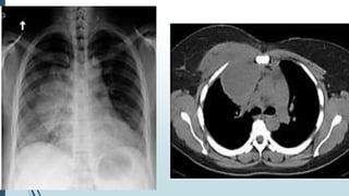
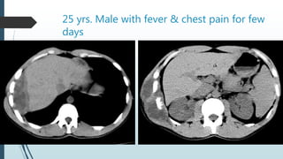
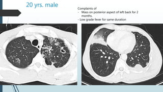
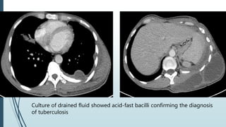
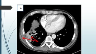
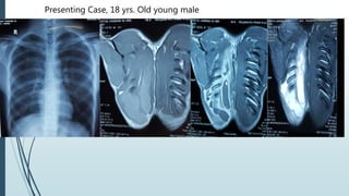
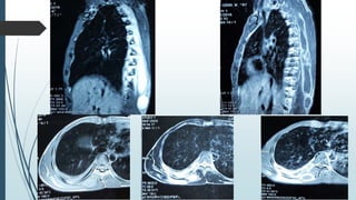
Empyema
Necessitans
Dr. Manmohan Bir Shrestha
For Radiology
Empyema Necessitans / Necessitasis
ď‚´Rare long term complication of empyema thoracis
ď‚´refers to extension of a pleural infection out of the thorax and
into the neighboring chest wall and surrounding soft tissues
ď‚´ extension of an empyema out of the pleural cavity.
ď‚´With the advent of antimicrobials, its incidence has
decreased
Pathology
ď‚´ It may either occur due the virulence of the organism or may be facilitated by
previous thoracic surgery (e.g. thoracotomy) or trauma allowing infection to
track through.
ď‚´ It occurs commonly to subcutaneous tissues of the chest wall, but can also
spread to involve other sites such as
ď‚´ esophagus
ď‚´ breast
ď‚´Peritoneal & retroperitoneal region
ď‚´ pericardial region
ď‚´paravertebral regions.
ď‚´ The resultant subcutaneous abscess may eventually rupture through the skin.
ď‚´ Can even cause osteonecrosis of the affected rib.
Causative organisms
 Mycobacterium tuberculosis – most common
ď‚´Actinomyces
ď‚´Blastomycosis
ď‚´Aspergillus
ď‚´Nocardia
ď‚´Mucormycosis
ď‚´Fusobacterium
Clinical feature - varies
ď‚´Sign & symptoms of pulmonary tuberculosis
ď‚´Chest pain
ď‚´Erythema & swelling over chest wall.
Imaging modalities
ď‚´Chest x-ray
ď‚´Ultrasound
ď‚´CT Scan
ď‚´MRI
Plain radiograph
ď‚´are often non-specific and sometimes even normal
ď‚´may suggest soft tissue density in chest wall
ď‚´can resemble pleural effusion/empyema thoracis
ď‚´can show features of consolidation
CT Scan
ď‚´Fluid density in pleural space & extending into
subcutaneous tissues in the chest wall or
elsewhere in abdomen
ď‚´Shows bone erosions well
Post contrast – rim enhancing fluid collections.
MRI
T1WI – Iso to hypointense
T2WI – hyperintense
Post contrast – rim enhancing
Case of 40 yrs. female patient
ď‚´Complaints
ď‚´Fever, chills & sweats for one week
ď‚´On examination
ď‚´Palpable, non-tender subcutaneous mass over the
right anterior chest wall.
ď‚´Treatment
ď‚´Surgery for the drainage of abscess
with right anterior thoracotomy
ď‚´Culture of drained fluid
ď‚´Growth of Methicillin Resistant
Staphylococcus aureus
25 yrs. Male with fever & chest pain for few
days
20 yrs. male Complaints of
- Mass on posterior aspect of left back for 2
months.
- Low grade fever for same duration
Culture of drained fluid showed acid-fast bacilli confirming the diagnosis
of tuberculosis
56 yrs. Old male
ď‚´Complaints
ď‚´3 weeks of progressive, localized, right-sided posterior chest wall
pain and swelling.
ď‚´No fevers or chills, productive cough, hemoptysis, change in weight
ď‚´On examination
ď‚´hard and fixed mass on the right posterior chest wall at the level of
the 11th rib with minimal tenderness to palpation.
Pre-operative Post-operative
Culture report confirmed actinomycosis.
Follow up after 6 weeks
Presenting Case, 18 yrs. Old young male
Empyema necessitans
Empyema necessitans

Empyema necessitans

  • 1.
  • 2.
    Empyema Necessitans /Necessitasis ď‚´Rare long term complication of empyema thoracis ď‚´refers to extension of a pleural infection out of the thorax and into the neighboring chest wall and surrounding soft tissues ď‚´ extension of an empyema out of the pleural cavity. ď‚´With the advent of antimicrobials, its incidence has decreased
  • 3.
    Pathology ď‚´ It mayeither occur due the virulence of the organism or may be facilitated by previous thoracic surgery (e.g. thoracotomy) or trauma allowing infection to track through. ď‚´ It occurs commonly to subcutaneous tissues of the chest wall, but can also spread to involve other sites such as ď‚´ esophagus ď‚´ breast ď‚´Peritoneal & retroperitoneal region ď‚´ pericardial region ď‚´paravertebral regions. ď‚´ The resultant subcutaneous abscess may eventually rupture through the skin. ď‚´ Can even cause osteonecrosis of the affected rib.
  • 4.
    Causative organisms  Mycobacteriumtuberculosis – most common Actinomyces Blastomycosis Aspergillus Nocardia Mucormycosis Fusobacterium
  • 5.
    Clinical feature -varies ď‚´Sign & symptoms of pulmonary tuberculosis ď‚´Chest pain ď‚´Erythema & swelling over chest wall.
  • 6.
  • 7.
    Plain radiograph ď‚´are oftennon-specific and sometimes even normal ď‚´may suggest soft tissue density in chest wall ď‚´can resemble pleural effusion/empyema thoracis ď‚´can show features of consolidation
  • 8.
    CT Scan Fluid densityin pleural space & extending into subcutaneous tissues in the chest wall or elsewhere in abdomen Shows bone erosions well Post contrast – rim enhancing fluid collections.
  • 9.
    MRI T1WI – Isoto hypointense T2WI – hyperintense Post contrast – rim enhancing
  • 10.
    Case of 40yrs. female patient ď‚´Complaints ď‚´Fever, chills & sweats for one week ď‚´On examination ď‚´Palpable, non-tender subcutaneous mass over the right anterior chest wall.
  • 12.
    ď‚´Treatment ď‚´Surgery for thedrainage of abscess with right anterior thoracotomy ď‚´Culture of drained fluid ď‚´Growth of Methicillin Resistant Staphylococcus aureus
  • 13.
    25 yrs. Malewith fever & chest pain for few days
  • 14.
    20 yrs. maleComplaints of - Mass on posterior aspect of left back for 2 months. - Low grade fever for same duration
  • 15.
    Culture of drainedfluid showed acid-fast bacilli confirming the diagnosis of tuberculosis
  • 16.
    56 yrs. Oldmale ď‚´Complaints ď‚´3 weeks of progressive, localized, right-sided posterior chest wall pain and swelling. ď‚´No fevers or chills, productive cough, hemoptysis, change in weight ď‚´On examination ď‚´hard and fixed mass on the right posterior chest wall at the level of the 11th rib with minimal tenderness to palpation.
  • 18.
  • 19.
    Culture report confirmedactinomycosis. Follow up after 6 weeks
  • 20.
    Presenting Case, 18yrs. Old young male