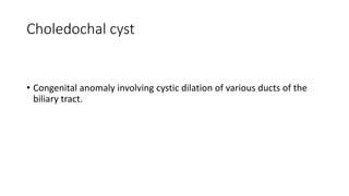
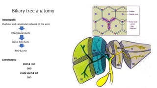
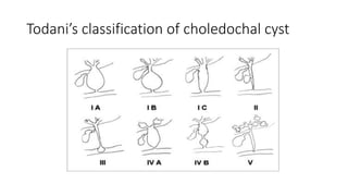
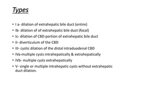
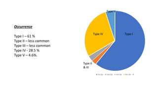
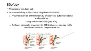
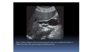
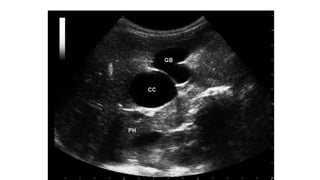
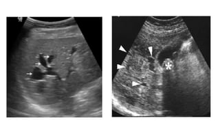
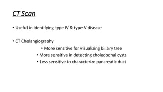
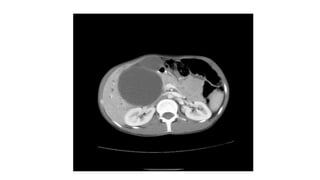
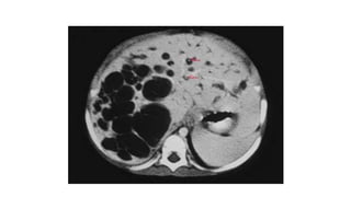
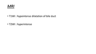
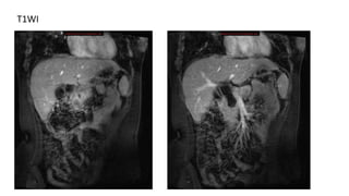
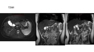
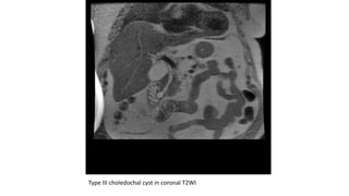
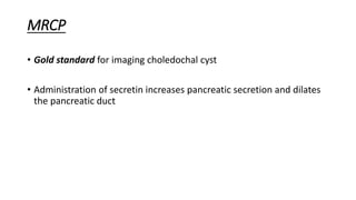
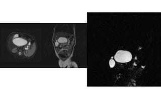
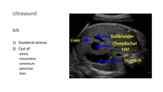
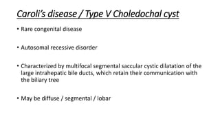
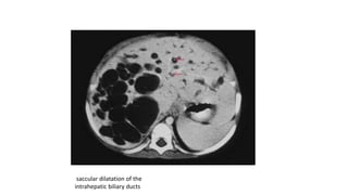
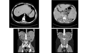
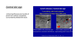
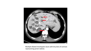
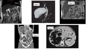
Choledochal cyst
Dr Manmohan Bir Shrestha
For RADIOLOGY
Objectives
Introduction
Biliary tree anatomy
Classification
Etiology
Clinical presentation
Laboratory investigations
Imaging modalities
Prenatal diagnosis
Caroli’s disease
Differentials
Treatment
Complications
Choledochal cyst
• Congenital anomaly involving cystic dilation of various ducts of the
biliary tract.
Epidemiology
• Incidence
• Western population-
1 in 100,000 to 150000 live births
• More common in Asia
• 1 in 1000 live births in Japan
• Sex prevalence
Female dominance-
• 3-4:1
Biliary tree anatomy
Intrahepatic
Ductular and canalicular network of the acini
Interlobular ducts
Septal bile ducts
RHD & LHD
Extrahepatic
RHD & LHD
CHD
Cystic duct & GB
CBD
Normal size of bile ducts
• CBD
• Adolescents & adults
• Upto 8mm
• Post cholecystectomy
• Upto 12 mm
• >60 years of age
• +1mm per decade
• Cystic duct
• Upto 1.8 mm
CHD at porta hepatis-
5 mm
Todani’s classification of choledochal cyst
Types
• I a- dilation of extrahepatic bile duct (entire)
• Ib- dilation of of extrahepatic bile duct (focal)
• Ic- dilation of CBD portion of extrahepatic bile duct
• II- diverticulum of the CBD
• III- cystic dilation of the distal intraduodenal CBD
• IVa-multiple cysts intrahepatically & extrahepatically
• IVb- multiple cysts extrahepatically
• V- single or multiple intrahepatic cysts without extrahepatic
duct dilation.
1st Qtr 2nd Qtr 3rd Qtr 4th Qtr
Type IV
Type V
Occurrence
Type I – 61 %
Type II – less common
Type III – less common
Type IV - 28.5 %
Type V – 4.6%.
Type I
Type II
& III
Types of union between CBD+MPD
• Normal junction (union inside duodenal
wall)
1) 2-10 mm short common channel with a
diameter of 3-5 mm(55-85%)
2) Separate entrances into
duodenum(42%)
3) 8-15 mm long common channel
• Abnormal junction (union outside
duodenal wall)
1) MPD inserting into CBD >15 MM
from entrance into duodenum
2) CBD entering into MPD
Etiology
• Weakness of the duct wall
• Pancreaticobiliary maljunction / Long common channel
o Proximal insertion of MPD into CBD or vice versa outside duodenal
wall producing
a long common channel (>15 mm)
o Reflux of pancreatic enzymes into CBD that causes damage to the
ductal wall and leads to cyst formation
Clinical presentation
• Triad of –
• Abdominal pain
• Jaundice
• Right upper quadrant palpable mass
Infantile form
• Present before 12 months of age
• Present with obstructive jaundice, acholic stools & hepatomegaly
• Signs of hepatic fibrosis may be present
Adult form
• Anytime after 12 months of age
• May present with fever, nausea, vomiting & jaundice.
• Undiagnosed cases may present with cholelithiasis.
Laboratory investigations
• Increased bilirubin
• Increased alkaline phosphatase
Histopathology
• Ducts are thickened (connective tissue+smooth muscle strands)
• Inflammatory reaction increases with age, may lead to ulceration in
mucosa & submucosa
• May lack a typical biliary mucosal lining
• Repeated destruction and regrowth causes epithelial metaplasia
• High risk of adenocarcinoma
Imaging modalities
• USG
• CT Scan
• CT cholangiography
• MRI
• MRCP
• ERCP
• PTC
• HIDA Scan
Objectives of imaging
• To classify
• Mention extension
• To visualize pancreaticobiliary junction
• To rule out association with other diseases
• To search for associated complications
• To rule out differentials
USG
• Ballooned/fusiform cyst beneath porta hepatis separate from GB
• *Communication with bile duct needs to be demonstrated*
• Abrupt change of caliber at junction of dilated segment to normal
ducts
• Intrahepatic bile duct dilatation secondary to stenosis.
CT Scan
• Useful in identifying type IV & type V disease
• CT Cholangiography
• More sensitive for visualizing biliary tree
• More sensitive in detecting choledochal cysts
• Less sensitive to characterize pancreatic duct
MRI
• T1WI : hypointense dilatation of bile duct
• T2WI : hyperintense
T1WI
T2WI
Type III choledochal cyst in coronal T2WI
MRCP
• Gold standard for imaging choledochal cyst
• Administration of secretin increases pancreatic secretion and dilates
the pancreatic duct
Type II ( diverticulum) choledochal cyst arising from lateral wall of CBD
ERCP
• More useful in type III choledochal cyst to exclude periampullary
mass
Fusiform choledochal cyst with a long common channel
& stricture at pancreatico biliary junction
Percutaneous transhepatic cholangiogram(PTC)
Cholangiogram – Caroli’s disease with characteristic strictures
and segmental intrahepatic duct dilations.
HIDA Scan
• The normal scan provides functional & morphological information
about the hepatic parenchyma in the first 10min ( after inj. of
radionuclide), extrahepatic biliary tree by 20 min and excretion into
the bowel by 1 hour.
• Useful in demonstrating biliary peritonitis after choledochal cyst
rupture
Prenatal diagnosis
Ultrasound
Earliest diagnosis at 25 weeks
MRI
Ultrasound
D/D
1) Duodenal atresia
2) Cyst of
-ovary
-mesentery
-omentum
-pancreas
-liver
Caroli’s disease / Type V Choledochal cyst
• Rare congenital disease
• Autosomal recessive disorder
• Characterized by multifocal segmental saccular cystic dilatation of the
large intrahepatic bile ducts, which retain their communication with
the biliary tree
• May be diffuse / segmental / lobar
• Types
1)Simple form
2)Caroli’s syndrome
• associated with periportal hepatic fibrosis
Etiology-
Ductal plate malformation
?perinatal hepatic artery
occlusion
?hypoplasia/aplasia of
fibromuscular wall
components
Association-
Benign renal tubular ectasia
Medullary sponge kidney(80%)
Infantile polycystic kidney
disease
Congenital hepatic fibrosis
saccular dilatation of the
intrahepatic biliary ducts
Central dot sign
-enhancing fibrovascular bundle of
portal vein radicles completely
surrounded by dilated bile ducts
Multiple dilated intrahepatic ducts with tiny dots of contrast
representing portal radicles
D/D of Caroli’s disease
Polycystic liver disease
No associated biliary duct dilatation
Rarely communicate with biliary ducts
Biliary hamartomas
Cyst size < 5mm (generally)
Do not communicate with biliary tree
Primary sclerosing cholangitis
Dilatation more fusiform & isolated
Associated inflammatory bowel disease
Recurrent pyogenic cholangitis
Saccular dilatation favors Caroli’s disease
Obstructive biliary dilatation
Type I
Type II
Type III
Type IV
Type V
D/D of Choledochal cyst
• Duodenal diverticulum
• Pancreatic pseudocyst / cystic tumors
• Other causes of biliary tree dilatation
Causes of Biliary Obstruction
Benign miscellaneous
Choledocholithiasis
Haemobilia
Congenital biliary disease
 Choledochal Cyst
 Biliary Atresia
Infectious cholangitis
Sclerosing cholangitis
Neoplasms
Cholangiocarcinoma
GB carcinoma
Locally invasive tumors
Ampullary tumors
Metastases
Extrinsic compression
Mirizzi syndrome
Pancreatitis
Adenopathy
Treatment
• Type I & IV
• Cyst excision & hepaticojejunostomy to a Roux-en-Y limb
• Type II
• Simple cyst resection
• Type III
• Marsupialization of the cyst ,
usually through transduodenal approach
• Type V
• Segmental resection of liver if disease is localized
• Liver transplantation
Complications
• Stones in GB, CBD, within cyst, in MPD, in intrahepatic biliary tree
• Malignant transformation
• Recurrent pancreatitis
• Cholangitis/Cholecystitis
• Cyst rupture with bile peritonitis
• Bleeding
• Biliary cirrhosis + portal hypertension
• Portal vein thrombosis
• Hepatic abscess
References
• Textbook of Radiology & Imaging - David Sutton 7th Edition
• Diagnostic Ultrasound - Carol M. Rumack 4th Edition
• Radiology Review Manual - Wolfgang Dahnert 7th Edition
• Bailey & Love’s Short Practice of Surgery - 7th Edition
• Paediatric Surgery - Arnold G. Coran - 7th Edition

Choledochal cyst

  • 1.
    Choledochal cyst Dr ManmohanBir Shrestha For RADIOLOGY
  • 2.
    Objectives Introduction Biliary tree anatomy Classification Etiology Clinicalpresentation Laboratory investigations Imaging modalities Prenatal diagnosis Caroli’s disease Differentials Treatment Complications
  • 3.
    Choledochal cyst • Congenitalanomaly involving cystic dilation of various ducts of the biliary tract.
  • 4.
    Epidemiology • Incidence • Westernpopulation- 1 in 100,000 to 150000 live births • More common in Asia • 1 in 1000 live births in Japan • Sex prevalence Female dominance- • 3-4:1
  • 5.
    Biliary tree anatomy Intrahepatic Ductularand canalicular network of the acini Interlobular ducts Septal bile ducts RHD & LHD Extrahepatic RHD & LHD CHD Cystic duct & GB CBD
  • 6.
    Normal size ofbile ducts • CBD • Adolescents & adults • Upto 8mm • Post cholecystectomy • Upto 12 mm • >60 years of age • +1mm per decade • Cystic duct • Upto 1.8 mm CHD at porta hepatis- 5 mm
  • 7.
  • 8.
    Types • I a-dilation of extrahepatic bile duct (entire) • Ib- dilation of of extrahepatic bile duct (focal) • Ic- dilation of CBD portion of extrahepatic bile duct • II- diverticulum of the CBD • III- cystic dilation of the distal intraduodenal CBD • IVa-multiple cysts intrahepatically & extrahepatically • IVb- multiple cysts extrahepatically • V- single or multiple intrahepatic cysts without extrahepatic duct dilation.
  • 9.
    1st Qtr 2ndQtr 3rd Qtr 4th Qtr Type IV Type V Occurrence Type I – 61 % Type II – less common Type III – less common Type IV - 28.5 % Type V – 4.6%. Type I Type II & III
  • 10.
    Types of unionbetween CBD+MPD • Normal junction (union inside duodenal wall) 1) 2-10 mm short common channel with a diameter of 3-5 mm(55-85%) 2) Separate entrances into duodenum(42%) 3) 8-15 mm long common channel • Abnormal junction (union outside duodenal wall) 1) MPD inserting into CBD >15 MM from entrance into duodenum 2) CBD entering into MPD
  • 11.
    Etiology • Weakness ofthe duct wall • Pancreaticobiliary maljunction / Long common channel o Proximal insertion of MPD into CBD or vice versa outside duodenal wall producing a long common channel (>15 mm) o Reflux of pancreatic enzymes into CBD that causes damage to the ductal wall and leads to cyst formation
  • 12.
    Clinical presentation • Triadof – • Abdominal pain • Jaundice • Right upper quadrant palpable mass Infantile form • Present before 12 months of age • Present with obstructive jaundice, acholic stools & hepatomegaly • Signs of hepatic fibrosis may be present Adult form • Anytime after 12 months of age • May present with fever, nausea, vomiting & jaundice. • Undiagnosed cases may present with cholelithiasis.
  • 13.
    Laboratory investigations • Increasedbilirubin • Increased alkaline phosphatase
  • 14.
    Histopathology • Ducts arethickened (connective tissue+smooth muscle strands) • Inflammatory reaction increases with age, may lead to ulceration in mucosa & submucosa • May lack a typical biliary mucosal lining • Repeated destruction and regrowth causes epithelial metaplasia • High risk of adenocarcinoma
  • 15.
    Imaging modalities • USG •CT Scan • CT cholangiography • MRI • MRCP • ERCP • PTC • HIDA Scan
  • 16.
    Objectives of imaging •To classify • Mention extension • To visualize pancreaticobiliary junction • To rule out association with other diseases • To search for associated complications • To rule out differentials
  • 17.
    USG • Ballooned/fusiform cystbeneath porta hepatis separate from GB • *Communication with bile duct needs to be demonstrated* • Abrupt change of caliber at junction of dilated segment to normal ducts • Intrahepatic bile duct dilatation secondary to stenosis.
  • 21.
    CT Scan • Usefulin identifying type IV & type V disease • CT Cholangiography • More sensitive for visualizing biliary tree • More sensitive in detecting choledochal cysts • Less sensitive to characterize pancreatic duct
  • 24.
    MRI • T1WI :hypointense dilatation of bile duct • T2WI : hyperintense
  • 25.
  • 26.
  • 27.
    Type III choledochalcyst in coronal T2WI
  • 28.
    MRCP • Gold standardfor imaging choledochal cyst • Administration of secretin increases pancreatic secretion and dilates the pancreatic duct
  • 30.
    Type II (diverticulum) choledochal cyst arising from lateral wall of CBD
  • 31.
    ERCP • More usefulin type III choledochal cyst to exclude periampullary mass Fusiform choledochal cyst with a long common channel & stricture at pancreatico biliary junction
  • 32.
    Percutaneous transhepatic cholangiogram(PTC) Cholangiogram– Caroli’s disease with characteristic strictures and segmental intrahepatic duct dilations.
  • 33.
    HIDA Scan • Thenormal scan provides functional & morphological information about the hepatic parenchyma in the first 10min ( after inj. of radionuclide), extrahepatic biliary tree by 20 min and excretion into the bowel by 1 hour. • Useful in demonstrating biliary peritonitis after choledochal cyst rupture
  • 34.
  • 35.
    Ultrasound D/D 1) Duodenal atresia 2)Cyst of -ovary -mesentery -omentum -pancreas -liver
  • 36.
    Caroli’s disease /Type V Choledochal cyst • Rare congenital disease • Autosomal recessive disorder • Characterized by multifocal segmental saccular cystic dilatation of the large intrahepatic bile ducts, which retain their communication with the biliary tree • May be diffuse / segmental / lobar
  • 37.
    • Types 1)Simple form 2)Caroli’ssyndrome • associated with periportal hepatic fibrosis
  • 38.
    Etiology- Ductal plate malformation ?perinatalhepatic artery occlusion ?hypoplasia/aplasia of fibromuscular wall components Association- Benign renal tubular ectasia Medullary sponge kidney(80%) Infantile polycystic kidney disease Congenital hepatic fibrosis
  • 40.
    saccular dilatation ofthe intrahepatic biliary ducts
  • 42.
    Central dot sign -enhancingfibrovascular bundle of portal vein radicles completely surrounded by dilated bile ducts
  • 43.
    Multiple dilated intrahepaticducts with tiny dots of contrast representing portal radicles
  • 44.
    D/D of Caroli’sdisease Polycystic liver disease No associated biliary duct dilatation Rarely communicate with biliary ducts Biliary hamartomas Cyst size < 5mm (generally) Do not communicate with biliary tree Primary sclerosing cholangitis Dilatation more fusiform & isolated Associated inflammatory bowel disease Recurrent pyogenic cholangitis Saccular dilatation favors Caroli’s disease Obstructive biliary dilatation
  • 45.
    Type I Type II TypeIII Type IV Type V
  • 46.
    D/D of Choledochalcyst • Duodenal diverticulum • Pancreatic pseudocyst / cystic tumors • Other causes of biliary tree dilatation
  • 47.
    Causes of BiliaryObstruction Benign miscellaneous Choledocholithiasis Haemobilia Congenital biliary disease  Choledochal Cyst  Biliary Atresia Infectious cholangitis Sclerosing cholangitis Neoplasms Cholangiocarcinoma GB carcinoma Locally invasive tumors Ampullary tumors Metastases Extrinsic compression Mirizzi syndrome Pancreatitis Adenopathy
  • 48.
    Treatment • Type I& IV • Cyst excision & hepaticojejunostomy to a Roux-en-Y limb • Type II • Simple cyst resection • Type III • Marsupialization of the cyst , usually through transduodenal approach • Type V • Segmental resection of liver if disease is localized • Liver transplantation
  • 49.
    Complications • Stones inGB, CBD, within cyst, in MPD, in intrahepatic biliary tree • Malignant transformation • Recurrent pancreatitis • Cholangitis/Cholecystitis • Cyst rupture with bile peritonitis • Bleeding • Biliary cirrhosis + portal hypertension • Portal vein thrombosis • Hepatic abscess
  • 50.
    References • Textbook ofRadiology & Imaging - David Sutton 7th Edition • Diagnostic Ultrasound - Carol M. Rumack 4th Edition • Radiology Review Manual - Wolfgang Dahnert 7th Edition • Bailey & Love’s Short Practice of Surgery - 7th Edition • Paediatric Surgery - Arnold G. Coran - 7th Edition