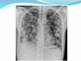
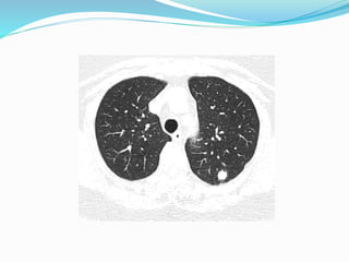
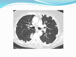
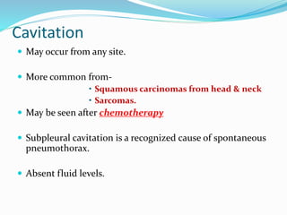
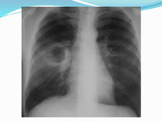
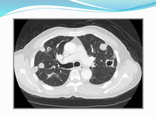
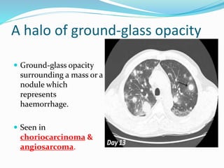
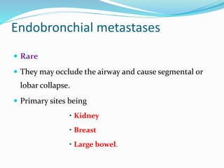
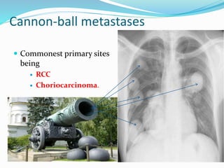
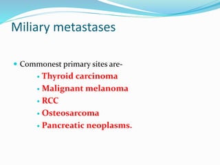
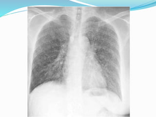
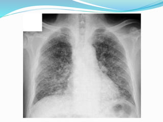
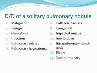
WELCOME
Date-15/6/2015
Dr. Manmohan Bir Shrestha
MD Resident, Phase-A
Department of Radiology & Imaging
Metastasis
 Metastasis are the tumor implants discontinuous with
primary tumor.
 It is the hallmark of malignancy.
 All cancers can metastasize with few exceptions. The
major exceptions are gliomas and basal cell carcinoma.
Pulmonary Metastases
 In about 75 % cases, presents as multiple pulmonary
nodules.
 Also present as solitary pulmonary nodule/ cavitation/
calcification.
 Approximately 3 % of asymptomatic pulmonary
nodules are metastases.
Site
 Usually bilateral, affecting both lungs equally with a
basal predominance.
 They are often peripheral and may be subpleural.
Route of spread
 Most commonly - haematogenous.
 Lymphatic spread - less common.
 Endobronchial spread - rare
Lungs – Filter like Organ
 Supplied by pulmonary artery containing
deoxygenated blood from right ventricle.
 This blood contains lymphatic fluid from the body
tissues which flows into the lungs.
Primary site
May originate at any site.
 Approximately 80 % of pulmonary metastases
arise from primary tumours of-
• Breast
• Skeleton
• Urogenital system.
Clinical features
 Cough
 Dyspnea or shortness of breath
 Chest pain
 Haemoptysis
 Hoarseness of voice
 Features of secondary pulmonary infection.
Radiological findings
Common Other patterns
 Nodules.  Cavitation
 Calcification
 Pneumonia-like consolidation
 A halo of ground glass opacity.
 Endobronchial
 Cannon-ball metastases
 Miliary metastases
 Lymphangitis carcinomatosa.
Nodules
 May be solitary or multiple.
 Rounded nodules of variable sizes ranging from few
millimeters to few centimeters with well-defined margin.
75 % - multiple pulmonary nodules.
 Commonest tumours producing solitary pulm. nodules are
carcinomas of –
• Colon
• Kidney
• Breast
• Testicular tumours
• Bone sarcomas
• Malignant melanoma.
Cavitation
 May occur from any site.
 More common from-
• Squamous carcinomas from head & neck
• Sarcomas.
 May be seen after chemotherapy
 Subpleural cavitation is a recognized cause of spontaneous
pneumothorax.
 Absent fluid levels.
Calcification
 Is seen in some cases.
 Most often in
• Osteogenic sarcoma
• Chondrosarcoma
• Mucinous adenocarcinoma.
Pneumonia-like consolidation
 Adenocarcinoma metastases may destroy adjacent
lung parenchyma, resulting in pneumonia-like
consolidation.
A halo of ground-glass opacity
 Ground-glass opacity
surrounding a mass or a
nodule which
represents
haemorrhage.
 Seen in
choriocarcinoma &
angiosarcoma.
Endobronchial metastases
 Rare
 They may occlude the airway and cause segmental or
lobar collapse.
 Primary sites being
• Kidney
• Breast
• Large bowel.
Cannon-ball metastases
 Commonest primary sites
being
 RCC
 Choriocarcinoma.
Miliary metastases
 Commonest primary sites are-
 Thyroid carcinoma
 Malignant melanoma
 RCC
 Osteosarcoma
 Pancreatic neoplasms.
Lymphangitis Carcinomatosa
 Results from haematogenous metastases invading and
occluding peripheral pulmonary lymphatics.
 Commonest primary sites are
 Lung
 Breast
 Stomach
 Pancreas
 Cervix
 prostate
Cont…
 Usually bilateral, but lung and breast cancer may
cause unilateral lymphangitis.
 Chest X-ray
 Coarse, linear, reticular and nodular basal shadowing
often with pleural effusions and hilar
lymphadenopathy.
Cont…
 HRCT
 Nodular thickening of the interlobular septa and
thickening of the centrilobular bronchovascular
bundles.
D/D of a solitary pulmonary nodule
1. Malignant
2. Benign
3. Granuloma
4. Infection
5. Pulmonary infarct
6. Pulmonary haematoma
7. Collagen diseases
8. Congenital
9. Impacted mucus
10. Amyloidosis
11. Intrapulmonary lymph
node
12. Pleural
13. Non-pulmonary
D/D of a multiple pulmonary nodule
1. Malignant
2. Benign
3. Infection
4. Inflammatory
5. Vascular
6. Miscellaneous.
D/D of a cavitating pulmonary lesion
1. Infections
2. Malignant
3. Abscess
4. Pulmonary infarct
5. Pulmonary haematoma
6. Pneumoconiosis
7. Developmental
8. Sarcoidosis
9. Bulles, blebs
10. Traumatic lung cysts
11. Pneumatocele.
Approaching to the pulm. metastases
 Pulmonary metastases –initial finding
 “Unknown Primary”.
 Clinical presentation and clinical evaluation.
Cont…
 Chest X-ray
 CT Scan
 PET Scan
Cont.
 Cytological examination of sputum
 Cytological examination of pleural fluid
 FNAC or excision biopsy of an enlarged lymph node
 Bronchoscopy-
• biopsy
• bronchoscopic alveolar lavage cytology.
Cont…
 USG guided FNAC of lesion
 CT guided FNAC of lesion
 Surgical lung biopsy or open lung biopsy
 HRCT.
Complications
1. Pneumothorax
2. Pleural effusion
3. Lung collapse.
Treatment modalities
 Chemotherapy
 Radiotherapy
 Placements of stents in the airway
 Surgical
 Palliative care.
THANK YOU

Pulmonary metastases

  • 1.
  • 2.
    Dr. Manmohan BirShrestha MD Resident, Phase-A Department of Radiology & Imaging
  • 3.
    Metastasis  Metastasis arethe tumor implants discontinuous with primary tumor.  It is the hallmark of malignancy.  All cancers can metastasize with few exceptions. The major exceptions are gliomas and basal cell carcinoma.
  • 4.
    Pulmonary Metastases  Inabout 75 % cases, presents as multiple pulmonary nodules.  Also present as solitary pulmonary nodule/ cavitation/ calcification.  Approximately 3 % of asymptomatic pulmonary nodules are metastases.
  • 5.
    Site  Usually bilateral,affecting both lungs equally with a basal predominance.  They are often peripheral and may be subpleural.
  • 6.
    Route of spread Most commonly - haematogenous.  Lymphatic spread - less common.  Endobronchial spread - rare
  • 7.
    Lungs – Filterlike Organ  Supplied by pulmonary artery containing deoxygenated blood from right ventricle.  This blood contains lymphatic fluid from the body tissues which flows into the lungs.
  • 8.
    Primary site May originateat any site.  Approximately 80 % of pulmonary metastases arise from primary tumours of- • Breast • Skeleton • Urogenital system.
  • 9.
    Clinical features  Cough Dyspnea or shortness of breath  Chest pain  Haemoptysis  Hoarseness of voice  Features of secondary pulmonary infection.
  • 10.
    Radiological findings Common Otherpatterns  Nodules.  Cavitation  Calcification  Pneumonia-like consolidation  A halo of ground glass opacity.  Endobronchial  Cannon-ball metastases  Miliary metastases  Lymphangitis carcinomatosa.
  • 11.
    Nodules  May besolitary or multiple.  Rounded nodules of variable sizes ranging from few millimeters to few centimeters with well-defined margin. 75 % - multiple pulmonary nodules.  Commonest tumours producing solitary pulm. nodules are carcinomas of – • Colon • Kidney • Breast • Testicular tumours • Bone sarcomas • Malignant melanoma.
  • 15.
    Cavitation  May occurfrom any site.  More common from- • Squamous carcinomas from head & neck • Sarcomas.  May be seen after chemotherapy  Subpleural cavitation is a recognized cause of spontaneous pneumothorax.  Absent fluid levels.
  • 18.
    Calcification  Is seenin some cases.  Most often in • Osteogenic sarcoma • Chondrosarcoma • Mucinous adenocarcinoma.
  • 19.
    Pneumonia-like consolidation  Adenocarcinomametastases may destroy adjacent lung parenchyma, resulting in pneumonia-like consolidation.
  • 20.
    A halo ofground-glass opacity  Ground-glass opacity surrounding a mass or a nodule which represents haemorrhage.  Seen in choriocarcinoma & angiosarcoma.
  • 21.
    Endobronchial metastases  Rare They may occlude the airway and cause segmental or lobar collapse.  Primary sites being • Kidney • Breast • Large bowel.
  • 22.
    Cannon-ball metastases  Commonestprimary sites being  RCC  Choriocarcinoma.
  • 23.
    Miliary metastases  Commonestprimary sites are-  Thyroid carcinoma  Malignant melanoma  RCC  Osteosarcoma  Pancreatic neoplasms.
  • 25.
    Lymphangitis Carcinomatosa  Resultsfrom haematogenous metastases invading and occluding peripheral pulmonary lymphatics.  Commonest primary sites are  Lung  Breast  Stomach  Pancreas  Cervix  prostate
  • 26.
    Cont…  Usually bilateral,but lung and breast cancer may cause unilateral lymphangitis.  Chest X-ray  Coarse, linear, reticular and nodular basal shadowing often with pleural effusions and hilar lymphadenopathy.
  • 28.
    Cont…  HRCT  Nodularthickening of the interlobular septa and thickening of the centrilobular bronchovascular bundles.
  • 29.
    D/D of asolitary pulmonary nodule 1. Malignant 2. Benign 3. Granuloma 4. Infection 5. Pulmonary infarct 6. Pulmonary haematoma 7. Collagen diseases 8. Congenital 9. Impacted mucus 10. Amyloidosis 11. Intrapulmonary lymph node 12. Pleural 13. Non-pulmonary
  • 30.
    D/D of amultiple pulmonary nodule 1. Malignant 2. Benign 3. Infection 4. Inflammatory 5. Vascular 6. Miscellaneous.
  • 31.
    D/D of acavitating pulmonary lesion 1. Infections 2. Malignant 3. Abscess 4. Pulmonary infarct 5. Pulmonary haematoma 6. Pneumoconiosis 7. Developmental 8. Sarcoidosis 9. Bulles, blebs 10. Traumatic lung cysts 11. Pneumatocele.
  • 32.
    Approaching to thepulm. metastases  Pulmonary metastases –initial finding  “Unknown Primary”.  Clinical presentation and clinical evaluation.
  • 33.
    Cont…  Chest X-ray CT Scan  PET Scan
  • 34.
    Cont.  Cytological examinationof sputum  Cytological examination of pleural fluid  FNAC or excision biopsy of an enlarged lymph node  Bronchoscopy- • biopsy • bronchoscopic alveolar lavage cytology.
  • 35.
    Cont…  USG guidedFNAC of lesion  CT guided FNAC of lesion  Surgical lung biopsy or open lung biopsy  HRCT.
  • 36.
    Complications 1. Pneumothorax 2. Pleuraleffusion 3. Lung collapse.
  • 37.
    Treatment modalities  Chemotherapy Radiotherapy  Placements of stents in the airway  Surgical  Palliative care.
  • 38.