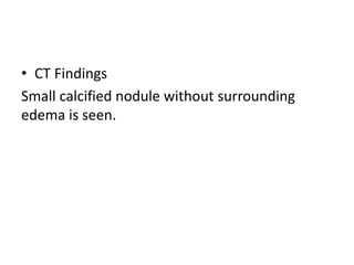
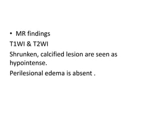
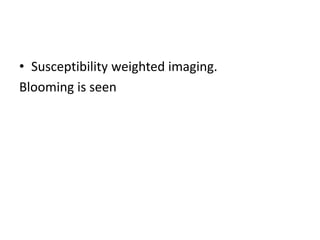
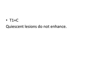
CNS INFECTIONS
Acquired pyogenic infections
• Meningitis
• Abscess
• Ventriculitis
• Empyema
Meningitis
Meningitis is acute and chronic inflammatory
infiltrate of meninges and CSF.
o Etiology
o Pathology
o Imaging
o Differential diagnosis.
• Etiology
o Haematogenous spread.
o Direct geographic extensions from sinusitis,
otitis or mastoiditis.
• Pathology
I. Cloudy CSF fills the subarchnoid spaces,
followed by development of variably dense
purulent exudate that covers the pial
surfaces.
II. Basal cisterns and subarachnoid spaces are
the most commonly involved by meningitis.
Imaging
• Imaging should be use in conjunction with
appropriate clinical and laboratory evaluation.
• CT findings
Non contrast CT
-mild ventricular enlargement with slightly
blurred margins.
- Cisterns and sulci appears effaced as they
become almost isodense with brain.
• Contrast enhanced CT.
Intense enhancement of inflammatory exudate
as it covers brain surfaces as it covers brain
surfaces extending into and filling the sulci.
• MR findings.
T1WI
Purulent exudate are isointense to underlying
brain.
• T2WI
Exudate are isointense to CSF.
• FLAIR
Exudates do not supressed on FLAIR.
• Diffusion weighted imaging (DWI)
Purulent subarachnoid spaces shows restriction
on diffusion.
• T1+C
Meningitis enhances uniformly and intensely.
Curvilinear pattern that follow gyri and sulci.
• Differential diagnosis.
Carcinomatous meningitis appears identical on
imaging so clinical information is essential.
Abscess
• Localized infection of brain parenchyma.
• Etiology
1. Haematogenous spread
2. Penetrating injury
3. Direct geographical extension from sinonasal
or otomastiod infections
• Pathology
4 Stages are recognised in evolution of cerebral
abscess.
1. Early cerebritis.
2. Late cerebritis.
3. Early capsule.
4. Late capsule.
Imaging
• Early cerebritis.
Infection is focal but not yet localized.
Begins 3-5 days after initial infection.
• CT findings.
A poorly marginated cortical/ subcrotical mass.
No enhancemet on CECT.
• MRI findings.
T1WI
Hypointense to isointense .
• T2WI/FLAIR
Hyperintense
• DWI
Restriction of diffusion is noted.
• Susceptibility weighted imaging (SWAN)
Punctate blooming is noted due to
haemorrhagic foci
• T1+C
Patchy enhancement may or may not be seen
• Late cerebritis.
Necrotic centre is surrounded by poorly
oraganised rim of inflammatory cells.
Begin at 4 days and last between 10 days to
2weeks.
• CT Findings.
NECT- better delineate central hypodense mass
with surrounding edema
CECT-Irregular rim enhancement.
• MR findings.
T1WI- Hypointense center and iso-mildly
hyperintense rim.
• T2WI
Central core is hyperintense and rim is relatively
hypointense.
• DWI
Strong restriction is noted.
• T1+C
Intense irregular rim enhancement is noted
• Early capsule
Necrotic core liquifies and proliferating
granulation tissue around the rim gradually
forms well delineated collagen capsule.
Stage around 2 weeks and may last for month or
two
• TWI/FLAIR
Well delineated round / ovoid mass with
liquefied hyperintense core.
• DWI
Necrotic center restrict strongly.
• T1+C
Rim enhancement rim is thinnest on deeper
side.
4. Late capsule
Central cavity gradually involutes and shrinks
Collagen deposition further thicken the wall.
This stage begins several weeks following
infection and last for several months.
• Contrast enhancement may persist for
months.
Differential diagnosis
• Early cerebritis are poorly defined may mimic
cerebral ischemia or neoplasm.
• Ring enhancing lesions , the most common
differential is infection vs
neoplasm(glioblastoma or metastasis)
Ventriculitis
• A collection of purulent material in the
ventricle is more likely due to intraventricular
rupture of brain abscess.
• Risk is increases if abscess is deep seated,
multiloculated or close to ventricular wall.
• Imaging
CT findings
Debris level in the dependent part of occipital
horn together with periventricular hypodensity.
• MR findings
T2WI
Ventricular debris appears hypointense with
layering in the occipital horn.
• T1+C
Some degree of ependymal enhancement can
be identified.
Debris fluid level is noted in the dependent part
of occipital horns.
• DWI
Diffusion shows diffusion restriction of layered
debris.
Differential diagnosis
• Primary malignant CNS neoplasm such as
GBM and primary CNS lymphoma can spread
TUBERCULOSIS
• Etiology
• Pathology
• Imaging
• Differential diagnosis.
• Etiology
Mycobacterium tuberculosis complex
o Vast majority caused by M. tuberculosis
o Other mycobacteria(m. bovis) rare.
Neurotuberculosis is secondary to hematogenous
spread extracranial infection, most frequently in
the lung.
Pathology
CNS TB has several distinct pathological manifestations
• TB meningitis (70-80%)
Exudative, proliferative and necrotizing component in sub
arachnoid cistern.
• Tuberculoma (TB granuloma)(20-30%)
Focal parenchymal infection with central caseating
necrosis.
• Pseudoabscesses
Found in 20% of patients coinfected with TB and HIV
IMAGING
• TB meningitis
Non contrast CT findings
-blurred ventricular margins
-effacement of basal cisterns by hyperdense
exudate.
• Contrast enhance CT findings
-Intense enhancement of basilar meninges and
subarachnoid spaces.
• MRI findings
T1WI basilar exudates are isointense with brain
giving the apperance of dirty CSF.
• FLAIR
Increased signal intensity in the sulci and
cisterns
• T1 C+FS
Marked linear and nodular enhancement is
noted.
• Vascular complications are common
complication of TBM
-flow voids of major arteries appears reduced.
-parenchyma adjacent to meningeal
inflammation may demonstrate necrosis.
-penetrating artery infarct with restricted
diffusion are common.
• Tuberculoma
Non contrast CT findings
-Isodense to slightly hyperdense round,
lobulated mass with variable perilesional
edema.
-Calcifications can be seen in healed ganuloma
• Contrast enhanced CT
Punctate solid ring like enhancement.
• MRI findings
T1WI
Appears hypointense or isointense to brain
parenchyma.
• T2WI
-Hypointense
-Liquified areas may be hyperinetnse with
hypointense rim(caseating tuberculoma)
• T1+C
Ring like enhancement with around the no
enhancing centre.
• MR spectroscopy
Helpful in characterizing and distinguishing the
tuberculomas from pyogenic abscess and
neoplasm.
A large lipid peak with abcesnce of other
metabolites and succinate is seen in 85-90%
cases
Differential diagnosis
• TBM
1. Pyogenic meningitis.
2. Carcinomatous meningitis
• Tuberculomas
1. NCC
2. Pygenic abscess.
3. Neoplasm.
Neurocysticercsis
• Most common parasitic infection in the world.
• CNS lesions developed in 60-70% of patients
with cysticercosis.
Etiology
Pathology
Imaging
Differential diagnosis
• Etiology
NCC caused by encysted larvae of pork
tapeworm taenia solium .
• Pathology
four stages of NCC development and regression are
recognised.
Patients may have multiple lesions at different stage
of evolution.
1. Vesicular stage
2. Colloid vesicular stage.
3. Granular nodular stage.
4. Nodular calcified stage.
Imaging
• Vesicular stage
Viable larvae appears as translucent, thin
walled, fluid filed cystic lesion with eccentrically
located scolex.
• Non contrast CT
Smooth thin walled cyst that is isodense to CSF.
• Contrast enhanced CT.
No enhancement , no surrounding edema.
• MR findings
Cyst is isointense to CSF on T1WI, T2WI and
FLAIR.
Scolex is hyperintense.
• T1+C
Enhancement is absent
Lack of perilesional edema.
• Colloid vesicular stage.
Larvae begin to degenerate
Cyst fluid become thick and turbid
A fibrous capsule develops and perilesional
edema becomes prominent.
• Non contrast CT Findings
Cyst fluid is hyperdense relative to CSF.
• Contrast enhanced CT
Ring enhancing capsule .
Moderate perilesional edema.
• MR findings
T1WI
Cyst fluid is hyperintense to CSF
• FLAIR
-Scolex appears hypeintense
-Moderate to marked surrounding perilesional
edema is noted.
• T1+C
Intense enhancement of cyst wall and often
shaggy.
• Granular nodular stage.
Collapse and retraction of cyst into
granulomatous nodule that will eventually
calcify.
• CT findings
Noncontrast CT
Mild residual edema.
• Contrast enhanced CT
Involuting, mildly to moderately enhancing
nodule
• Nodular calcified stage.
Entire lesion become a fibrocalcified nodule
• CT Findings
Small calcified nodule without surrounding
edema is seen.
• MR findings
T1WI & T2WI
Shrunken, calcified lesion are seen as
hypointense.
Perilesional edema is absent .
• Susceptibility weighted imaging.
Blooming is seen
• T1+C
Quiescent lesions do not enhance.
Differential diagnosis
• Subarachnoid/ cisternal NCC resembles TB
meningitis
• A giant parenchymal colliodal- vesicular NCC
mimimcs neoplasm, tuberculoma or
toxoplasmosis.

Cns infections