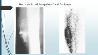
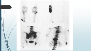
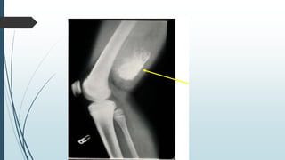
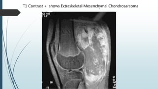
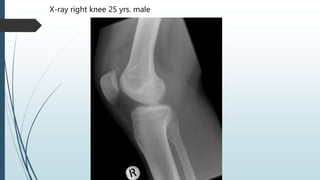
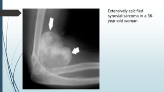
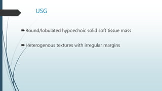
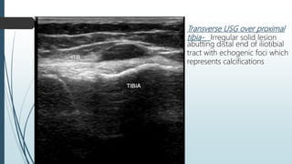
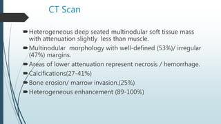
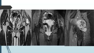
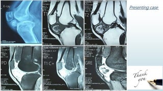
Mesenchymal
Chondrosarcoma
Dr. Manmohan Bir Shrestha
Radiology & ImagingFOR RADIOLOGY
Extraskeletal Mesenchymal Chondrosarcoma
Synovial Sarcoma
Chondrosarcoma
2nd most frequent primary malignant tumor of bone.
of cartilaginous origin
tumor matrix formation is entirely chondroid in nature.
2 Extraskeletal varieties (attributes 2 % of all soft tissue
sarcomas)
Myxoid
Mesenchymal
Extraskeletal Mesenchymal Chondrosarcoma
Rare
50% arise in soft tissues
Sex Prevalence – M:F= 1:1
Bimodal age distribution
3rd decade
Tumors of head & neck
Common in meninges & periorbital region
5th decade
Tumors of thigh, trunk & calf
Clinical feature
Localized swelling, slow-growing but some has
aggressive nature
Localized pain
Joint immobility if near joint
Some patient may present with metastases
Histology
More or less differentiated cartilage cells
Proliferation of small mesenchymal cells
Hemangiopericytoma like vascular pattern
Metastases
 to lungs & lymph nodes.
Plain radiograph & CT Scan
Soft tissue mass with matrix mineralization
(50-100%) rings +arcs/flocculent
+ stippled calcification/dense mineralization
MRI
T1WI – isointense to muscle
T2WI – fat signal intensity
Signal voids from calcification
After contrast – homogenous/heterogenous
enhancement
Radionuclide scan
Increased vascularity in blood pool phase.
Hard mass in middle-aged man’s calf for 8 years
T1 Contrast + shows Extraskeletal Mesenchymal Chondrosarcoma
Treatment
Surgical resection
Chemotherapy
Radiotherapy
Prognosis
25% 10 year survival rate
Synovial Sarcoma
Definition :
Slow growing expansile malignant tumor originating with
extensive malignant potential.
Synonyms :
Malignant Synovioma
Tendosynovial Sarcoma
Synovioblastic Sarcoma
Synovial Endothelioma
Incidence :
4th most common soft tissue sarcoma
(after malignant fibrous histiocytoma,
liposarcoma and rhabdomyosarcoma)
2-10% of all primary malignancies worldwide.
Age : Mean age 35 yrs.
(84% between 15-50 yrs. )
Sex : M/F = 1-1.2/1
Site :
 in synovial lining/bursa /tendon sheath adjacent to joint
 Uncommonly intraarticular (5-10%)
 arise near joints, it is rare for them to arise from the joint itself and
despite their name, they do not arise from synovial structures, e.g.
joints, tendon sheaths and bursae.
Location :
 Lower extremity (2/3rd 0f the cases)
 Popliteal fossa, hip, foot & ankle
 Upper extremity (1/3rd of the cases)
 Elbow, wrist & hands.
 Pelvis (8%)
 Trunk (7%)
 Head & neck (5%)
 Retroperitoneum(0.3%)
Clinical features
 Slow growing Soft tissue mass or
swelling associated with local pain.
 Metastases present in 16-25% at presentation.
(can cause cannon ball metastases to lungs)
Imaging modalities
Plain x-ray
CT scan
MRI
USG
Radionuclide scanning
Plain X-ray
Large spheroid well-defined soft tissue mass.
Amorphous punctate calcification/ ossification (often eccentric
or peripheral )
Involvement of adjacent bone (11-20%)
Periosteal reaction
Invasion of cortex with wide zone of transition
Juxta articular osteoporosis
Lesion about 1 cm away from joint cartilage
*** Calcifications in other soft-tissue sarcomas are uncommon
X-ray right knee 25 yrs. male
Extensively calcified
synovial sarcoma in a 36-
year-old woman
USG
Round/lobulated hypoechoic solid soft tissue mass
Heterogenous textures with irregular margins
Transverse USG over proximal
tibia- Irregular solid lesion
abutting distal end of iliotibial
tract with echogenic foci which
represents calcifications
CT Scan
Heterogeneous deep seated multinodular soft tissue mass
with attenuation slightly less than muscle.
Multinodular morphology with well-defined (53%)/ irregular
(47%) margins.
Areas of lower attenuation represent necrosis / hemorrhage.
Calcifications(27-41%)
Bone erosion/ marrow invasion.(25%)
Heterogeneous enhancement (89-100%)
MRI
T1WI -Prominently heterogeneous soft-tissue mass
with signal intensity similar to or slightly higher than
muscle.
Triple sign on T2WI in 35-57%
Hypo –calcified/fibrotic tissue
Iso – solid cellular elements
Hyper – hemorrhage/necrosis
Fluid-fluid levels due to previous hemorrhage.
Bone marrow invasion /Cortical erosion (21%)
Neurovascular encasement
Contrast Enhanced MRI
 Prominent heterogeneous (83%)/homogeneous enhancement(17%)
 Peripheral / Nodular enhancement for necrotic tissue
 Serpentine vascular channels.
 Initially rapid progressive increase in signal intensity followed by wash
out(60%) / late sustained increase (40%)
20 yrs. female with left hip pain , x-ray shows lucency over left greater
trochanter
Radionuclide Scan
Prominently increased uptake in blood flow & blood
pool phase.
Heterogeneous mild uptake due to mixture of viable
& necrotic tissue, perhaps associated with
calcification.
Metastases
 Lungs (94%)
 Lymph nodes (4-18%)
 Bone (8-11%)
* Metastasis is present in 16-25% cases at presentation.
Treatment
Local excision /amputation.
Radiotherapy.
Chemotherapy.
Prognosis
Local recurrence (30-50%)
5 yrs. survival rate (36-76%)
10 yrs. survival rate (20-63%)
Poor prognostic factors
Tumor size > 5 cm.
Presence of poorly differentiated areas.
Summary
Incidence
Synovial sarcoma > Mesenchymal Chondrosarcoma.
Contents (Both)
Soft tissue component
Calcified areas
Mineralization
Cystic areas
Necrosis/hemorrhage
Site
Around joints or soft tissue
Age
Mesenchymal chondrosarcoma bimodal (3rd & 5th decade)
Synovial Sarcoma (15-50, mean 35 years)
Histopathological differentiation remains specific
Presenting caseX-ray
T1 T2
PD T1 C+ GRE

Extraskeletal Mesenchymal Chondrosarcoma

  • 1.
    Mesenchymal Chondrosarcoma Dr. Manmohan BirShrestha Radiology & ImagingFOR RADIOLOGY
  • 2.
  • 4.
    Chondrosarcoma 2nd most frequentprimary malignant tumor of bone. of cartilaginous origin tumor matrix formation is entirely chondroid in nature. 2 Extraskeletal varieties (attributes 2 % of all soft tissue sarcomas) Myxoid Mesenchymal
  • 5.
    Extraskeletal Mesenchymal Chondrosarcoma Rare 50%arise in soft tissues Sex Prevalence – M:F= 1:1 Bimodal age distribution 3rd decade Tumors of head & neck Common in meninges & periorbital region 5th decade Tumors of thigh, trunk & calf
  • 6.
    Clinical feature Localized swelling,slow-growing but some has aggressive nature Localized pain Joint immobility if near joint Some patient may present with metastases
  • 7.
    Histology More or lessdifferentiated cartilage cells Proliferation of small mesenchymal cells Hemangiopericytoma like vascular pattern Metastases  to lungs & lymph nodes.
  • 8.
    Plain radiograph &CT Scan Soft tissue mass with matrix mineralization (50-100%) rings +arcs/flocculent + stippled calcification/dense mineralization
  • 9.
    MRI T1WI – isointenseto muscle T2WI – fat signal intensity Signal voids from calcification After contrast – homogenous/heterogenous enhancement Radionuclide scan Increased vascularity in blood pool phase.
  • 10.
    Hard mass inmiddle-aged man’s calf for 8 years
  • 13.
    T1 Contrast +shows Extraskeletal Mesenchymal Chondrosarcoma
  • 14.
  • 16.
    Synovial Sarcoma Definition : Slowgrowing expansile malignant tumor originating with extensive malignant potential. Synonyms : Malignant Synovioma Tendosynovial Sarcoma Synovioblastic Sarcoma Synovial Endothelioma
  • 17.
    Incidence : 4th mostcommon soft tissue sarcoma (after malignant fibrous histiocytoma, liposarcoma and rhabdomyosarcoma) 2-10% of all primary malignancies worldwide. Age : Mean age 35 yrs. (84% between 15-50 yrs. ) Sex : M/F = 1-1.2/1
  • 18.
    Site :  insynovial lining/bursa /tendon sheath adjacent to joint  Uncommonly intraarticular (5-10%)  arise near joints, it is rare for them to arise from the joint itself and despite their name, they do not arise from synovial structures, e.g. joints, tendon sheaths and bursae. Location :  Lower extremity (2/3rd 0f the cases)  Popliteal fossa, hip, foot & ankle  Upper extremity (1/3rd of the cases)  Elbow, wrist & hands.  Pelvis (8%)  Trunk (7%)  Head & neck (5%)  Retroperitoneum(0.3%)
  • 19.
    Clinical features  Slowgrowing Soft tissue mass or swelling associated with local pain.  Metastases present in 16-25% at presentation. (can cause cannon ball metastases to lungs)
  • 20.
    Imaging modalities Plain x-ray CTscan MRI USG Radionuclide scanning
  • 21.
    Plain X-ray Large spheroidwell-defined soft tissue mass. Amorphous punctate calcification/ ossification (often eccentric or peripheral ) Involvement of adjacent bone (11-20%) Periosteal reaction Invasion of cortex with wide zone of transition Juxta articular osteoporosis Lesion about 1 cm away from joint cartilage *** Calcifications in other soft-tissue sarcomas are uncommon
  • 22.
    X-ray right knee25 yrs. male
  • 23.
  • 24.
    USG Round/lobulated hypoechoic solidsoft tissue mass Heterogenous textures with irregular margins
  • 25.
    Transverse USG overproximal tibia- Irregular solid lesion abutting distal end of iliotibial tract with echogenic foci which represents calcifications
  • 26.
    CT Scan Heterogeneous deepseated multinodular soft tissue mass with attenuation slightly less than muscle. Multinodular morphology with well-defined (53%)/ irregular (47%) margins. Areas of lower attenuation represent necrosis / hemorrhage. Calcifications(27-41%) Bone erosion/ marrow invasion.(25%) Heterogeneous enhancement (89-100%)
  • 27.
    MRI T1WI -Prominently heterogeneoussoft-tissue mass with signal intensity similar to or slightly higher than muscle. Triple sign on T2WI in 35-57% Hypo –calcified/fibrotic tissue Iso – solid cellular elements Hyper – hemorrhage/necrosis Fluid-fluid levels due to previous hemorrhage. Bone marrow invasion /Cortical erosion (21%) Neurovascular encasement
  • 28.
    Contrast Enhanced MRI Prominent heterogeneous (83%)/homogeneous enhancement(17%)  Peripheral / Nodular enhancement for necrotic tissue  Serpentine vascular channels.  Initially rapid progressive increase in signal intensity followed by wash out(60%) / late sustained increase (40%)
  • 29.
    20 yrs. femalewith left hip pain , x-ray shows lucency over left greater trochanter
  • 32.
    Radionuclide Scan Prominently increaseduptake in blood flow & blood pool phase. Heterogeneous mild uptake due to mixture of viable & necrotic tissue, perhaps associated with calcification.
  • 33.
    Metastases  Lungs (94%) Lymph nodes (4-18%)  Bone (8-11%) * Metastasis is present in 16-25% cases at presentation.
  • 34.
  • 35.
    Prognosis Local recurrence (30-50%) 5yrs. survival rate (36-76%) 10 yrs. survival rate (20-63%) Poor prognostic factors Tumor size > 5 cm. Presence of poorly differentiated areas.
  • 36.
    Summary Incidence Synovial sarcoma >Mesenchymal Chondrosarcoma. Contents (Both) Soft tissue component Calcified areas Mineralization Cystic areas Necrosis/hemorrhage Site Around joints or soft tissue Age Mesenchymal chondrosarcoma bimodal (3rd & 5th decade) Synovial Sarcoma (15-50, mean 35 years) Histopathological differentiation remains specific
  • 37.