MANAGEMENT of
ACUTE PANCREATITIS
Dr. Aishwarya Bhattacharya
• Acute pancreatitis – disease of high morbidity and motality
• Mortality • Mild cases : ~1%
• Severe cases : (10-30)%
EVIDENCE BASED
APPROACH
GUIDELINES
• Atlanta
• British Society of Gastroenterology
• American College of Gastroenterology
• International Association of Pancreas
• Santorini Conference
• World Congress of Gastroenterology
Risk factor assessment
Clinical risk stratification
Monitoring response
To initial therapy
3D Approach towards management
of acute pancreatitis
• Risk stratification Mild cases – general ward
Severe cases – Always in ICU setting
Strategy tailoring according to :
•Severity
•Risk factors (eg: age,obesity)
•Presence of SIRS
•Routine lab values(Hct, Ser.creatinine )
NONOPERATIVE MANAGEMENT IS THE MAINSTAY
PRINCIPLES of Management :
Fluid resuscitation
Nutritional Support
Symptomatic Treatment
Management of Metabolic Complications
Prophylactic Antibiotic Coverage
Monitoring and Reassessment
Role of ERCP
Role of surgery
FLUID RESUSCITATION
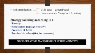
• Approach : Aggressive fluid resuscitation
• Amount of fluid required : (250-500) ml/hr [acc. to AGC guidelines ]
or
(5-10) ml/kg/hr [acc. to IAP guidelines ]
• Ideal fluid : Isotonic crystalloids – RINGER LACTATE
In severe volume depletion -20ml/kg over 30 min
followed by
3ml/kg/hr for (8-12) hrs
• Goal : Reduction in BUN
IAP suggested resuscitation goals :
* HR < 120 bpm
*MAP : ( 65-85 )mm of Hg
*Urine output > ( 0.5-1) ml/kg/hr
*Hematocrit ( 35-44 )% ( one of the best indicators of survival )
• Importance :
* Prevention of acute pancreatitis induced hypovolaemic shock
* Inadequate resuscitation – increased chance of necrosis
* Most beneficial over first 12-24 hrs
• EXCEPTIONS : Pre-existing CARDIOVASCULAR and RENAL comorbidities
Acute pancreatitis
Third space fluid loss
HYPOVOLEMIC SHOCK
Reduced pancreatic microcirculation Acute renal insufficency
Pancreatic Necrosis
MULTIPLE ORGAN FAILURE
( Early inflammatory phase )
NUTRITIONAL SUPPORT
• Different school of thoughts –
1. Continue oral feeding
2. Nil per oral
3. Nasojejunal tube feeding
4. Nasogastric tube feeding
5. Total parenteral nutrition
1. Oral feeding : continuation of oral feeding may not be
possible due to - * Aggravasion of pain after oral intake
* Nausea and recurrent vomiting
* Preexisting abdominal distension caused by ileus
• In mild AP, oral feedings can be started immediately if
• In mild AP, initiation of feeding with a low-fat solid diet
appears as safe as a clear liquid diet (Level II evidence)
There is no nausea and vomiting, and
Abdominal pain has resolved
(level II evidence)
2.Nil per oral :The traditional school of thought
Rationale for:
1. Avoidance of oral intake prevents stimulation of exocrine pancreatic
functions Pancreatic rest
2. Patient often unable to retain oral feed.
3. Ileus resulting from pancreatitis.
Rationale against:
1. Acute pancreatitis – inflammatory stress -
2. Prolonged avoidance of enteral feeding – altered gut mucosal integrity –
increased chance of infection.
3. Total Parenteral Nutrition :
Rationale for :
• Maintenance of proper nutrition avoiding gastrointestinal complications
Rationale against :
• Increased chance of altered gut mucosal integrity
• Acts as a portal for introduction of additional infection
• Increased expenses
4. Nasogastric and 5. Nasojejunal Tube Feeding :
• Maintenance of Nutrition Enterally avoiding the gastrointestinal complications
Of both NPM and TPN
• Low expenses
Rationale against :
• Not applicable in patiens with Ileus
Latest Recommendations :
• Strict limitation of enteral nutrition is unnecessary
 Nasojejunal tube feeding not better than Nasogastric tube feeding
 Jejunal tube feeding only in patients unable to resume enteral feed early
 TPN not required unless severely debilitated patient
 In case TPN or tube feeding required , resume oral feed as soon as pain disappears
and patient is able to retain feed ( generally 3-7 days in mild disease)
 Suggested addition of Lactobacillus sp. Preparations to enteral feed may reduce
infective complications of acute pancreatitis
Symptomatic Treatment
• Pain control : - Essential for quality patient care
- Ensures patient comfort , pulmonary toilet , sedation
- commonly used : Diclofenac , Acetamenophen ,
Tramadol ,
• Controlling Emesis – ondransetron mostly used
• Mobilization of patient
Management of metabolic complications
• Hypocalcemia : (500-2000)mg IV one time , rate not to exceed
(0.5-2) ml/min ( under continuous cardiac monitoring )
• Hyperglycemia : Insulin
• Hypoglycemia : glucose containing fluid infusion
• Diabetic Ketoacidosis
• Avoid prophylactic antibiotic doses – use ONLY for DEFINED INFECTIONS
( INFECTIVE NECROSIS or EXTRAPANCREATIC inf)
• Infection : Source : Gut flora
Organisms : Escherichia coli , Klebsiella pneumonia , Enterococcus sp.
INDICATIONS : 1. Infective necrosis
2. Sterile necrosis > 50%
3. Extrapancreatic infections
Prophylactic Antibiotic Therapy
• “….broad-spectrum antibiotics should be used early in the course of necrotizing
pancreatitis particularly in patients with signs of organ failure
or systemic sepsis.” – Maingot’s Abdominal Operations 12th Edition
The role of antibiotics in acute pancreatitis
•
Cholangitis, catheter-acquired infections, bacteremia, urinary tract
infections, pneumonia (strong recommendation, Level I evidence)
Routine use of prophylactic antibiotics in
patients with severe acute pancreatitis is not
recommended
(strong recommendation, Level II evidence).
Selection of antibiotics :
i. Either CT guided FNA for aspiration of pus, Gram stain and
culture should be done for determining apt antibiotic
or
ii. Emperical antibiotic therapy should be started after attaining proper specimen for
C/S
• Preferred antibiotics : 1. Carbapenem ( Imipenem+cilastatin)
2. Quinolones
3. Metronidazole
4. 3rd generation cephalosporines
• Secondary fungal infection (mainly Candida sp. ) - Fluconazole
Fig : FNA needle
Monitoring and reassessment
• Careful monitoring – Mild acute pancreatitis – general ward setting
- Severe acute pacreatitis – ICU setting
• Parameters in use : 1. Vitals
2. Laboratory Values : Hct, TLC , serum creatinine ,RBG ,
serum Na,K,Ca , Plateles , Bilirubin
3. Follow up of symptoms
4. Others : PaO2 , FiO2 , Arterial pH , Urine output , GCS
Scoring systems in use : APACHE II
Marshall ( for Organ Failure )
SOFA ( Mortality prediction in asso. With MODS )
These scores can be used as : * Individual scores for each organ ( for
Organ dysfunction )
* Sum of Scores on single ICU day
* Sum of worst scores during ICU
stay
* Better stratification of mortality risk
*Dynamic procedure
*NOT RESTRICTED BY ADMISSION VALUES
ERCP in acute pancreatitis
• Patients with acute pancreatitis and concurrent acute cholangitis should undergo
ERCP within 24 h of admission (strong recommendation, Level I evidence).
• ERCP is not needed in most patients with gallstone pancreatitis who lack laboratory
or clinical evidence of ongoing biliary obstruction (strong recommendation, Level II
evidence).
In the absence of cholangitis and / or jaundice, MRCP or endoscopic
ultrasound (EUS) rather than diagnostic ERCP should be used to screen
for choledocholithiasis if highly suspected.
• ERCP : Diagnostic and potentially therapeutic
When is early ERCP indicated
• Concomitant cholangitis (Evidence Level I)
• Significant persistent biliary obstruction (bilirubin > 5 mg/ dl) (Evidence A)
• ERCP in severe biliary pancreatitis without biliary sepsis or obstruction
(Evidence Level I)
When is early ERCP NOT indicated
• Mild pancreatitis of suspected or proven biliary etiology in the
absence of the biliary obstruction (Evidence Level I)
• DRAWBACK : Post ERCP Pancreatitis
Conditional recommendations of Pancreatic ducts/stents
Or Post-procedure Rectal NSAID Suppositories
Role of Surgery
• Surgical interventions addressed to : a. Aetiology
b. Complications
• Surgery in acute pancreatitis :
Emergency Elective Prevention of recurrence
• Infected Necrosis
• Haemorrhage
• Pancreatic abcess
• Fulminant
pancreatitis
• Abdominal
compartment syn.
• Colonic perforation
• Pseudocyst
• Pancreatic fistula
• cholecystectomy
• In stable patients with infected
necrosis
• In symptomatic patients with
infected necrosis:
• Surgical, radiologic, and / or
endoscopic drainage
 Should be delayed preferably for
more than 4 weeks
 To allow liquefication of the
contents and the development of a
fibrous wall around the necrosis
(walled-off necrosis) (Level II
evidence).
Minimally invasive methods of
necrosectomy are preferred to
open necrosectomy (Level II
evidence).
Surgery in Sterile Pancreatic Necrosis
Surgery in selected cases
• Massive pancreatic necrosis (>50%) with a deteriorating clinical
course (Evidence level I)
• Patients with progression of organ dysfunction (Level II)
• No signs of the improvement (Level II)
Management Algorithm in a
patient of
ACUTE PANCREATITIS
Mistakes in the management of acute pancreatitis and
how to avoid them
• Mistake 1 | Failing to adequately assess fluid status
• Mistake 2 | Delaying ERCP in patients with acute pancreatitis and cholangitis
• Mistake 3 | Delaying cholecystectomy in patients with biliary pancreatitis
• Mistake 4 | Early surgical or endoscopic intervention for acute necrotizing
pancreatitis
• Mistake 5 | Administering prophylactic antibiotics
• Mistake 6 | Recommending unnecessary bowel rest
• Mistake 7 | Performing routine cross-sectional imaging on admission
THANK you

MANAGEMENT OF ACUTE PANCREATITIS

  • 2.
  • 3.
    • Acute pancreatitis– disease of high morbidity and motality • Mortality • Mild cases : ~1% • Severe cases : (10-30)% EVIDENCE BASED APPROACH
  • 4.
    GUIDELINES • Atlanta • BritishSociety of Gastroenterology • American College of Gastroenterology • International Association of Pancreas • Santorini Conference • World Congress of Gastroenterology
  • 5.
    Risk factor assessment Clinicalrisk stratification Monitoring response To initial therapy 3D Approach towards management of acute pancreatitis
  • 6.
    • Risk stratificationMild cases – general ward Severe cases – Always in ICU setting Strategy tailoring according to : •Severity •Risk factors (eg: age,obesity) •Presence of SIRS •Routine lab values(Hct, Ser.creatinine ) NONOPERATIVE MANAGEMENT IS THE MAINSTAY
  • 7.
    PRINCIPLES of Management: Fluid resuscitation Nutritional Support Symptomatic Treatment Management of Metabolic Complications Prophylactic Antibiotic Coverage Monitoring and Reassessment Role of ERCP Role of surgery
  • 8.
    FLUID RESUSCITATION • Approach: Aggressive fluid resuscitation • Amount of fluid required : (250-500) ml/hr [acc. to AGC guidelines ] or (5-10) ml/kg/hr [acc. to IAP guidelines ] • Ideal fluid : Isotonic crystalloids – RINGER LACTATE In severe volume depletion -20ml/kg over 30 min followed by 3ml/kg/hr for (8-12) hrs
  • 9.
    • Goal :Reduction in BUN IAP suggested resuscitation goals : * HR < 120 bpm *MAP : ( 65-85 )mm of Hg *Urine output > ( 0.5-1) ml/kg/hr *Hematocrit ( 35-44 )% ( one of the best indicators of survival ) • Importance : * Prevention of acute pancreatitis induced hypovolaemic shock * Inadequate resuscitation – increased chance of necrosis * Most beneficial over first 12-24 hrs • EXCEPTIONS : Pre-existing CARDIOVASCULAR and RENAL comorbidities
  • 10.
    Acute pancreatitis Third spacefluid loss HYPOVOLEMIC SHOCK Reduced pancreatic microcirculation Acute renal insufficency Pancreatic Necrosis MULTIPLE ORGAN FAILURE ( Early inflammatory phase )
  • 11.
    NUTRITIONAL SUPPORT • Differentschool of thoughts – 1. Continue oral feeding 2. Nil per oral 3. Nasojejunal tube feeding 4. Nasogastric tube feeding 5. Total parenteral nutrition
  • 12.
    1. Oral feeding: continuation of oral feeding may not be possible due to - * Aggravasion of pain after oral intake * Nausea and recurrent vomiting * Preexisting abdominal distension caused by ileus • In mild AP, oral feedings can be started immediately if • In mild AP, initiation of feeding with a low-fat solid diet appears as safe as a clear liquid diet (Level II evidence) There is no nausea and vomiting, and Abdominal pain has resolved (level II evidence)
  • 13.
    2.Nil per oral:The traditional school of thought Rationale for: 1. Avoidance of oral intake prevents stimulation of exocrine pancreatic functions Pancreatic rest 2. Patient often unable to retain oral feed. 3. Ileus resulting from pancreatitis. Rationale against: 1. Acute pancreatitis – inflammatory stress - 2. Prolonged avoidance of enteral feeding – altered gut mucosal integrity – increased chance of infection.
  • 14.
    3. Total ParenteralNutrition : Rationale for : • Maintenance of proper nutrition avoiding gastrointestinal complications Rationale against : • Increased chance of altered gut mucosal integrity • Acts as a portal for introduction of additional infection • Increased expenses 4. Nasogastric and 5. Nasojejunal Tube Feeding : • Maintenance of Nutrition Enterally avoiding the gastrointestinal complications Of both NPM and TPN • Low expenses Rationale against : • Not applicable in patiens with Ileus
  • 15.
    Latest Recommendations : •Strict limitation of enteral nutrition is unnecessary  Nasojejunal tube feeding not better than Nasogastric tube feeding  Jejunal tube feeding only in patients unable to resume enteral feed early  TPN not required unless severely debilitated patient  In case TPN or tube feeding required , resume oral feed as soon as pain disappears and patient is able to retain feed ( generally 3-7 days in mild disease)  Suggested addition of Lactobacillus sp. Preparations to enteral feed may reduce infective complications of acute pancreatitis
  • 16.
    Symptomatic Treatment • Paincontrol : - Essential for quality patient care - Ensures patient comfort , pulmonary toilet , sedation - commonly used : Diclofenac , Acetamenophen , Tramadol , • Controlling Emesis – ondransetron mostly used • Mobilization of patient
  • 17.
    Management of metaboliccomplications • Hypocalcemia : (500-2000)mg IV one time , rate not to exceed (0.5-2) ml/min ( under continuous cardiac monitoring ) • Hyperglycemia : Insulin • Hypoglycemia : glucose containing fluid infusion • Diabetic Ketoacidosis
  • 18.
    • Avoid prophylacticantibiotic doses – use ONLY for DEFINED INFECTIONS ( INFECTIVE NECROSIS or EXTRAPANCREATIC inf) • Infection : Source : Gut flora Organisms : Escherichia coli , Klebsiella pneumonia , Enterococcus sp. INDICATIONS : 1. Infective necrosis 2. Sterile necrosis > 50% 3. Extrapancreatic infections Prophylactic Antibiotic Therapy • “….broad-spectrum antibiotics should be used early in the course of necrotizing pancreatitis particularly in patients with signs of organ failure or systemic sepsis.” – Maingot’s Abdominal Operations 12th Edition
  • 19.
    The role ofantibiotics in acute pancreatitis • Cholangitis, catheter-acquired infections, bacteremia, urinary tract infections, pneumonia (strong recommendation, Level I evidence) Routine use of prophylactic antibiotics in patients with severe acute pancreatitis is not recommended (strong recommendation, Level II evidence).
  • 20.
    Selection of antibiotics: i. Either CT guided FNA for aspiration of pus, Gram stain and culture should be done for determining apt antibiotic or ii. Emperical antibiotic therapy should be started after attaining proper specimen for C/S • Preferred antibiotics : 1. Carbapenem ( Imipenem+cilastatin) 2. Quinolones 3. Metronidazole 4. 3rd generation cephalosporines • Secondary fungal infection (mainly Candida sp. ) - Fluconazole Fig : FNA needle
  • 21.
    Monitoring and reassessment •Careful monitoring – Mild acute pancreatitis – general ward setting - Severe acute pacreatitis – ICU setting • Parameters in use : 1. Vitals 2. Laboratory Values : Hct, TLC , serum creatinine ,RBG , serum Na,K,Ca , Plateles , Bilirubin 3. Follow up of symptoms 4. Others : PaO2 , FiO2 , Arterial pH , Urine output , GCS Scoring systems in use : APACHE II Marshall ( for Organ Failure ) SOFA ( Mortality prediction in asso. With MODS )
  • 22.
    These scores canbe used as : * Individual scores for each organ ( for Organ dysfunction ) * Sum of Scores on single ICU day * Sum of worst scores during ICU stay * Better stratification of mortality risk *Dynamic procedure *NOT RESTRICTED BY ADMISSION VALUES
  • 23.
    ERCP in acutepancreatitis • Patients with acute pancreatitis and concurrent acute cholangitis should undergo ERCP within 24 h of admission (strong recommendation, Level I evidence). • ERCP is not needed in most patients with gallstone pancreatitis who lack laboratory or clinical evidence of ongoing biliary obstruction (strong recommendation, Level II evidence). In the absence of cholangitis and / or jaundice, MRCP or endoscopic ultrasound (EUS) rather than diagnostic ERCP should be used to screen for choledocholithiasis if highly suspected. • ERCP : Diagnostic and potentially therapeutic
  • 24.
    When is earlyERCP indicated • Concomitant cholangitis (Evidence Level I) • Significant persistent biliary obstruction (bilirubin > 5 mg/ dl) (Evidence A) • ERCP in severe biliary pancreatitis without biliary sepsis or obstruction (Evidence Level I)
  • 25.
    When is earlyERCP NOT indicated • Mild pancreatitis of suspected or proven biliary etiology in the absence of the biliary obstruction (Evidence Level I) • DRAWBACK : Post ERCP Pancreatitis Conditional recommendations of Pancreatic ducts/stents Or Post-procedure Rectal NSAID Suppositories
  • 26.
    Role of Surgery •Surgical interventions addressed to : a. Aetiology b. Complications • Surgery in acute pancreatitis : Emergency Elective Prevention of recurrence • Infected Necrosis • Haemorrhage • Pancreatic abcess • Fulminant pancreatitis • Abdominal compartment syn. • Colonic perforation • Pseudocyst • Pancreatic fistula • cholecystectomy
  • 27.
    • In stablepatients with infected necrosis • In symptomatic patients with infected necrosis: • Surgical, radiologic, and / or endoscopic drainage  Should be delayed preferably for more than 4 weeks  To allow liquefication of the contents and the development of a fibrous wall around the necrosis (walled-off necrosis) (Level II evidence). Minimally invasive methods of necrosectomy are preferred to open necrosectomy (Level II evidence).
  • 28.
    Surgery in SterilePancreatic Necrosis Surgery in selected cases • Massive pancreatic necrosis (>50%) with a deteriorating clinical course (Evidence level I) • Patients with progression of organ dysfunction (Level II) • No signs of the improvement (Level II)
  • 29.
    Management Algorithm ina patient of ACUTE PANCREATITIS
  • 30.
    Mistakes in themanagement of acute pancreatitis and how to avoid them • Mistake 1 | Failing to adequately assess fluid status • Mistake 2 | Delaying ERCP in patients with acute pancreatitis and cholangitis • Mistake 3 | Delaying cholecystectomy in patients with biliary pancreatitis • Mistake 4 | Early surgical or endoscopic intervention for acute necrotizing pancreatitis • Mistake 5 | Administering prophylactic antibiotics • Mistake 6 | Recommending unnecessary bowel rest • Mistake 7 | Performing routine cross-sectional imaging on admission
  • 31.