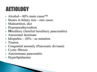
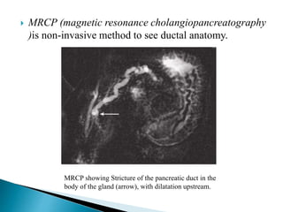
 It is persistent progressive irreversible damage of the
pancreas due to chronic inflammation.

 Alcohol—80% main cause™
 Stones in biliary tree—rare cause
 Malnutrition, diet
 Hyperparathyroidism
 ™Hereditary (familial hereditary pancreatitis)
 Autosomal dominant
 Idiopathic—20%—as mutation
 Trauma
 Congenital anomaly (Pancreatic divisum)
 Cystic fibrosis
 Autoimmune pancreatitis
 Hyperlipidaemia
 T – Toxic alcohol/tobacco/dietary/drug.
Metabolichypercalcaemia/lipidemia/lipoprotein lipase deficiency.
 I – Idiopathic—early/late onset/tropical –30%
 G – Genetic mutations—CFTR/SPINK 1.
 A – Autoimmune primary/with Sjögren/Crohn’s disease.
 R – Recurrent and severe acute/ischemic.
 O – Obstructive—pancreas divisum/annular pancreas/stenotic
papilla/duodenal obstruction/trauma/pancreatic ductal stones/
choledochocele
 Chronic pancreatitis is more common in males, common in
Kerala (induced by diet, rich in Tapioca).
 Alcohol reduces pancreatic blood flow, alters cell viability,
releases the free radicals, creates pancreatic ischaemia, and
activates the pancreatic stellate cells which produce abundant
extracellular matrix and collagen.
 Genetic predisposition may be the cause of idiopathic
pancreatitis.
 Mutation in pancreatic secretory trypsin
inhibitor causes activation of trypsin causing pancreatitis.
 Hereditary pancreatitis is an autosomal disorder with mutation
in trypsinogen gene in chromosome 7.
 It causes recurrent painful episodes of acute pancreatitis in
childhood, leading to chronic pancreatitis and pancreatic cancer
in adulthood.
 Oxidative stress hypothesis—Reactive by-products of hepatic
mixed function oxidase activity damage the pancreas through
chronic reflux of bile into the pancreatic duct.
 The toxic-metabolic theory—Alcohol is directly toxic to the acinar
cell where it brings changes in intracellular metabolism causing
pancreatic lipid accumulation, fatty degeneration, cellular necrosis,
and eventual widespread fibrosis.
 Stone and duct obstruction theory—Alcohol increases the lithogenicity
of pancreatic juice, leading to stone formation. Chronic
contact of stones with duct epithelial cells produces ulceration,
scarring, atrophy, fibrosis and chronic obstruction of the acini.
 The necrosis-fibrosis theory—Acute and chronic pancreatitis represents
a spectrum of disease. Inflammation from acute pancreatitis
leads to scarring, extrinsic compression of the pancreatic ductules,
obstruction, stasis, atrophy and stone formation.
 The genetic defect of hereditary pancreatitis produces recurrent
acute pancreatitis in early childhood, leading to chronic pancreatitis
in early adulthood.
 Cellular mechanisms of pancreatic fibrogenesis is due to pancreatic
stellate cells which are stimulated and activated by alcohol, oxidative
stress, cytokines of acute pancreatitis.
Transforming growth factor beta 1(TGF ᵦ1) is an important mediator of
pancreatic fibrosis.
 The sentinel acute pancreatitis event (SAPE) hypothesis—An
episode of acute pancreatitis, the sentinel event, sets the stage for
the attraction of collagen-secreting stellate cells.
 Heavy, prolonged alcohol use is the most common cause of chronic
pancreatitis. Alcohol-related chronic pancreatitis is associated with
more severe pain, more extensive calcification and ductal changes,
and more rapid progression to endocrine and exocrine insufficiency.
 Often recurrent episodes of acute pancreatitis for several years are
seen in these patients. Some cofactors amplify the effect of alcohol
in these patients. Prevalence of some genetic mutations linked with
pancreatitis like cystic fibrosis transmembrane regulator (CFTR),
serine protease inhibitor Kazal type-1 (SPINK1) has been noted in
alcoholic pancreatitis. A high-fat diet and smoking affect pancreatic
bicarbonate and water secretion adversely, inducing oxidative stress
and increases the rate of pancreatic calcification.
 Tropical pancreatitis is endemic in some regions of India (Kerala),
Africa, and South America. Episodic abdominal pain begins in
childhood and is followed by rapid progression to endocrine and
exocrine insufficiency with pancreatic calculi in non-alcoholic
individuals.
 Dietary toxins (cyanogens in the cassava plant,
tapioca) and micronutrients like zinc, copper, and selenium,
vitamin A deficiencies, genetic factors, ductal abnormalities are
the probable causes of tropical pancreatitis.
 Linnamarin and its methyl derivative, in acid pH of stomach
releases hydrocyanic acid which is cytotoxic in presence of
rhodanase releases thiocyanates causing depletion of methionine,
damaging pancreas → pancreatitis. Gross look is small, firm
fibrotic/adipose type.
 Early-onset idiopathic chronic pancreatitis manifests with severe
abdominal pain in childhood, with relatively few structural and
functional changes.
 Late-onset idiopathic chronic pancreatitis manifests in middle
and late adulthood, often with minimal pain and pronounced
exocrine insufficiency.
• Focal necrosis
•™Segmental or diffuse
fibrosis
•Parenchymal calcification
or ductal stones
•Stricture and/or dilatation
of the duct
•There is loss of exocrine function initially and endocrine
functions eventually.
•Ductular metaplasia and acinar atrophy along with fibrosis
and cyst formation develops.
 Early—pancreatic oedema—chronic inflammation—normal
secretory function.
 Moderate—early fibrosis; only few acinar cells—exocrine
dysfunction.
 Late—fibrosis—loss of secretory function—diabetes
mellitus.
 Complications develop secondary to healing and fibrosis;
deposition of inspissated proteinaceous material in the duct;
over expression of CTGF and TGF-B1 that stimulate
extracellular matrix
 I: Based on main duct dilated or not:
◦ Large duct disease—main pancreatic duct is dilated.
◦ Small duct disease—main pancreatic duct, is normal or smaller
in size.
 II: Staging/classification of chronic pancreatitis (Stages A, B,
C)
A new classification of chronic pancreatitis, based on
combination of clinical signs, morphology and function, is
presented (2009 Büchler et al).
 It is the early stage of chronic pancreatitis where
complications have not yet appeared and the clinical
exocrine and endocrine function is preserved.
 Subclinical signs (impaired glucose tolerance,
reduced exocrine function but without steatorrhoea)
might already be apparent.
 Stage A is accepted under the following conditions:
Pain of any type and degree and/or attacks of acute
pancreatitis, no complications, no steatorrhoea, and no
insulin-dependant diabetes mellitus.
 It is the intermediate stage where chronic pancreatitis
has led to complications but clinical exocrine and
endocrine function is still preserved.
 The type of complication is specified (e.g. stage B, bile
duct).
 Stage B is accepted under the following conditions:
Patients with complications but without steatorrhoea or
diabetes mellitus.
 Stage C is the end stage of chronic pancreatitis, where pancreatic
fibrosis has led to loss of clinical exocrine and/or endocrine pancreatic
function (steatorrhoea and/or diabetes mellitus).
 Complications of chronic pancreatitis might or might not be present.
The type of exocrine and/or endocrine pancreatic function loss is
specified (e.g.stage C, steatorrhoea).
 Stage C can be sub-classified into three categories:
C1: Patients with endocrine function impairment.
C2: Patients with exocrine function impairment.
C3: Patients with exocrine/endocrine function impairment and/or
complications—as they are defined.
 Stage C is accepted under the following conditions—Patients
with clinical manifestation of end-stage functional impairment with
or without complications.
 Pain in epigastric region (80%)
◦ It is persistent and severe, which radiates to back.
◦ This pain is due to irritation of retropancreatic nerves,
or due to ductal dilatation and stasis, or due to chronic
inflammation itself.
 Two patterns of pain have been described
◦ Type A pain is short relapsing episodes lasting days to weeks,
with pain-free intervals.
◦ Type B pain is prolonged, severe, unrelenting pain.
 There is often a gradual diminish in pain over years
due to “pancreatic burnout” by extensive calcifications,
exocrine and endocrine insufficiency.
Patient will occupy leaning forward position to relieve
severe pain of pancreatitis.
 Exocrine dysfunction: Diarrhoea, asthenia, loss of weight
and appetite, steatorrhoea (signifies severe pancreatic
insufficiency) (90%), malabsorption.
 Endocrine dysfunction: Diabetes mellitus. Pancreatic
diabetes may often be typically brittle because of
concomitant glucagon deficiency and requires insulin.
 Mild jaundice is due to narrowing of retropancreatic bile
duct and cholangitis.
 Mass per abdomen, just above the umbilicus, tender,
nodular, hard, felt on deep palpation, not moving with
respiration, not mobile, resonant on percussion.
1. Pancreatic calcification
2. Steatorrhoea
3. Diabetes mellitus
 Triad is found in less than one-third of patients with
CP
 Mallet-Guys sign: In right knee-chest position, if left
hypochondrium is palpated tenderness can be evoked in
case of chronic-relapsing
pancreatitis. In this position, bowel loops are being
shifted to right so
as to have a direct palpation of pancreas.
 Note:
• Chronic pancreatitis can lead to carcinoma
pancreas.
 Stage A: 85%, recurrent/acute episodic pain with
weight loss.™
 Stage B: Severe prolonged progressive pain with
impaired pancreatic function with cholestasis,
pseudocyst, sinistral portal hypertension.
 Stage C: Severe exocrine/endocrine deficiency, less
severe pain, complications like pseudocyst and
obstruction.
 Carcinoma of head of the pancreas.
 Retroperitoneal tumour.
 CT scan abdomen: It is 95% reliable; to see
pseudocyst, calcification, ductal stones, duct stricture
and dilatation, vasculature, fibrosis, surrounding
structures, CBD status.
 It shows 90% sensitivity; 95% specificity
CT scan abdomen showing features of chronic pancreatitis—
calcification, ductal changes, and cyst formation.
 ERCP(endoscopic retrograde cholangiopancreatography)
is useful in chronic pancreatitis to see dilatations, strictures
and altered ductal anatomy. It is mainly to assess
structural pathology of the pancreas.
ERCP showing pancreatic duct
 MRCP (magnetic resonance cholangiopancreatography
)is non-invasive method to see ductal anatomy.
MRCP showing Stricture of the pancreatic duct in the
body of the gland (arrow), with dilatation upstream.
 Endosonography (EUS) to see possible malignant site
and to take FNAC.
 Site, duct status, stricture, stones, parenchyma,
pseudocyst, CBD status, nodes are identified in EUS.
 Positive five parameters suggest chronic pancreatitis
 Secretin cholecystokinin test is the gold standard for
assessing pancreatic function.
 Pancreolauryl test.
 LFT, prothrombin time is needed to manage the patient.
 Serum trypsinogen and fecal elastase tests— to identify
exocrine insufficiency.
 Plain X-ray abdomen may show ductal calculi or
parenchymal calcifications.

Chronic pancreatitis

  • 2.
     It ispersistent progressive irreversible damage of the pancreas due to chronic inflammation. 
  • 3.
     Alcohol—80% maincause™  Stones in biliary tree—rare cause  Malnutrition, diet  Hyperparathyroidism  ™Hereditary (familial hereditary pancreatitis)  Autosomal dominant  Idiopathic—20%—as mutation  Trauma  Congenital anomaly (Pancreatic divisum)  Cystic fibrosis  Autoimmune pancreatitis  Hyperlipidaemia
  • 4.
     T –Toxic alcohol/tobacco/dietary/drug. Metabolichypercalcaemia/lipidemia/lipoprotein lipase deficiency.  I – Idiopathic—early/late onset/tropical –30%  G – Genetic mutations—CFTR/SPINK 1.  A – Autoimmune primary/with Sjögren/Crohn’s disease.  R – Recurrent and severe acute/ischemic.  O – Obstructive—pancreas divisum/annular pancreas/stenotic papilla/duodenal obstruction/trauma/pancreatic ductal stones/ choledochocele
  • 5.
     Chronic pancreatitisis more common in males, common in Kerala (induced by diet, rich in Tapioca).  Alcohol reduces pancreatic blood flow, alters cell viability, releases the free radicals, creates pancreatic ischaemia, and activates the pancreatic stellate cells which produce abundant extracellular matrix and collagen.  Genetic predisposition may be the cause of idiopathic pancreatitis.  Mutation in pancreatic secretory trypsin inhibitor causes activation of trypsin causing pancreatitis.  Hereditary pancreatitis is an autosomal disorder with mutation in trypsinogen gene in chromosome 7.  It causes recurrent painful episodes of acute pancreatitis in childhood, leading to chronic pancreatitis and pancreatic cancer in adulthood.
  • 6.
     Oxidative stresshypothesis—Reactive by-products of hepatic mixed function oxidase activity damage the pancreas through chronic reflux of bile into the pancreatic duct.  The toxic-metabolic theory—Alcohol is directly toxic to the acinar cell where it brings changes in intracellular metabolism causing pancreatic lipid accumulation, fatty degeneration, cellular necrosis, and eventual widespread fibrosis.  Stone and duct obstruction theory—Alcohol increases the lithogenicity of pancreatic juice, leading to stone formation. Chronic contact of stones with duct epithelial cells produces ulceration, scarring, atrophy, fibrosis and chronic obstruction of the acini.  The necrosis-fibrosis theory—Acute and chronic pancreatitis represents a spectrum of disease. Inflammation from acute pancreatitis leads to scarring, extrinsic compression of the pancreatic ductules, obstruction, stasis, atrophy and stone formation.
  • 7.
     The geneticdefect of hereditary pancreatitis produces recurrent acute pancreatitis in early childhood, leading to chronic pancreatitis in early adulthood.  Cellular mechanisms of pancreatic fibrogenesis is due to pancreatic stellate cells which are stimulated and activated by alcohol, oxidative stress, cytokines of acute pancreatitis. Transforming growth factor beta 1(TGF ᵦ1) is an important mediator of pancreatic fibrosis.  The sentinel acute pancreatitis event (SAPE) hypothesis—An episode of acute pancreatitis, the sentinel event, sets the stage for the attraction of collagen-secreting stellate cells.  Heavy, prolonged alcohol use is the most common cause of chronic pancreatitis. Alcohol-related chronic pancreatitis is associated with more severe pain, more extensive calcification and ductal changes, and more rapid progression to endocrine and exocrine insufficiency.
  • 8.
     Often recurrentepisodes of acute pancreatitis for several years are seen in these patients. Some cofactors amplify the effect of alcohol in these patients. Prevalence of some genetic mutations linked with pancreatitis like cystic fibrosis transmembrane regulator (CFTR), serine protease inhibitor Kazal type-1 (SPINK1) has been noted in alcoholic pancreatitis. A high-fat diet and smoking affect pancreatic bicarbonate and water secretion adversely, inducing oxidative stress and increases the rate of pancreatic calcification.  Tropical pancreatitis is endemic in some regions of India (Kerala), Africa, and South America. Episodic abdominal pain begins in childhood and is followed by rapid progression to endocrine and exocrine insufficiency with pancreatic calculi in non-alcoholic individuals.
  • 9.
     Dietary toxins(cyanogens in the cassava plant, tapioca) and micronutrients like zinc, copper, and selenium, vitamin A deficiencies, genetic factors, ductal abnormalities are the probable causes of tropical pancreatitis.  Linnamarin and its methyl derivative, in acid pH of stomach releases hydrocyanic acid which is cytotoxic in presence of rhodanase releases thiocyanates causing depletion of methionine, damaging pancreas → pancreatitis. Gross look is small, firm fibrotic/adipose type.  Early-onset idiopathic chronic pancreatitis manifests with severe abdominal pain in childhood, with relatively few structural and functional changes.  Late-onset idiopathic chronic pancreatitis manifests in middle and late adulthood, often with minimal pain and pronounced exocrine insufficiency.
  • 10.
    • Focal necrosis •™Segmentalor diffuse fibrosis •Parenchymal calcification or ductal stones •Stricture and/or dilatation of the duct •There is loss of exocrine function initially and endocrine functions eventually. •Ductular metaplasia and acinar atrophy along with fibrosis and cyst formation develops.
  • 11.
     Early—pancreatic oedema—chronicinflammation—normal secretory function.  Moderate—early fibrosis; only few acinar cells—exocrine dysfunction.  Late—fibrosis—loss of secretory function—diabetes mellitus.  Complications develop secondary to healing and fibrosis; deposition of inspissated proteinaceous material in the duct; over expression of CTGF and TGF-B1 that stimulate extracellular matrix
  • 12.
     I: Basedon main duct dilated or not: ◦ Large duct disease—main pancreatic duct is dilated. ◦ Small duct disease—main pancreatic duct, is normal or smaller in size.  II: Staging/classification of chronic pancreatitis (Stages A, B, C) A new classification of chronic pancreatitis, based on combination of clinical signs, morphology and function, is presented (2009 Büchler et al).
  • 13.
     It isthe early stage of chronic pancreatitis where complications have not yet appeared and the clinical exocrine and endocrine function is preserved.  Subclinical signs (impaired glucose tolerance, reduced exocrine function but without steatorrhoea) might already be apparent.  Stage A is accepted under the following conditions: Pain of any type and degree and/or attacks of acute pancreatitis, no complications, no steatorrhoea, and no insulin-dependant diabetes mellitus.
  • 14.
     It isthe intermediate stage where chronic pancreatitis has led to complications but clinical exocrine and endocrine function is still preserved.  The type of complication is specified (e.g. stage B, bile duct).  Stage B is accepted under the following conditions: Patients with complications but without steatorrhoea or diabetes mellitus.
  • 15.
     Stage Cis the end stage of chronic pancreatitis, where pancreatic fibrosis has led to loss of clinical exocrine and/or endocrine pancreatic function (steatorrhoea and/or diabetes mellitus).  Complications of chronic pancreatitis might or might not be present. The type of exocrine and/or endocrine pancreatic function loss is specified (e.g.stage C, steatorrhoea).  Stage C can be sub-classified into three categories: C1: Patients with endocrine function impairment. C2: Patients with exocrine function impairment. C3: Patients with exocrine/endocrine function impairment and/or complications—as they are defined.  Stage C is accepted under the following conditions—Patients with clinical manifestation of end-stage functional impairment with or without complications.
  • 16.
     Pain inepigastric region (80%) ◦ It is persistent and severe, which radiates to back. ◦ This pain is due to irritation of retropancreatic nerves, or due to ductal dilatation and stasis, or due to chronic inflammation itself.  Two patterns of pain have been described ◦ Type A pain is short relapsing episodes lasting days to weeks, with pain-free intervals. ◦ Type B pain is prolonged, severe, unrelenting pain.  There is often a gradual diminish in pain over years due to “pancreatic burnout” by extensive calcifications, exocrine and endocrine insufficiency.
  • 17.
    Patient will occupyleaning forward position to relieve severe pain of pancreatitis.
  • 18.
     Exocrine dysfunction:Diarrhoea, asthenia, loss of weight and appetite, steatorrhoea (signifies severe pancreatic insufficiency) (90%), malabsorption.  Endocrine dysfunction: Diabetes mellitus. Pancreatic diabetes may often be typically brittle because of concomitant glucagon deficiency and requires insulin.  Mild jaundice is due to narrowing of retropancreatic bile duct and cholangitis.  Mass per abdomen, just above the umbilicus, tender, nodular, hard, felt on deep palpation, not moving with respiration, not mobile, resonant on percussion.
  • 19.
    1. Pancreatic calcification 2.Steatorrhoea 3. Diabetes mellitus  Triad is found in less than one-third of patients with CP
  • 20.
     Mallet-Guys sign:In right knee-chest position, if left hypochondrium is palpated tenderness can be evoked in case of chronic-relapsing pancreatitis. In this position, bowel loops are being shifted to right so as to have a direct palpation of pancreas.  Note: • Chronic pancreatitis can lead to carcinoma pancreas.
  • 21.
     Stage A:85%, recurrent/acute episodic pain with weight loss.™  Stage B: Severe prolonged progressive pain with impaired pancreatic function with cholestasis, pseudocyst, sinistral portal hypertension.  Stage C: Severe exocrine/endocrine deficiency, less severe pain, complications like pseudocyst and obstruction.
  • 22.
     Carcinoma ofhead of the pancreas.  Retroperitoneal tumour.
  • 23.
     CT scanabdomen: It is 95% reliable; to see pseudocyst, calcification, ductal stones, duct stricture and dilatation, vasculature, fibrosis, surrounding structures, CBD status.  It shows 90% sensitivity; 95% specificity
  • 24.
    CT scan abdomenshowing features of chronic pancreatitis— calcification, ductal changes, and cyst formation.
  • 25.
     ERCP(endoscopic retrogradecholangiopancreatography) is useful in chronic pancreatitis to see dilatations, strictures and altered ductal anatomy. It is mainly to assess structural pathology of the pancreas. ERCP showing pancreatic duct
  • 26.
     MRCP (magneticresonance cholangiopancreatography )is non-invasive method to see ductal anatomy. MRCP showing Stricture of the pancreatic duct in the body of the gland (arrow), with dilatation upstream.
  • 27.
     Endosonography (EUS)to see possible malignant site and to take FNAC.  Site, duct status, stricture, stones, parenchyma, pseudocyst, CBD status, nodes are identified in EUS.  Positive five parameters suggest chronic pancreatitis
  • 28.
     Secretin cholecystokinintest is the gold standard for assessing pancreatic function.  Pancreolauryl test.  LFT, prothrombin time is needed to manage the patient.  Serum trypsinogen and fecal elastase tests— to identify exocrine insufficiency.  Plain X-ray abdomen may show ductal calculi or parenchymal calcifications.