Chronic pancreatitis
Dr. Lala Robin, MS. Gen. Surgery,
Senior Resident,
CMC
Definition
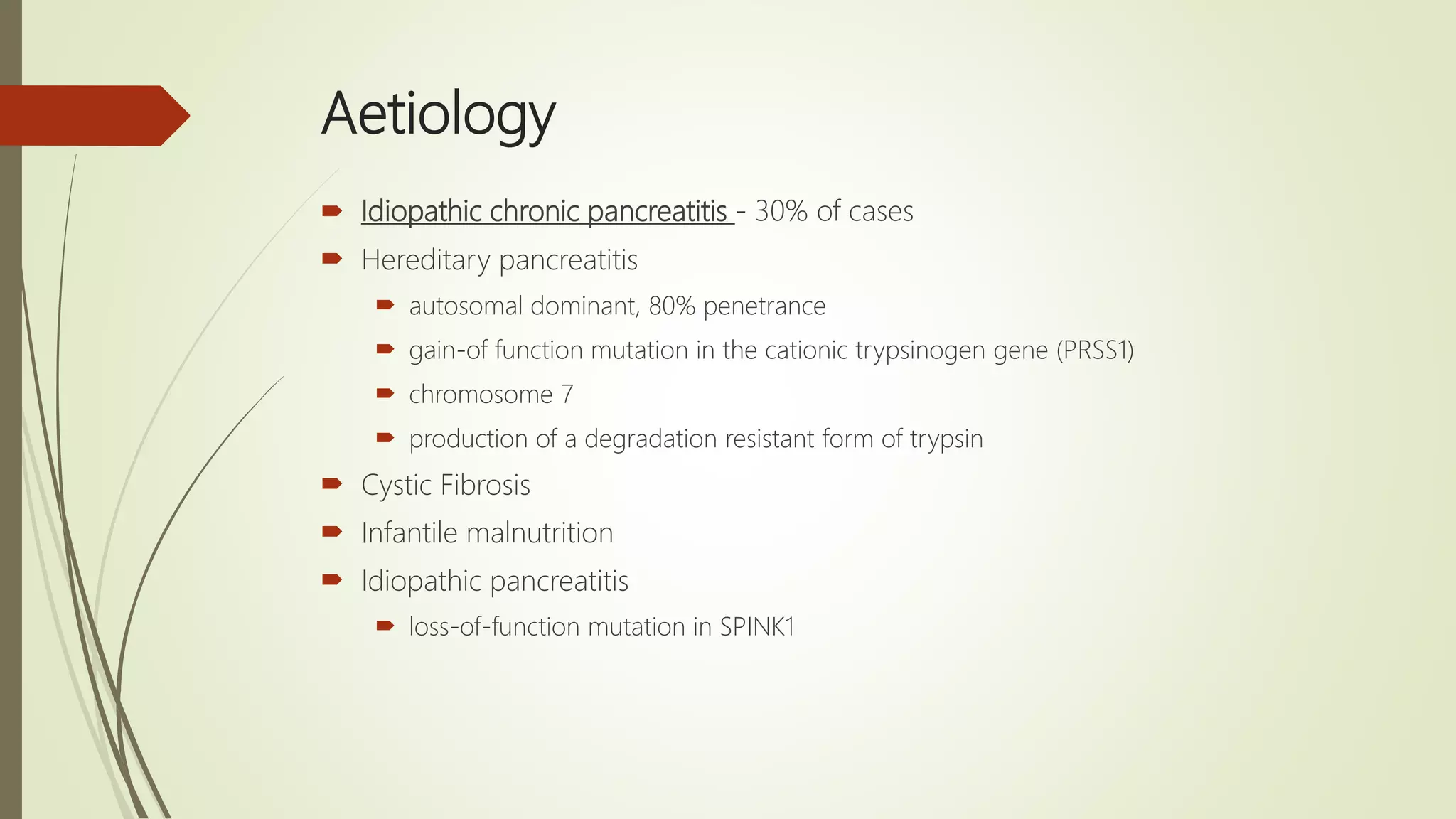
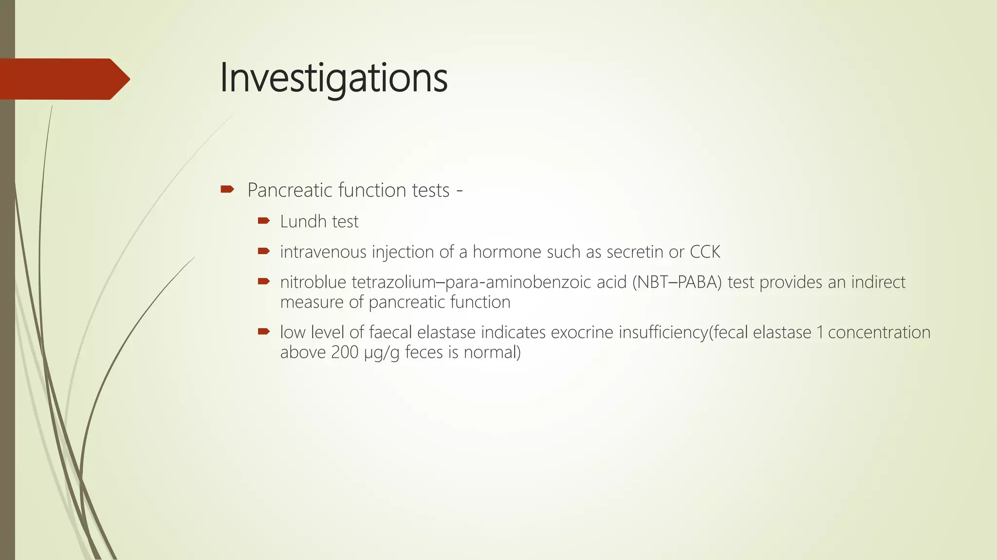
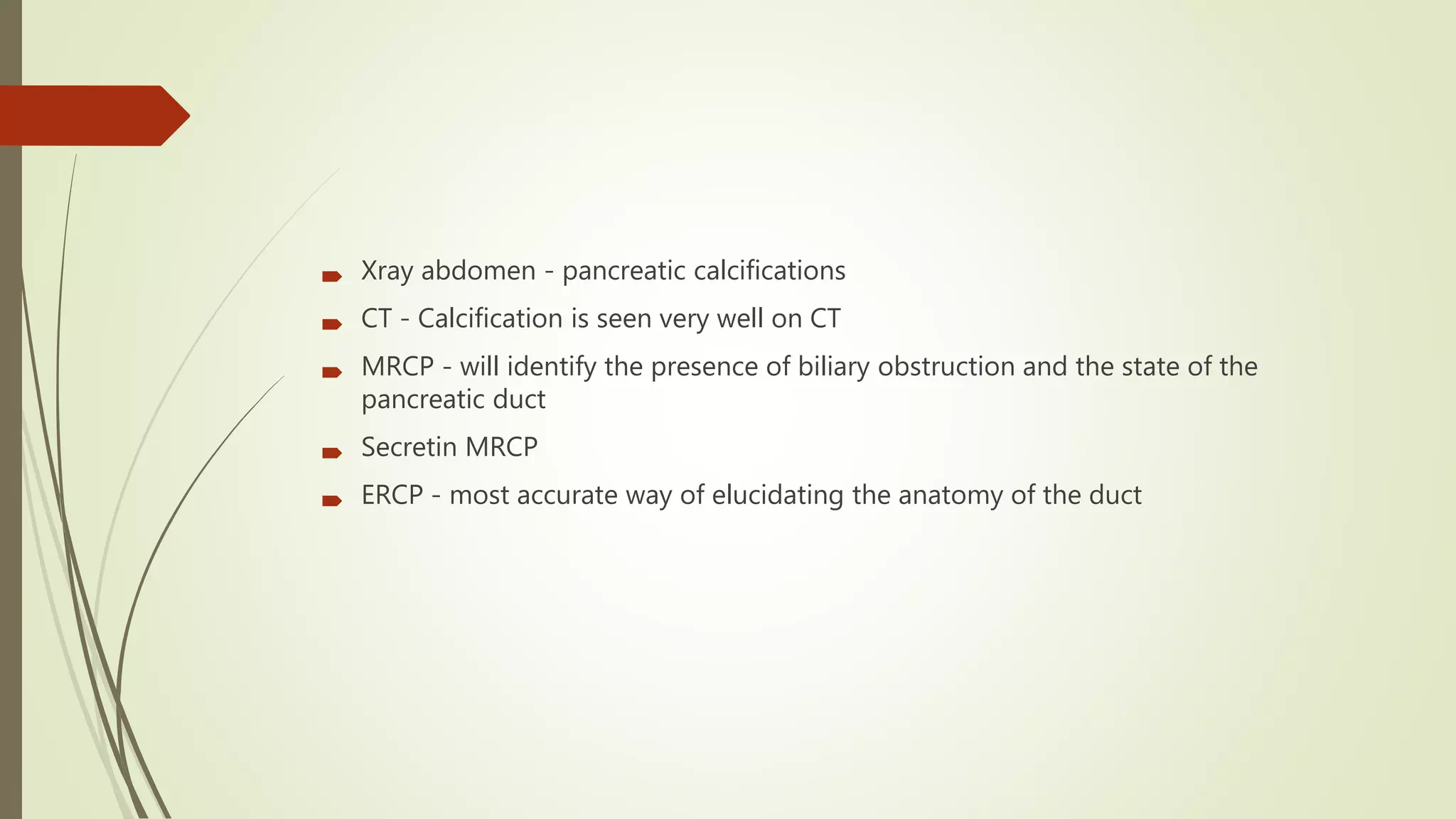
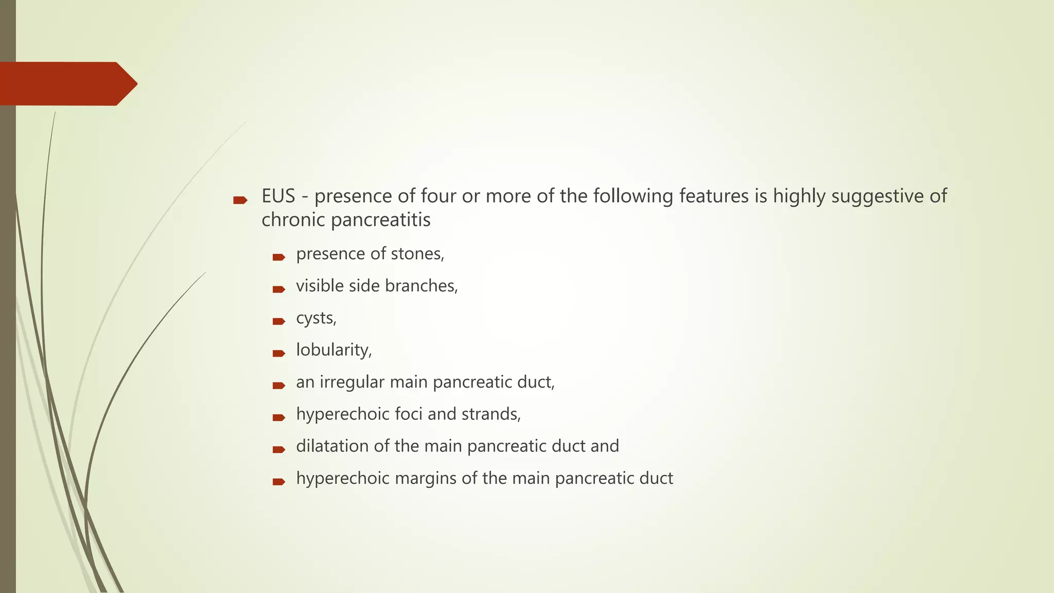
 Chronic pancreatitis is a progressive inflammatory disease in which there is
irreversible destruction of pancreatic tissue.
 Its clinical course is characterised by severe pain and, in the later stages, exocrine
and endocrine pancreatic insufficiency.
 Chronic pancreatitis is defined as a continuing inflammatory disease of the
pancreas characterised by irreversible morphological change typically causing
pain and/or permanent loss of function
 occurs more frequently in men (male: female ratio of 4:1)
 mean age of onset is about 40 years
Aetiology
 Alcohol - 60–70% of cases
 Tobacco smoking
 Hyperlipidaemia
 Hypercalcaemia
 Pancreatic duct obstruction
 stricture formation after trauma , acute pancreatitis
 pancreatic cancer
 Congenital ---> pancreas divisum and annular pancreas with papillary stenosis
Aetiology
 Idiopathic chronic pancreatitis - 30% of cases
 Hereditary pancreatitis
 autosomal dominant, 80% penetrance
 gain-of function mutation in the cationic trypsinogen gene (PRSS1)
 chromosome 7
 production of a degradation resistant form of trypsin
 Cystic Fibrosis
 Infantile malnutrition
 Idiopathic pancreatitis
 loss-of-function mutation in SPINK1
Aetiology
 Tropical pancreatitis
 begins at a young age
 high incidence of diabetes mellitus and stone formation
 Cassava intake, malnutrition, exposure to hydrocarbons
 Autoimmune pancreatitis
 diffuse and irregular narrowing of the main pancreatic duct
 levels of the immunoglobulin subtype IgG4 are elevated
 autoimmune cholangiopathy
Pathology
 pancreas enlarges and becomes hard as a result of fibrosis
 ducts become distorted and dilated with areas of both stricture formation and
ectasia
 ductular metaplasia and atrophy of acini, hyperplasia of duct epithelium and
interlobular fibrosis
Clinical features
 Pain
 head of the pancreas --> epigastric and right subcostal pain
 left side of the pancreas --> left subcostal and back pain
 Weight loss
 analgesic dependence
 inability to work
 Loss of exocrine function --> steatorrhea, diabetes
Investigations
 Pancreatic function tests -
 Lundh test
 intravenous injection of a hormone such as secretin or CCK
 nitroblue tetrazolium–para-aminobenzoic acid (NBT–PABA) test provides an indirect
measure of pancreatic function
 low level of faecal elastase indicates exocrine insufficiency(fecal elastase 1 concentration
above 200 µg/g feces is normal)
 Xray abdomen - pancreatic calcifications
 CT - Calcification is seen very well on CT
 MRCP - will identify the presence of biliary obstruction and the state of the
pancreatic duct
 Secretin MRCP
 ERCP - most accurate way of elucidating the anatomy of the duct
 EUS - presence of four or more of the following features is highly suggestive of
chronic pancreatitis
 presence of stones,
 visible side branches,
 cysts,
 lobularity,
 an irregular main pancreatic duct,
 hyperechoic foci and strands,
 dilatation of the main pancreatic duct and
 hyperechoic margins of the main pancreatic duct
Medical treatment of chronic pancreatitis
 Treat the addiction
 Help the patient to stop alcohol consumption and tobacco smoking
 Involve a dependency counsellor or a psychologist
 Alleviate abdominal pain
 Eliminate obstructive factors (duodenum, bile duct, pancreatic duct)
 Escalate analgesia in a stepwise fashion
 Nutritional and pharmacological measures
 Diet: low in fat and high in protein and carbohydrates
 Pancreatic enzyme supplementation with meals
 Correct malabsorption of the fat-soluble vitamins and vitamin B12
 Micronutrient therapy with methionine, vitamins C & E, selenium (may reduce pain
slow disease progression)
 Steroids (only in autoimmune pancreatitis, for relief of symptoms)
 Medium-chain triglycerides in patients with severe fat malabsorption (they are directly
absorbed by the small intestine without the need for digestion)
 Reducing gastric secretions may help
 Treat diabetes mellitus
Endoscopic Interventions
 Endoscopic pancreatic sphincterotomy
 stent across the stricture (stent should be left in for no more than 4–6 weeks)
 Pancreatic ductal stone removal by ERCP and ESWL
 Percutaneous or transgastric drainage of pseudocysts under ultrasound or CT
guidance
SURGERY IN CHRONIC PANCREATITIS
 Aim of surgery is to overcome obstruction and remove any mass lesions.

 mass in the head of the pancreas --> pancreatoduodenectomy or a Beger
procedure (duodenum-preserving resection of the pancreatic head)
 duct is markedly dilated --> longitudinal pancreatojejunostomy (Puestow) or Frey
procedure
 Patients with intractable pain and diffuse disease --> Total pancreatectomy and
islet autotransplantation
Prognosis
 gradual decline in their professional, social and personal lives.
 Development of pancreatic cancer is a risk in those who have had the disease for
more than 20 years
THANK YOU

Chronic pancreatitis

  • 1.
    Chronic pancreatitis Dr. LalaRobin, MS. Gen. Surgery, Senior Resident, CMC
  • 2.
    Definition  Chronic pancreatitisis a progressive inflammatory disease in which there is irreversible destruction of pancreatic tissue.  Its clinical course is characterised by severe pain and, in the later stages, exocrine and endocrine pancreatic insufficiency.  Chronic pancreatitis is defined as a continuing inflammatory disease of the pancreas characterised by irreversible morphological change typically causing pain and/or permanent loss of function  occurs more frequently in men (male: female ratio of 4:1)  mean age of onset is about 40 years
  • 3.
    Aetiology  Alcohol -60–70% of cases  Tobacco smoking  Hyperlipidaemia  Hypercalcaemia  Pancreatic duct obstruction  stricture formation after trauma , acute pancreatitis  pancreatic cancer  Congenital ---> pancreas divisum and annular pancreas with papillary stenosis
  • 5.
    Aetiology  Idiopathic chronicpancreatitis - 30% of cases  Hereditary pancreatitis  autosomal dominant, 80% penetrance  gain-of function mutation in the cationic trypsinogen gene (PRSS1)  chromosome 7  production of a degradation resistant form of trypsin  Cystic Fibrosis  Infantile malnutrition  Idiopathic pancreatitis  loss-of-function mutation in SPINK1
  • 6.
    Aetiology  Tropical pancreatitis begins at a young age  high incidence of diabetes mellitus and stone formation  Cassava intake, malnutrition, exposure to hydrocarbons  Autoimmune pancreatitis  diffuse and irregular narrowing of the main pancreatic duct  levels of the immunoglobulin subtype IgG4 are elevated  autoimmune cholangiopathy
  • 7.
    Pathology  pancreas enlargesand becomes hard as a result of fibrosis  ducts become distorted and dilated with areas of both stricture formation and ectasia  ductular metaplasia and atrophy of acini, hyperplasia of duct epithelium and interlobular fibrosis
  • 8.
    Clinical features  Pain head of the pancreas --> epigastric and right subcostal pain  left side of the pancreas --> left subcostal and back pain  Weight loss  analgesic dependence  inability to work  Loss of exocrine function --> steatorrhea, diabetes
  • 9.
    Investigations  Pancreatic functiontests -  Lundh test  intravenous injection of a hormone such as secretin or CCK  nitroblue tetrazolium–para-aminobenzoic acid (NBT–PABA) test provides an indirect measure of pancreatic function  low level of faecal elastase indicates exocrine insufficiency(fecal elastase 1 concentration above 200 µg/g feces is normal)
  • 10.
     Xray abdomen- pancreatic calcifications  CT - Calcification is seen very well on CT  MRCP - will identify the presence of biliary obstruction and the state of the pancreatic duct  Secretin MRCP  ERCP - most accurate way of elucidating the anatomy of the duct
  • 13.
     EUS -presence of four or more of the following features is highly suggestive of chronic pancreatitis  presence of stones,  visible side branches,  cysts,  lobularity,  an irregular main pancreatic duct,  hyperechoic foci and strands,  dilatation of the main pancreatic duct and  hyperechoic margins of the main pancreatic duct
  • 14.
    Medical treatment ofchronic pancreatitis  Treat the addiction  Help the patient to stop alcohol consumption and tobacco smoking  Involve a dependency counsellor or a psychologist  Alleviate abdominal pain  Eliminate obstructive factors (duodenum, bile duct, pancreatic duct)  Escalate analgesia in a stepwise fashion
  • 15.
     Nutritional andpharmacological measures  Diet: low in fat and high in protein and carbohydrates  Pancreatic enzyme supplementation with meals  Correct malabsorption of the fat-soluble vitamins and vitamin B12  Micronutrient therapy with methionine, vitamins C & E, selenium (may reduce pain slow disease progression)  Steroids (only in autoimmune pancreatitis, for relief of symptoms)  Medium-chain triglycerides in patients with severe fat malabsorption (they are directly absorbed by the small intestine without the need for digestion)  Reducing gastric secretions may help  Treat diabetes mellitus
  • 16.
    Endoscopic Interventions  Endoscopicpancreatic sphincterotomy  stent across the stricture (stent should be left in for no more than 4–6 weeks)  Pancreatic ductal stone removal by ERCP and ESWL  Percutaneous or transgastric drainage of pseudocysts under ultrasound or CT guidance
  • 17.
    SURGERY IN CHRONICPANCREATITIS  Aim of surgery is to overcome obstruction and remove any mass lesions.   mass in the head of the pancreas --> pancreatoduodenectomy or a Beger procedure (duodenum-preserving resection of the pancreatic head)  duct is markedly dilated --> longitudinal pancreatojejunostomy (Puestow) or Frey procedure  Patients with intractable pain and diffuse disease --> Total pancreatectomy and islet autotransplantation
  • 18.
    Prognosis  gradual declinein their professional, social and personal lives.  Development of pancreatic cancer is a risk in those who have had the disease for more than 20 years
  • 19.