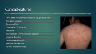
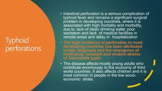
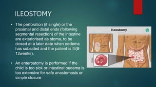
Typhoid Perforation
By:
Muhammad Wasil Khan & Ramsha Mazhar
Cause + Transmission +Risk Factors
• Cause: Gram Negative bacteria
1. Salmonella typhi
2. Salmonella paratyphi (A,B or C)
• Transmission: Humans are the only reservoir thus
spread via:
1. Direct Contact
2. Contaminated food/water
Risk Factors:
1. Children and Young
Adults
2. Poor Sanitation
3. Over Crowding
ClinicalFeatures
Fever (Step-wise) temperature goes up, plateaus and
then goes up again.
Abdominal Pain
Diarrhea or Constipation
Headache
Rose spots (1-5mm balnchable papules)
Intestinal Bleeding
Hepatospleenomegaly
Intestinal Perforations
Typhoid Encephalopathy
Diagnosisand
Treatment
• Diagnosis:
 Clinical– 3 days or more of fever and Gi
symptoms + Endemic Area
 Blood culture
 Stool Culture
• Treatment:
1. First Line– Floroquinolones,
Azithromycin, Ceftriaxone or
Carbapenems
2. Severe illness– Dexamethasone
Typhoid
Intestinal
Perforation
• The bacterium lives in the intestines and
bloodstream of humans. It spreads
between individuals by direct contact with
the faeces of an infected person.
S. typhi enters through the mouth and
spends 1 to 3 weeks in the intestine.
After this, it makes its way through the
intestinal wall and into the bloodstream.
• From the bloodstream, it spreads into
other tissues and organs. The immune
system of the host can do little to fight
back because S. typhi can live within the
host’s cells, safe from the immune
system.
Pathophysiology
• The virulence of Salmonella is determined by typhoid toxin, Vi antigen
(polysaccharide capsule), liposaccharide O antigen, and flagellar H antigen.
• The main role of the Vi antigen is to act as an antiphagocytic agent preventing the action of
macrophages, thus shielding the O antigen from antibodies that confer the serum
resistance.
• The flagellar H antigen provides bacterial mobility and adherence upon the gut wall
mucosa.
• Invasion of the gut wall is assisted by flagella, and the type III secretion system is capable
of transferring bacterial protein into enterocytes and M cells (specialized epithelial cells that
serve as antigen-presenting cells in gut mucosa or lymphoid tissue) or by direct penetration
of mucosa.
• Bacteria attached to M cells are absorbed by pinched off cytoplasm containing bacteria
and extruded into the luminal space. In this process, M cells are damaged, and the basal
lamina is exposed. It provides easy access to pathogens for the invasion, which worsens
the condition
• Bacteria induce proliferation of Payer patches via recruitment of lymphocytes and
mononuclear cells and induce necrosis and eventually, ulceration that complicates
the symptoms.
• In general, hemorrhage and perforation occur in the terminal ileum secondary to
necrosis of Peyer's patches at 2-3 weeks after the onset of the disease.
• Perforation of terminal ileum is a cause for acute obscure peritonitis, heralded
by exacerbation of abdominal pain associated with tenderness, rigidity and
guarding, most pronounced over right iliac fossa.
Typhoid
perforations
• Intestinal perforation is a serious complication of
typhoid fever and remains a significant surgical
problem in developing countries, where it is
associated with high mortality and morbidity,
due to lack of clean drinking water, poor
sanitation and lack of medical facilities in
remote areas and delay in hospitalization
• The high incidence of perforation in most
developing countries has been attributed
to late diagnosis and the emergence of
multi-drug resistant and virulent strains
of Salmonella typhi.
• The disease affects mostly young adults who
contribute enormously to the economy of third
world countries .It also affects children and it is
most common in people in the low socio-
economic strata.
PATHOLOGY
 ULCERS; shallow irregular oval ulcers disposed
longitudinally on the antimesenteric border of
the ileum esp. terminal ileum.
 Perforation may be small or wide up to 2.5cm.
Most within 45cm from the ileocecal junction.
They are multiple in 20% of patients.
 Histologically, tissue around perforation shows
infiltration by lymphocyte ,macrophages and few
neutrophils. The macrophages may ingest
RBCs to produce typhoid cells.
CVS
 Tachypnea
 Hypotension
 Shock
CHEST
 Respiratory function is compromised by chest infection, which
is worsened by the marked abdominal distention(if present).
 Crepitation may be heard, sometimes bilaterally, indicating
that pneumonia has set in and is worsening the condition
ABDOMEN
 Generalized tenderness
 Rebound tenderness
 Guarding
 Rigidity
 Diminish or absent bowel sounds
 Tenderness and fullness in the recto-vesical or recto-
uterine pouch, suggesting a pelvic collection of pus.
DIAGNOSIS
Work up: CBC, LFT,
UCE
Erect chest
radiograph or a
lateral decubitus film
(if ill) -- free gas in
the peritoneal cavity.
Abdominal X ray
Ultrasound
CT scan (if X ray
shows any
obstruction)
Blood C/S
MULTIPLE ILEUM
PERFORATION
ILEAL
PERFORATION
Correction of Fluid and Electrolyte Deficits
Nasogastric Decompression
Urethral Catheter
Reversal of Hypoxia
Blood Transfusion
Antibiotic Therapy
DEFINITIVE TREATMENT
The definitive treatment for intestinal perforation is operative
to evacuate faecal contamination and prevent further
contamination.
LAPAROTOMY + SURGICAL OPTIONS;
Simple closure of perforations
Segmental resection of affected intestine
Enterostomy
SIMPLE CLOSURE
• Single perforation,
• If perforations are far apart
• If the number of perforations are so numerous
that resection may result in a short gut.
• The edge of the perforation is excised
circumferentially (the excised edge is sent to the
lab for histopathology).
RESECTION & ANASTOMOSIS
Large solitary perforation
Multiple perforation in close vicinity to each
other.
Adjacent bowel is friable/ near perforation
A limited right hemicolectomy may be necessary
if the most distal perforation is too close to the
ileocaecal junction for safe anastomosis ( <3 cm).
ILEOSTOMY
• The perforation (if single) or the
proximal and distal ends (following
segmental resection) of the intestine
are exteriorised as stoma, to be
closed at a later date when oedema
has subsided and the patient is fit(8-
12weeks).
• An enterostomy is performed if the
child is too sick or intestinal oedema is
too extensive for safe anastomosis or
simple closure
Prolonged ileus
Surgical site infection
Anastomotic leakage
Enterocutaneous fistula
Intraperitoneal abscess
Adhesion intestinal obstruction
Reperforation
Pleural effusion
Differential
Diagnosis
Acute Cholecystitis and Biliary Colic
Acute Pancreatitis
Constipation
Crohn Disease
Endometriosis
Inflammatory Bowel Disease
Peptic Ulcer Disease
Pelvic Inflammatory Disease
THANK YOU!

Typhoid Perforation

  • 1.
  • 2.
    Cause + Transmission+Risk Factors • Cause: Gram Negative bacteria 1. Salmonella typhi 2. Salmonella paratyphi (A,B or C) • Transmission: Humans are the only reservoir thus spread via: 1. Direct Contact 2. Contaminated food/water Risk Factors: 1. Children and Young Adults 2. Poor Sanitation 3. Over Crowding
  • 3.
    ClinicalFeatures Fever (Step-wise) temperaturegoes up, plateaus and then goes up again. Abdominal Pain Diarrhea or Constipation Headache Rose spots (1-5mm balnchable papules) Intestinal Bleeding Hepatospleenomegaly Intestinal Perforations Typhoid Encephalopathy
  • 5.
    Diagnosisand Treatment • Diagnosis:  Clinical–3 days or more of fever and Gi symptoms + Endemic Area  Blood culture  Stool Culture • Treatment: 1. First Line– Floroquinolones, Azithromycin, Ceftriaxone or Carbapenems 2. Severe illness– Dexamethasone
  • 6.
    Typhoid Intestinal Perforation • The bacteriumlives in the intestines and bloodstream of humans. It spreads between individuals by direct contact with the faeces of an infected person. S. typhi enters through the mouth and spends 1 to 3 weeks in the intestine. After this, it makes its way through the intestinal wall and into the bloodstream. • From the bloodstream, it spreads into other tissues and organs. The immune system of the host can do little to fight back because S. typhi can live within the host’s cells, safe from the immune system.
  • 7.
    Pathophysiology • The virulenceof Salmonella is determined by typhoid toxin, Vi antigen (polysaccharide capsule), liposaccharide O antigen, and flagellar H antigen. • The main role of the Vi antigen is to act as an antiphagocytic agent preventing the action of macrophages, thus shielding the O antigen from antibodies that confer the serum resistance. • The flagellar H antigen provides bacterial mobility and adherence upon the gut wall mucosa. • Invasion of the gut wall is assisted by flagella, and the type III secretion system is capable of transferring bacterial protein into enterocytes and M cells (specialized epithelial cells that serve as antigen-presenting cells in gut mucosa or lymphoid tissue) or by direct penetration of mucosa. • Bacteria attached to M cells are absorbed by pinched off cytoplasm containing bacteria and extruded into the luminal space. In this process, M cells are damaged, and the basal lamina is exposed. It provides easy access to pathogens for the invasion, which worsens the condition
  • 8.
    • Bacteria induceproliferation of Payer patches via recruitment of lymphocytes and mononuclear cells and induce necrosis and eventually, ulceration that complicates the symptoms. • In general, hemorrhage and perforation occur in the terminal ileum secondary to necrosis of Peyer's patches at 2-3 weeks after the onset of the disease. • Perforation of terminal ileum is a cause for acute obscure peritonitis, heralded by exacerbation of abdominal pain associated with tenderness, rigidity and guarding, most pronounced over right iliac fossa.
  • 9.
    Typhoid perforations • Intestinal perforationis a serious complication of typhoid fever and remains a significant surgical problem in developing countries, where it is associated with high mortality and morbidity, due to lack of clean drinking water, poor sanitation and lack of medical facilities in remote areas and delay in hospitalization • The high incidence of perforation in most developing countries has been attributed to late diagnosis and the emergence of multi-drug resistant and virulent strains of Salmonella typhi. • The disease affects mostly young adults who contribute enormously to the economy of third world countries .It also affects children and it is most common in people in the low socio- economic strata.
  • 10.
    PATHOLOGY  ULCERS; shallowirregular oval ulcers disposed longitudinally on the antimesenteric border of the ileum esp. terminal ileum.  Perforation may be small or wide up to 2.5cm. Most within 45cm from the ileocecal junction. They are multiple in 20% of patients.  Histologically, tissue around perforation shows infiltration by lymphocyte ,macrophages and few neutrophils. The macrophages may ingest RBCs to produce typhoid cells.
  • 11.
  • 12.
    CHEST  Respiratory functionis compromised by chest infection, which is worsened by the marked abdominal distention(if present).  Crepitation may be heard, sometimes bilaterally, indicating that pneumonia has set in and is worsening the condition
  • 13.
    ABDOMEN  Generalized tenderness Rebound tenderness  Guarding  Rigidity  Diminish or absent bowel sounds  Tenderness and fullness in the recto-vesical or recto- uterine pouch, suggesting a pelvic collection of pus.
  • 14.
    DIAGNOSIS Work up: CBC,LFT, UCE Erect chest radiograph or a lateral decubitus film (if ill) -- free gas in the peritoneal cavity. Abdominal X ray Ultrasound CT scan (if X ray shows any obstruction) Blood C/S
  • 15.
  • 17.
    Correction of Fluidand Electrolyte Deficits Nasogastric Decompression Urethral Catheter Reversal of Hypoxia Blood Transfusion Antibiotic Therapy
  • 18.
    DEFINITIVE TREATMENT The definitivetreatment for intestinal perforation is operative to evacuate faecal contamination and prevent further contamination. LAPAROTOMY + SURGICAL OPTIONS; Simple closure of perforations Segmental resection of affected intestine Enterostomy
  • 19.
    SIMPLE CLOSURE • Singleperforation, • If perforations are far apart • If the number of perforations are so numerous that resection may result in a short gut. • The edge of the perforation is excised circumferentially (the excised edge is sent to the lab for histopathology).
  • 20.
    RESECTION & ANASTOMOSIS Largesolitary perforation Multiple perforation in close vicinity to each other. Adjacent bowel is friable/ near perforation A limited right hemicolectomy may be necessary if the most distal perforation is too close to the ileocaecal junction for safe anastomosis ( <3 cm).
  • 21.
    ILEOSTOMY • The perforation(if single) or the proximal and distal ends (following segmental resection) of the intestine are exteriorised as stoma, to be closed at a later date when oedema has subsided and the patient is fit(8- 12weeks). • An enterostomy is performed if the child is too sick or intestinal oedema is too extensive for safe anastomosis or simple closure
  • 22.
    Prolonged ileus Surgical siteinfection Anastomotic leakage Enterocutaneous fistula Intraperitoneal abscess Adhesion intestinal obstruction Reperforation Pleural effusion
  • 23.
    Differential Diagnosis Acute Cholecystitis andBiliary Colic Acute Pancreatitis Constipation Crohn Disease Endometriosis Inflammatory Bowel Disease Peptic Ulcer Disease Pelvic Inflammatory Disease
  • 24.