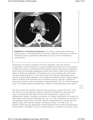
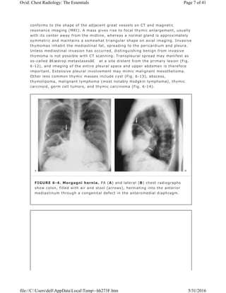
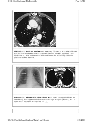
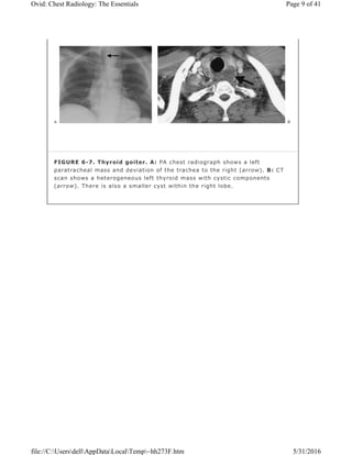
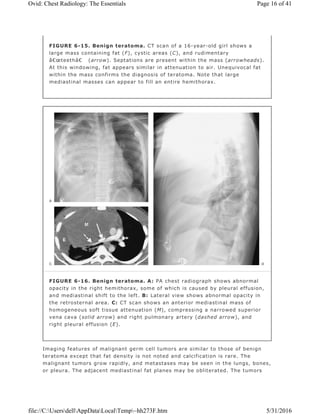
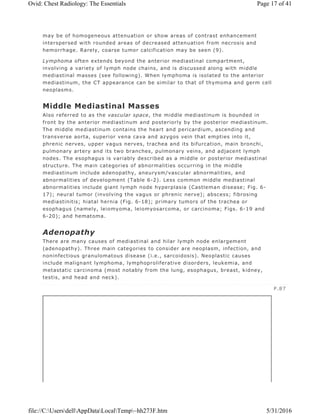
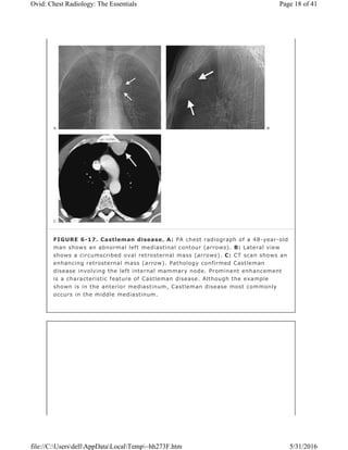
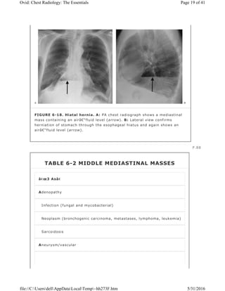
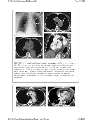
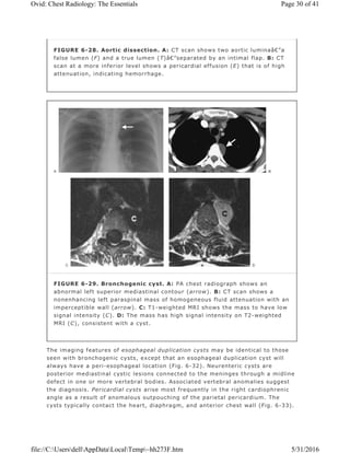
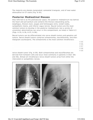
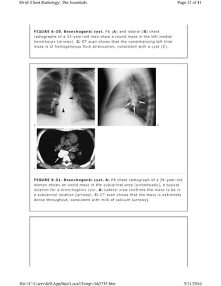
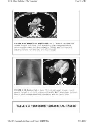
Editors: Collins, Jannette; Stern, Eric J.
Title: Chest Radiology: The Essentials, 2nd Edition
Copyright ©2008 Lippincott Williams & Wilkins
> Table of Contents > Chapter 6 - Mediastinal Masses
Chapter 6
Mediastinal Masses
Learning Objectives
1. State the anatomic boundaries of the anterior, middle, posterior, and
superior mediastinum.
2. Name the four most common causes of an anterior mediastinal mass;
localize a mass in the anterior mediastinum on a chest radiograph and
chest computed tomographic (CT) scan.
3. Name the three most common causes of a middle mediastinal mass; localize
a mass in the middle mediastinum on a chest radiograph and chest CT scan.
4. Name the most common cause of a posterior mediastinal mass; localize a
mass in the posterior mediastinum on a chest radiograph and chest CT
scan.
5. Name five etiologies of bilateral hilar lymph node enlargement.
6. State the three most common locations (Garland triad) of thoracic lymph
node enlargement in sarcoidosis.
7. Recognize a cystic mass in the mediastinum on chest CT and suggest the
possible diagnosis of a bronchogenic, pericardial, thymic, or esophageal
duplication cyst.
8. Describe and recognize the findings of fibrosing mediastinitis on chest CT.
The mediastinum includes the organs located between the two lungs. Because
the mediastinum is invested by the medial parietal pleura, a mass isolated to the
mediastinum will generally have a smooth contour (created by the pleural
surface). Lung parenchymal masses, on the other hand, are not surrounded by
pleura and may therefore have an irregular contour. Mediastinal masses can be
recognized on chest radiographs when there is an abnormal contour to the
normal mediastinal structures. On the right, these normal structures include,
from superior to inferior, the brachiocephalic vessels, superior vena cava, azygos
arch, ascending aorta, and right atrium. On the left, the mediastinal contour,
from superior to inferior, is made up of the brachiocephalic vessels, aortic arch,
main pulmonary artery, and left ventricle. On the lateral chest radiograph, the
mediastinum extends from the inner margin of the sternum to the inner margins
of the posterior ribs.
Page 1 of 41Ovid: Chest Radiology: The Essentials
5/31/2016file://C:UsersdellAppDataLocalTemp~hh273F.htm
The mediastinum can be divided into compartments, and many classification
systems have been devised to simplify the differential diagnosis when an
abnormality is seen in one or more compartments. A system based on anatomic
subdivisions (the superior, anterior, middle, and posterior mediastinal
compartments) will be used in this chapter (Fig. 6-1). Approximately 60% of all
mediastinal masses arise in the anterior mediastinum, 25% appear in the
posterior mediastinum, and 15% occur in the middle mediastinum (1). When an
abnormality is not isolated to one mediastinal compartment, as is often the case
with large mediastinal masses, the list of diagnostic possibilities can be
determined by localizing the abnormality to the mediastinal compartment serving
as the “epicenter†of the abnormality or by considering all abnormalities
that occur in the compartments involved. Associated radiologic findings can also
help to narrow the list of diagnostic possibilities; these include deviation of the
trachea (commonly seen with thyroid masses); presence of axillary, abdominal,
and retroperitoneal adenopathy (suggesting the diagnosis of lymphoma); or
posterior rib erosion or destruction (consistent with a posterior mediastinal
mass, such as a neurogenic tumor).
Superior Mediastinum
The superior mediastinum is bounded above by the thoracic inlet and below by a
line from the sternomanubrial joint to the fourth thoracic vertebral body. The
other mediastinal compartments lie inferior to this line. Abnormalities in the
superior mediastinum include masses that extend down into the upper
mediastinum from the neck, such as thyroid goiters or cystic hygromas;
mediastinal adenopathy; and vascular abnormalities, such as aneurysms.
P.79
Page 2 of 41Ovid: Chest Radiology: The Essentials
5/31/2016file://C:UsersdellAppDataLocalTemp~hh273F.htm
Anterior Mediastinum
Also referred to as the prevascular space, the anterior mediastinum is bounded
above by the superior mediastinum, laterally by pleura, anteriorly by the
sternum, and posteriorly by pericardium and great vessels. This compartment
contains areolar tissue, lymph nodes, lymphatic vessels, thymus gland, thyroid
gland, parathyroid glands, and internal mammary arteries and veins.
Masses occurring in the anterior mediastinum are listed in Table 6-1. In addition
to the “4 Ts†(thymoma, thyroid mass, teratoma, and terrible lymphoma),
which account for the great majority of lesions, there are many other less
common causes of an anterior mediastinal mass. These include vascular
tortuosity or aneurysm, cardiac tumors or prominent pericardiac fat (Fig. 6-2),
cystic hygroma, bronchogenic cyst, pericardial cyst, hemangioma,
lymphangioma, parathyroid adenoma (Fig. 6-3), various other mesenchymal
FIGURE 6-1. Mediastinal compartments. Lateral chest radiograph shows
the boundaries delineating the superior (S), anterior (A), middle (M), and
posterior (P) mediastinal compartments.
Page 3 of 41Ovid: Chest Radiology: The Essentials
5/31/2016file://C:UsersdellAppDataLocalTemp~hh273F.htm
tumors (e.g., fibroma or lipoma), sternal tumor, primary lung tumor invading the
anterior mediastinum, Morgagni hernia (Fig. 6-4), abscess (Fig. 6-5), and
mediastinal lipomatosis (Fig. 6-6).
FIGURE 6-2. Prominent pericardial fat. A: Posteroanterior (PA) chest
radiograph shows a round mass at the right cardiophrenic angle (arrow) that
is less opaque than the adjacent heart. B: Lateral view shows that the mass
is projected over the anterior inferior heart (arrows) in the typical location
of pericardiac fat. C: CT shows this mass to be of fat attenuation (F), similar
to that of the subcutaneous fat.
TABLE 6-1 ANTERIOR MEDIASTINAL MASSES
“4 Tsâ€
Thymoma (generally over age 40)
Teratoma (and other germ cell tumors, generally under age 40)
Page 4 of 41Ovid: Chest Radiology: The Essentials
5/31/2016file://C:UsersdellAppDataLocalTemp~hh273F.htm
An intrathoracic thyroid mass is usually a benign multi-nodular goiter that
originates in the neck and extends downward into the mediastinum through the
thoracic inlet. This continuity is an important diagnostic feature on chest
radiography. Many thyroid masses displace or narrow the trachea (Fig. 6-7).
Another useful sign of a thyroid mass is the relative high attenuation value of
the thyroid tissue, at least 20 Hounsfield units above that seen in adjacent
muscles on both precontrast and postcontrast computed tomographic (CT)
images (2). CT scans can show cystic components. Calcification is common and
usually caused by benign disease when it is dense, amorphous, and well defined
with a nodular, curvilinear, or circular configuration. Distinguishing between
benign and malignant thyroid masses on chest radiography or CT scanning is not
possible unless the tumor has clearly spread beyond the
thyroid gland (indicating malignancy). Malignant thyroid masses can also have
calcifications, generally with a configuration of fine dots grouped in a cloudlike
formation (3,4,5). However, the patterns of benign and malignant calcifications
serve as general guidelines, and malignant medullary thyroid carcinoma can
contain well-defined, occasionally ring-shaped, dense calcifications that are
similar in appearance to the calcifications seen with benign thyroid goiter.
Radionuclide imaging is a very sensitive and specific method of determining the
thyroid nature of an intrathoracic mass (Fig. 6-8), but CT provides more
information about the mass.
Thyroid (goiter or neoplasm, look for tracheal deviation)
Terrible lymphoma
P.80
Page 5 of 41Ovid: Chest Radiology: The Essentials
5/31/2016file://C:UsersdellAppDataLocalTemp~hh273F.htm
Thymomas are tumors consisting of thymic epithelial cells and reactive
lymphocytes, with noninvasive or invasive patterns of growth (6). The presence
or absence of tumor spread beyond the capsule (usually determined surgically),
rather than the histologic appearance within the thymus, determines whether a
tumor is benign or malignant. Thymomas occur at an average age of 50 years
and with equal frequency in men and women (6). They are associated with a
variety of autoimmune diseases, most notably myasthenia gravis. Approximately
40% of patients with thymoma have myasthenia gravis, and the incidence of
thymoma in patients with this disease is approximately 15% (7). Most thymomas
arise in the upper anterior mediastinum, anterior to the ascending aorta above
the right ventricular outflow tract and main pulmonary artery (Fig. 6-9). They
can extend into the adjacent middle or posterior mediastinum, and they can
occur or extend into the lower third of the mediastinum, as low as the
cardiophrenic angles. Punctate, curvilinear, or ringlike calcification is common in
both benign and invasive thymomas (8). On a CT scan, thymomas are usually of
homogeneous attenuation and show uniform enhancement, but rarely they can
appear cystic with discrete nodular components (Figs. 6-10 and 6-11). In
patients under age 40, diagnosing a small thymoma can be difficult because the
normal gland is variable in size. A normal thymus, in contrast to a thymic mass,
FIGURE 6-3. Parathyroid adenoma. CT shows a nonspecific enhancing
mass (arrow) in the aortopulmonary window. Although commonly an anterior
mediastinal mass, parathyroid adenomas can occur in any mediastinal
compartment.
P.81
P.82
Page 6 of 41Ovid: Chest Radiology: The Essentials
5/31/2016file://C:UsersdellAppDataLocalTemp~hh273F.htm
conforms to the shape of the adjacent great vessels on CT and magnetic
resonance imaging (MRI). A mass gives rise to focal thymic enlargement, usually
with its center away from the midline, whereas a normal gland is approximately
symmetric and maintains a somewhat triangular shape on axial imaging. Invasive
thymomas inhabit the mediastinal fat, spreading to the pericardium and pleura.
Unless mediastinal invasion has occurred, distinguishing benign from invasive
thymoma is not possible with CT scanning. Transpleural spread may manifest as
so-called “drop metastases†at a site distant from the primary lesion (Fig.
6-12), and imaging of the entire pleural space and upper abdomen is therefore
important. Extensive pleural involvement may mimic malignant mesothelioma.
Other less common thymic masses include cyst (Fig. 6-13), abscess,
thymolipoma, malignant lymphoma (most notably Hodgkin lymphoma), thymic
carcinoid, germ cell tumors, and thymic carcinoma (Fig. 6-14).
FIGURE 6-4. Morgagni hernia. PA (A) and lateral (B) chest radiographs
show colon, filled with air and stool (arrows), herniating into the anterior
mediastinum through a congenital defect in the anteromedial diaphragm.
Page 7 of 41Ovid: Chest Radiology: The Essentials
5/31/2016file://C:UsersdellAppDataLocalTemp~hh273F.htm
FIGURE 6-5. Anterior mediastinal abscess. CT scan of a 54-year-old man
who recently underwent aortic valve replacement shows a loculated fluid
collection (A) with an enhancing rim anterior to the ascending aorta and
posterior to the sternum.
FIGURE 6-6. Mediastinal lipomatosis. A: PA chest radiograph shows an
abnormally wide upper mediastinum with straight margins (arrows). B: CT
scan shows abundant mediastinal fat (F).
Page 8 of 41Ovid: Chest Radiology: The Essentials
5/31/2016file://C:UsersdellAppDataLocalTemp~hh273F.htm
FIGURE 6-7. Thyroid goiter. A: PA chest radiograph shows a left
paratracheal mass and deviation of the trachea to the right (arrow). B: CT
scan shows a heterogeneous left thyroid mass with cystic components
(arrow). There is also a smaller cyst within the right lobe.
Page 9 of 41Ovid: Chest Radiology: The Essentials
5/31/2016file://C:UsersdellAppDataLocalTemp~hh273F.htm
FIGURE 6-8. Papillary thyroid carcinoma. A: PA chest radiograph of a
48-year-old woman shows a right paratracheal mass and deviation of the
trachea to the left (arrow). B: Technetium pertechnetate–enhanced
thyroid scan shows a “cold†nodule in the right lobe of the thyroid
gland (arrow). Nearly all thyroid cancers are nonfunctioning or “coldâ€
nodules on nuclear medicine thyroid scans. R, right lobe of thyroid; L, left
lobe of thyroid.
Page 10 of 41Ovid: Chest Radiology: The Essentials
5/31/2016file://C:UsersdellAppDataLocalTemp~hh273F.htm
A teratoma is a neoplasm derived from more than one embryonic germ layer.
Other germ cell tumors include benign dermoid cyst, malignant seminoma (the
most common germ cell tumor), teratocarcinoma, embryonal carcinoma,
endodermal sinus tumor (yolk sac tumor), choriocarcinoma, and mixtures of
these types. Germ cell tumors in the mediastinum arise from primitive rest cells
and generally are not metastatic from gonadal tumors. Some malignant germ cell
tumors secrete beta-human chorionic gonadotropin and alpha-fetoprotein, which
can be used to diagnose and monitor progression of disease.
Benign teratomas are found in patients of all ages but are most common in
adolescents and young adults (Fig. 6-15). They usually produce a well-defined,
FIGURE 6-9. Benign thymoma. A: PA chest radiograph of a 70-year-old
woman shows a rounded mass overlying the right heart border. The
visualized margins are well circumscribed (arrows). B: Lateral chest
radiograph suggests that the mass is located in the anterior mediastinum
(arrows). C: CT scan shows a peanut-shaped mass of fairly homogeneous
attenuation (arrows). The mass is located anterior to the ascending aorta
(A), above the right ventricular outflow tract, a typical location for
thymomas.
Page 11 of 41Ovid: Chest Radiology: The Essentials
5/31/2016file://C:UsersdellAppDataLocalTemp~hh273F.htm
rounded, or lobulated mass in the anterior mediastinum. They grow slowly,
although rapid increase in size may occur as a result of hemorrhage, producing
imaging features suggestive of a malignant mass.
Calcification, ossification, teeth, or fat may be visible on a chest radiograph and
on CT scans. CT scans may show cystic components and/or a fat–fluid level. A
cyst wall with curvilinear calcification is often present. Unequivocal fat within the
mass confirms the diagnosis of teratoma, but the absence of fat or calcium does
not exclude a teratoma (Fig. 6-16).
P.83
P.84
P.85
P.86
FIGURE 6-10. Benign cystic thymoma. A: PA chest radiograph shows an
abnormal left mediastinal contour (arrow). B: Lateral view shows a round,
circumscribed mass with a high-attenuation rim in the anterior mediastinum
(arrows). C: CT scan shows a mass of soft tissue attenuation (T) with dense
rim calcification anterior to the ascending aorta.
Page 12 of 41Ovid: Chest Radiology: The Essentials
5/31/2016file://C:UsersdellAppDataLocalTemp~hh273F.htm
FIGURE 6-11. Benign thymoma. A: PA chest radiograph shows an
abnormal left mediastinal contour (arrow). B: CT scan shows soft tissue
anterior to the ascending aorta in the expected location of the thymus gland
(arrow). C: CT scan inferior to (B) shows that the thymic soft tissue (T) is
focally enlarged on the left.
Page 13 of 41Ovid: Chest Radiology: The Essentials
5/31/2016file://C:UsersdellAppDataLocalTemp~hh273F.htm
FIGURE 6-12. Malignant thymoma. A: CT scan shows a lobulated mass
(T) of homogeneous soft tissue attenuation in the anterior mediastinum. B:
CT scan at a level inferior to (A) shows course calcification (arrow) within
the mass. C: A “drop†metastasis (arrow) is seen along the right
hemidiaphragm.
FIGURE 6-13. Thymic cyst. CT scan of a 60-year-old man shows a
circumscribed, rim-calcified oval mass of homogeneous fluid attenuation (C)
in the expected location of the thymus gland.
Page 14 of 41Ovid: Chest Radiology: The Essentials
5/31/2016file://C:UsersdellAppDataLocalTemp~hh273F.htm
FIGURE 6-14. Thymic carcinoma. A: PA chest radiograph shows an
abnormal left mediastinal contour (arrow). B: Lateral view shows abnormal
opacity in the retrosternal area (arrows). C: CT scan shows a lobulated mass
of homogeneous soft tissue attenuation (T) in the anterior mediastinum. D:
Coronal positron emission tomographic scan shows normal activity in the
heart (H) and abnormal activity in the thymic mass (T).
Page 15 of 41Ovid: Chest Radiology: The Essentials
5/31/2016file://C:UsersdellAppDataLocalTemp~hh273F.htm
Imaging features of malignant germ cell tumors are similar to those of benign
teratoma except that fat density is not noted and calcification is rare. The
malignant tumors grow rapidly, and metastases may be seen in the lungs, bones,
or pleura. The adjacent mediastinal fat planes may be obliterated. The tumors
FIGURE 6-15. Benign teratoma. CT scan of a 16-year-old girl shows a
large mass containing fat (F), cystic areas (C), and rudimentary
“teeth†(arrow). Septations are present within the mass (arrowheads).
At this windowing, fat appears similar in attenuation to air. Unequivocal fat
within the mass confirms the diagnosis of teratoma. Note that large
mediastinal masses can appear to fill an entire hemithorax.
FIGURE 6-16. Benign teratoma. A: PA chest radiograph shows abnormal
opacity in the right hemithorax, some of which is caused by pleural effusion,
and mediastinal shift to the left. B: Lateral view shows abnormal opacity in
the retrosternal area. C: CT scan shows an anterior mediastinal mass of
homogeneous soft tissue attenuation (M), compressing a narrowed superior
vena cava (solid arrow) and right pulmonary artery (dashed arrow), and
right pleural effusion (E).
Page 16 of 41Ovid: Chest Radiology: The Essentials
5/31/2016file://C:UsersdellAppDataLocalTemp~hh273F.htm
may be of homogeneous attenuation or show areas of contrast enhancement
interspersed with rounded areas of decreased attenuation from necrosis and
hemorrhage. Rarely, coarse tumor calcification may be seen (9).
Lymphoma often extends beyond the anterior mediastinal compartment,
involving a variety of lymph node chains, and is discussed along with middle
mediastinal masses (see following). When lymphoma is isolated to the anterior
mediastinum, the CT appearance can be similar to that of thymoma and germ cell
neoplasms.
Middle Mediastinal Masses
Also referred to as the vascular space, the middle mediastinum is bounded in
front by the anterior mediastinum and posteriorly by the posterior mediastinum.
The middle mediastinum contains the heart and pericardium, ascending and
transverse aorta, superior vena cava and azygos vein that empties into it,
phrenic nerves, upper vagus nerves, trachea and its bifurcation, main bronchi,
pulmonary artery and its two branches, pulmonary veins, and adjacent lymph
nodes. The esophagus is variably described as a middle or posterior mediastinal
structure. The main categories of abnormalities occurring in the middle
mediastinum include adenopathy, aneurysm/vascular abnormalities, and
abnormalities of development (Table 6-2). Less common middle mediastinal
abnormalities include giant lymph node hyperplasia (Castleman disease; Fig. 6-
17); neural tumor (involving the vagus or phrenic nerve); abscess; fibrosing
mediastinitis; hiatal hernia (Fig. 6-18); primary tumors of the trachea or
esophagus (namely, leiomyoma, leiomyosarcoma, or carcinoma; Figs. 6-19 and
6-20); and hematoma.
Adenopathy
There are many causes of mediastinal and hilar lymph node enlargement
(adenopathy). Three main categories to consider are neoplasm, infection, and
noninfectious granulomatous disease (i.e., sarcoidosis). Neoplastic causes
include malignant lymphoma, lymphoproliferative disorders, leukemia, and
metastatic carcinoma (most notably from the lung, esophagus, breast, kidney,
testis, and head and neck).
P.87
Page 17 of 41Ovid: Chest Radiology: The Essentials
5/31/2016file://C:UsersdellAppDataLocalTemp~hh273F.htm
FIGURE 6-17. Castleman disease. A: PA chest radiograph of a 48-year-old
man shows an abnormal left mediastinal contour (arrows). B: Lateral view
shows a circumscribed oval retrosternal mass (arrows). C: CT scan shows an
enhancing retrosternal mass (arrow). Pathology confirmed Castleman
disease involving the left internal mammary node. Prominent enhancement
is a characteristic feature of Castleman disease. Although the example
shown is in the anterior mediastinum, Castleman disease most commonly
occurs in the middle mediastinum.
Page 18 of 41Ovid: Chest Radiology: The Essentials
5/31/2016file://C:UsersdellAppDataLocalTemp~hh273F.htm
FIGURE 6-18. Hiatal hernia. A: PA chest radiograph shows a mediastinal
mass containing an air–fluid level (arrow). B: Lateral view confirms
herniation of stomach through the esophageal hiatus and again shows an
air–fluid level (arrow).
P.88
TABLE 6-2 MIDDLE MEDIASTINAL MASSES
“3 Asâ€
Adenopathy
Infection (fungal and mycobacterial)
Neoplasm (bronchogenic carcinoma, metastases, lymphoma, leukemia)
Sarcoidosis
Aneurysm/vascular
Page 19 of 41Ovid: Chest Radiology: The Essentials
5/31/2016file://C:UsersdellAppDataLocalTemp~hh273F.htm
Lymphoma is classified as either Hodgkin or non-Hodgkin lymphoma (Figs. 6-21
and 6-22). The onset of Hodgkin lymphoma most commonly occurs in the second
or third decade, with a secondary peak in the fifth or sixth decades of life. Non-
Hodgkin lymphoma occurs in all age groups. The main feature of malignant
lymphomas on chest radiography or CT scanning is mediastinal and hilar
lymphadenopathy, in some cases with accompanying pulmonary, pleural, or chest
wall involvement. The enlarged nodes can calcify, especially after therapy, in
irregular, eggshell, or diffuse patterns. The appearances of intrathoracic
adenopathy on chest radiographs and CT scans are similar in Hodgkin and non-
Hodgkin lymphoma, but the frequencies and distributions of the abnormalities
differ. Any intrathoracic nodal group may be enlarged in patients with lymphoma.
In general, the anterior mediastinal and paratracheal nodes are the most
frequently involved, with tracheobronchial and subcarinal nodes also commonly
enlarged. The majority of cases of Hodgkin lymphoma show enlargement of two
or more nodal groups, whereas only one nodal group is involved in about half the
cases of non-Hodgkin lymphoma. Hilar adenopathy is rare without accompanying
mediastinal adenopathy. The enlarged nodes may be discrete or matted together,
and the margins can be well defined or ill defined. Low-density areas can be
seen, resulting from cystic degeneration. Hodgkin lymphoma can arise primarily
in the thymus. Parenchymal involvement of the lung at initial presentation is
unusual. Parenchymal involvement in Hodgkin disease is almost always
accompanied by intrathoracic adenopathy (except after irradiation), whereas in
non-Hodgkin lymphoma, isolated pulmonary involvement occurs more than 50%
of the time (10). The most common pattern of parenchymal involvement is one
or more discrete nodules, which can cavitate. Another common pattern is round
or segmental, focal or patchy areas of dense airspace opacity often with air
bronchograms, mimicking pneumonia.
Abnormalities of development
Bronchogenic cyst
Pericardial cyst
Esophageal duplication cyst
(Neurenteric cyst—posterior mediastinum)
Page 20 of 41Ovid: Chest Radiology: The Essentials
5/31/2016file://C:UsersdellAppDataLocalTemp~hh273F.htm
Pleural effusions are seen on CT scans in 50% of patients with lymphoma (11).
FIGURE 6-19. Esophageal carcinoma. A: CT scan of a 54-year-old woman
with dysphagia shows a circumferential mass involving the upper esophagus
(arrow). Note the lack of air within the thick-walled esophagus. B: CT scan
at a level inferior to (A), with lung windowing, shows a parenchymal
metastasis (arrow).
FIGURE 6-20. Esophageal carcinoma. CT scan of a 63-year-old man
shows a distal esophageal mass (E). The man also had a cavitary squamous
cell carcinoma in the right lower lobe (arrow).
Page 21 of 41Ovid: Chest Radiology: The Essentials
5/31/2016file://C:UsersdellAppDataLocalTemp~hh273F.htm
Most pleural effusions are unilateral exudates, occasionally chylous in nature,
and they can be large. Both pleural and pericardial effusions can occur and have
nodular solid components. Chest wall invasion and rib destruction are seen on
occasion.
The most frequent infections that give rise to intrathoracic adenopathy are
caused by mycobacterial disease (most notably tuberculosis) and fungal disease
(particularly histoplasmosis; Fig. 6-23), each of which can occur without evident
pneumonia. Some patients develop a chronic progressive immune
response to dead Histoplasma capsular antigens, resulting in a condition known
as fibrosing mediastinitis. In this condition, nonmalignant fibrous tissue encases
and obliterates vasculature (arteries, veins, lymphatics) and airways in the
mediastinum (Fig. 6-24). Subcarinal and right paratracheal nodes are most
commonly involved. Calcification of nodes and simultaneous encasement of
airways and vasculature are characteristic CT findings of this disease.
Intrathoracic nodal enlargement can also be seen in tularemia, whooping cough,
anthrax, plague, and mycoplasmal, viral, and other more common bacterial
infections.
P.89
FIGURE 6-21. Hodgkin lymphoma. A: CT scan of a 59-year-old man with
shortness of breath shows a homogeneous mass of soft tissue attenuation
(M) compressing the left atrium (LA), nearly encasing the aorta (A), and
abutting the esophagus (dashed arrow) and azygous vein (solid arrow).
There is also a right pleural effusion (E). B: CT scan at a more inferior level
shows retrocrural lymphadenopathy (A).
Page 22 of 41Ovid: Chest Radiology: The Essentials
5/31/2016file://C:UsersdellAppDataLocalTemp~hh273F.htm
Sarcoidosis is a frequent cause of intrathoracic adenopathy in young adults.
When multiple node groups are involved and adenopathy is symmetrically
distributed in the hila and mediastinum in young asymptomatic adults,
sarcoidosis is the likely cause. The hilar lymph nodes are frequently potato
shaped and clear of the cardiac borders, a feature that is often useful in
distinguishing sarcoidosis from lymphoma (Fig. 6-25).
Enlargement of paratracheal and bilateral hilar lymph nodes (the Garland triad,
or the “1-2-3 sign†) is a nonspecific pattern of adenopathy that is common
in patients with sarcoidosis (Fig. 6-26).
FIGURE 6-22. Hodgkin lymphoma. A: CT scan shows right supraclavicular
lymphadenopathy (A) compressing the right jugular vein (arrow). B: CT scan
at a more inferior level shows confluent anterior mediastinal and right
paratracheal lymphadenopathy (A). C: Another CT scan shows bilateral
axillary lymphadenopathy (A).
P.90
P.91
Page 23 of 41Ovid: Chest Radiology: The Essentials
5/31/2016file://C:UsersdellAppDataLocalTemp~hh273F.htm
FIGURE 6-23. Pulmonary histoplasmosis. A: PA chest radiograph shows
right paratracheal (solid arrow) and bilateral hilar (dashed arrows)
lymphadenopathy. B: CT scan shows bulky right paratracheal
lymphadenopathy (arrow). C: CT scan at a more inferior level shows
bilateral hilar (solid arrows) and subcarinal (dashed arrow)
lymphadenopathy. The appearance is indistinguishable from that of
sarcoidosis.
Page 24 of 41Ovid: Chest Radiology: The Essentials
5/31/2016file://C:UsersdellAppDataLocalTemp~hh273F.htm
Adenopathy is more easily identified and quantified via CT scanning than chest
radiography. The CT signs of adenopathy are (a) an increase in size of individual
nodes, (b) invasion of surrounding mediastinal fat, (c) nodal masses, and (d)
diffuse soft tissue density throughout the mediastinum obliterating the
mediastinal fat. There are numerous studies regarding normal lymph node size
on CT scans (12,13,14). In general, nodes greater than 10 mm in short-axis
diameter are considered abnormal although nonspecific and not necessarily
malignant. Likewise, nodes less than 10 mm in short-axis diameter can be
FIGURE 6-24. Fibrosing mediastinitis. A: PA chest radiograph of a 26-
year-old woman with dysphagia, dilated chest and neck veins, dyspnea on
exertion, and fatigue shows abnormal opacity obliterating the right heart
margin (indicating a right middle lobe process) associated with elevation of
the right hemidiaphragm and abnormal right paratracheal opacity. B: Lateral
view confirms abnormal opacity in the right middle lobe. C: CT scan shows
fibrous tissue encasing and almost obliterating the superior vena cava (solid
arrow) and coarse mediastinal calcifications (dashed arrows). D: CT scan of
a more inferior level shows calcified fibrous tissue (dashed arrow) encasing
the right lower lobe pulmonary artery, along with densely calcified
subcarinal fibrous tissue (solid arrow).
Page 25 of 41Ovid: Chest Radiology: The Essentials
5/31/2016file://C:UsersdellAppDataLocalTemp~hh273F.htm
pathologic.
Aneurysm/Vascular Abnormalities
As a structure in the middle mediastinum, the aorta can also give rise to
abnormalities in this compartment. It is very important to distinguish an aortic
aneurysm from other mediastinal masses, particularly if biopsy is being
considered. The aorta commonly becomes atherosclerotic and ectatic with
advancing
age, and it can become aneurysmal. Rupture is the feared complication of aortic
aneurysm (Fig. 6-27). Most atherosclerotic aneurysms are fusiform in shape,
although some are saccular. Fusiform aneurysms usually arise in the aortic arch
or descending thoracic aorta. Saccular aneurysms usually arise from the
descending aorta, or occasionally from the aortic arch, but they are unusual in
the ascending aorta. Aortic aneurysms can be distinguished from other
mediastinal masses by recognizing their conformity to the aorta and by the
presence of curvilinear calcification in the wall of the aneurysm. Traumatic aortic
pseudoaneurysm is discussed in Chapter 8. Mycotic aneurysms of the aorta occur
in patients with predisposing causes: intravenous drug abusers, patients with
valvular disease or congenital disorders of the heart or aorta, patients who have
undergone previous cardiac or aortic surgery, patients with adjacent pyogenic
infection, and those who are immunocompromised. Mycotic aneurysms are
FIGURE 6-25. Sarcoidosis. PA (A) and lateral (B) chest radiographs of an
asymptomatic 25-year-old man show bilateral hilar adenopathy (straight
arrows). The enlarged nodes are potato shaped and clear of the cardiac
borders, a feature that can help distinguish them from lymphomatous
enlargement of hilar nodes. On the lateral view, subcarinal adenopathy is
also seen (curved arrows).
P.92
Page 26 of 41Ovid: Chest Radiology: The Essentials
5/31/2016file://C:UsersdellAppDataLocalTemp~hh273F.htm
usually saccular in shape, enlarge rapidly, and lack calcification of the wall.
Aortic dissections are collections of blood within the media of the aortic wall that
communicate with the true aortic lumen through one or more tears in the intima.
Most dissections begin within an intimal tear, and bleeding splits the aortic
media. Two classification systems are used to describe aortic dissection. The
DeBakey classification divides aortic dissection into three types (15). Type I
refers to dissections that start in the ascending aorta and extend into the
descending aorta; type II, to dissections confined to the ascending aorta; and
type III, to dissections that start just beyond the left subclavian artery and are
confined to the descending aorta. The Stanford classification refers to type A
(involving the ascending aorta) and type B (confined to the descending aorta)
(16). The diagnostic feature of aortic dissection on contrast-enhanced CT
scanning is two lumina separated by an intimal flap (Fig. 6-28). The intimal flap
is seen as a curvilinear low-attenuation area within the opacified aorta. A false
lumen usually fills and empties in a delayed fashion compared with a true lumen.
The false lumen may be partially or totally filled by thrombus and therefore may
not opacify. The true lumen is usually compressed by the false lumen.
Displacement of calcified atheromatous plaques by the dissection can be
FIGURE 6-26. Sarcoidosis. A: CT scan of a 20-year-old man shows
enlarged paratracheal and aortopulmonary lymph nodes (A). B: CT scan at a
more inferior level shows bilateral hilar (arrows) lymphadenopathy.
Enlargement of paratracheal and bilateral hilar lymph nodes is commonly
seen with sarcoidosis and is referred to as the “1-2-3†sign or Garland
triad. C: CT scan with lung windowing shows small, ill-defined nodules in a
bronchovascular distribution (arrows); this is typical of the parenchymal
findings seen with sarcoidosis.
Page 27 of 41Ovid: Chest Radiology: The Essentials
5/31/2016file://C:UsersdellAppDataLocalTemp~hh273F.htm
demonstrated on precontrast CT scans when contrast enhancement of the two
lumina cannot be achieved (as with a thrombosed false lumen). Aortic dissection
is further discussed along with other aortic pathology in Chapter 19.
Abnormalities of Development
Developmental mediastinal cysts include bronchogenic cysts, esophageal
duplication cysts, neurenteric cysts, and pericardial cysts. The first three are
also referred to as bronchopulmonary foregut malformations, signifying their
origin from the embryologic foregut as the result of abnormal ventral budding of
the tracheobronchial tree (17). Mediastinal cysts containing cartilage are
classified as bronchogenic, and those with gastric epithelium as enteric.
Neurenteric and some esophageal duplication cysts arise within the posterior
mediastinum, but they will be discussed here with middle mediastinal masses.
Bronchogenic cysts have a fibrous capsule, often contain cartilage, are lined with
respiratory epithelium, and contain mucoid material. Most arise in the
mediastinum or hilar areas, but they can also arise within the lung parenchyma.
These cysts can rapidly increase in size as a result of hemorrhage, infection, or
distension with air, indicating communication with the airways. They are seen on
chest radiographs as well-defined
solitary masses in the mediastinum or hilum (Figs. 6-29 and 6-30) and are
usually found in close proximity to the major airways. The single most frequent
site is between the carina and the esophagus. Calcification, either rim
calcification or milk of calcium within the cyst, has been described (18). CT scans
usually show a simple cystic mass with an imperceptible or thin smooth wall. The
CT attenuation is generally that of water (-10 to +10 Hounsfield units) but can
be higher (as high as 120 Hounsfield units) when filled with milk of calcium or
proteinaceous material that accumulates after infection or hemorrhage (Fig. 6-
31). The cysts can be unilocular or multilocular. Curvilinear calcification of the
wall can be seen.
P.93
P.94
Page 28 of 41Ovid: Chest Radiology: The Essentials
5/31/2016file://C:UsersdellAppDataLocalTemp~hh273F.htm
FIGURE 6-27. Leaking thoracic aortic aneurysm. A: PA chest radiograph
of a 77-year-old man with chest pain shows a widened mediastinum and
abnormal left mediastinal contour (arrows). B: CT scan shows a focal
aneurysm of the aortic arch (arrow). Note the interruption in dense mural
calcification. C: CT scan at a more inferior level shows obliteration of the
normal aortic contours and adjacent left pleural effusion. D: Sagittal
reformatted CT scan shows the focal aneurysm (arrow) and adjacent fluid
collection.
Page 29 of 41Ovid: Chest Radiology: The Essentials
5/31/2016file://C:UsersdellAppDataLocalTemp~hh273F.htm
The imaging features of esophageal duplication cysts may be identical to those
seen with bronchogenic cysts, except that an esophageal duplication cyst will
always have a peri-esophageal location (Fig. 6-32). Neurenteric cysts are
posterior mediastinal cystic lesions connected to the meninges through a midline
defect in one or more vertebral bodies. Associated vertebral anomalies suggest
the diagnosis. Pericardial cysts arise most frequently in the right cardiophrenic
angle as a result of anomalous outpouching of the parietal pericardium. The
cysts typically contact the heart, diaphragm, and anterior chest wall (Fig. 6-33).
FIGURE 6-28. Aortic dissection. A: CT scan shows two aortic lumina—a
false lumen (F) and a true lumen (T)—separated by an intimal flap. B: CT
scan at a more inferior level shows a pericardial effusion (E) that is of high
attenuation, indicating hemorrhage.
FIGURE 6-29. Bronchogenic cyst. A: PA chest radiograph shows an
abnormal left superior mediastinal contour (arrow). B: CT scan shows a
nonenhancing left paraspinal mass of homogeneous fluid attenuation with an
imperceptible wall (arrow). C: T1-weighted MRI shows the mass to have low
signal intensity (C). D: The mass has high signal intensity on T2-weighted
MRI (C), consistent with a cyst.
Page 30 of 41Ovid: Chest Radiology: The Essentials
5/31/2016file://C:UsersdellAppDataLocalTemp~hh273F.htm
The majority are sharply marginated, somewhat triangular, and of near-water
attenuation on CT scans (Fig. 6-34).
Posterior Mediastinal Masses
Also referred to as the postvascular space, the posterior mediastinum lies behind
the heart and pericardium and contains the thoracic descending aorta,
esophagus, thoracic duct, azygos and hemiazygos veins, lymph nodes,
sympathetic chains, and inferior vagus nerves. Neural tumors are the most
common tumors to develop in the posterior mediastinum, but a variety of
uncommon abnormalities can occur in this compartment, as listed in Table 6-3
(Figs. 6-35, 6-36, 6-37, 6-38).
Neural tumors can be differentiated into nerve sheath tumors and ganglion cell
tumors. Nerve sheath tumors comprise schwannomas, neurofibromas, and their
malignant counterparts. The schwannoma is the most common intrathoracic
nerve sheath tumor (Fig. 6-39). Both schwannomas and neurofibromas are
derived from Schwann cells and occur most commonly in patients in their 30s
and 40s. Almost all intrathoracic nerve sheath tumors arise from either the
intercostal or sympathetic nerves.
P.95
P.96
P.97
P.98
P.99
Page 31 of 41Ovid: Chest Radiology: The Essentials
5/31/2016file://C:UsersdellAppDataLocalTemp~hh273F.htm
FIGURE 6-30. Bronchogenic cyst. PA (A) and lateral (B) chest
radiographs of a 23-year-old man show a round mass in the left medial
hemithorax (arrows). C: CT scan shows that the nonenhancing left hilar
mass is of homogeneous fluid attenuation, consistent with a cyst (C).
FIGURE 6-31. Bronchogenic cyst. A: PA chest radiograph of a 36-year-old
woman shows an ovoid mass in the subcarinal area (arrowheads), a typical
location for a bronchogenic cyst. B: Lateral view confirms the mass to be in
a subcarinal location (arrows). C: CT scan shows that the mass is extremely
dense throughout, consistent with milk of calcium (arrows).
Page 32 of 41Ovid: Chest Radiology: The Essentials
5/31/2016file://C:UsersdellAppDataLocalTemp~hh273F.htm
FIGURE 6-32. Esophageal duplication cyst. CT scan of a 45-year-old
woman shows a subcarinal cystic structure (C) of homogeneous fluid
attenuation in contact with the esophagus (arrow). The appearance is
indistinguishable from that of a bronchogenic cyst.
FIGURE 6-33. Pericardial cyst. A: PA chest radiograph shows a round
opacity (arrow) at the right cardiophrenic angle. B: CT scan shows the mass
(PC) to be of homogeneous fluid attenuation with rim calcification.
TABLE 6-3 POSTERIOR MEDIASTINAL MASSES
Page 33 of 41Ovid: Chest Radiology: The Essentials
5/31/2016file://C:UsersdellAppDataLocalTemp~hh273F.htm
Common
Neural tumors
Neurogenic (neuroblastoma, ganglioneuroma, ganglioneuroblastoma)
Nerve root tumors (schwannoma, neurofibroma, malignant
schwannoma)
Less common
Paraganglionic cell tumors (chemodectoma, pheochromocytoma)
Spinal tumor (metastases, primary bone tumor)
Lymphoma
Invasive thymoma
Mesenchymal tumor (fibroma, lipoma, leiomyoma, hemangioma,
lymphangioma)
Abscess
Pancreatic pseudocyst
Esophageal varices
Hematoma
Traumatic pseudomeningocele
Bochdalek hernia
Extramedullary hematopoiesis
Page 34 of 41Ovid: Chest Radiology: The Essentials
5/31/2016file://C:UsersdellAppDataLocalTemp~hh273F.htm
Page 35 of 41Ovid: Chest Radiology: The Essentials
5/31/2016file://C:UsersdellAppDataLocalTemp~hh273F.htm
FIGURE 6-34. Pericardial cyst. A: PA chest radiograph shows a smoothly
marginated, rounded mass adjacent to the right heart border (arrows). B:
Lateral chest radiograph shows the mass to be in the anterior or middle
mediastinum (arrowheads). C: CT scan shows a mass of homogeneous fluid
attenuation abutting the right pericardial border, typical of the appearance
of a pericardial cyst (arrows).
Page 36 of 41Ovid: Chest Radiology: The Essentials
5/31/2016file://C:UsersdellAppDataLocalTemp~hh273F.htm
FIGURE 6-35. Achalasia. A: PA chest radiograph of an 86-year-old woman
with dysphagia shows a widened contour to the upper mediastinum (arrows)
and air within the dilated air-filled esophagus (arrowheads). B: Lateral chest
radiograph shows an air–fluid level within the dilated esophagus
(arrowheads) and anterior deviation of the trachea (curved arrows).
FIGURE 6-36. Descending thoracic aortic aneurysm. A: PA chest
radiograph of a 69-year-old woman shows a rounded mass in continuity with
the descending aorta (straight arrows). Incidental note of calcified
granuloma in the left upper lobe (curved arrow). B: Lateral chest radiograph
shows curvilinear rim calcification within the wall of the aneurysm (arrows).
Page 37 of 41Ovid: Chest Radiology: The Essentials
5/31/2016file://C:UsersdellAppDataLocalTemp~hh273F.htm
FIGURE 6-37. Extramedullary hematopoiesis. CT scan of a 20-year-old
woman with thalassemia shows bilateral paraspinal soft tissue masses
(arrows) and expansion of the adjacent ribs.
FIGURE 6-38. Mediastinal pancreatic pseudocysts. A: CT scan of a 39-
year-old man with acute and chronic pancreatitis shows cystic areas within
the pancreas (arrow) and pancreatic calcifications. B: CT scan at a more
superior level shows rim-enhancing pancreatic pseudocysts (PC) within the
posterior mediastinum and a large right pleural effusion (E).
Page 38 of 41Ovid: Chest Radiology: The Essentials
5/31/2016file://C:UsersdellAppDataLocalTemp~hh273F.htm
The ganglion cell tumors include neuroblastoma (malignant), ganglioneuroma
(benign), and ganglioneuroblastoma (intermediate between benign and
malignant). The adrenal gland is the most common primary site for these
tumors, with the mediastinum being the second most common site.
Ganglioneuroma can occur between the ages of 1 and 50, while neuroblastoma
and ganglioneuroblastoma occur during childhood, generally under the age of 20
(19).
Nerve sheath and ganglion cell tumors are seen as well-defined masses with a
smooth or lobulated outline on imaging studies. Some are very large and can
occupy most of a hemithorax. Calcification can be seen in all types. In
neuroblastoma, the calcification is usually finely stippled, whereas in
ganglioneuroblastoma and ganglioneuroma it is denser and coarser. The bone
FIGURE 6-39. Benign schwannoma. PA (A) and lateral (B) chest
radiographs of a 9-year-old girl show a circumscribed mass in the right apex
(arrow). C: Axial T1-weighted MRI shows the mass (S) is paraspinal in
location and has no continuity with the spinal canal. D: Coronal MRI, with
intravenous contrast, shows that the mass (M) has high signal intensity.
Page 39 of 41Ovid: Chest Radiology: The Essentials
5/31/2016file://C:UsersdellAppDataLocalTemp~hh273F.htm
adjacent to the tumor shows a scalloped edge, with preservation but thickening
of the bony cortex. The ribs can be thinned and splayed apart, and the
intervertebral foramina can appear widened. On CT scans, many tumors have
mixed attenuation, including low-attenuation regions, that enhance on images
taken after administration of intravascular contrast material.
Paragangliomas are tumors of the paraganglionic cells and can be benign or
malignant chemodectomas or pheochromocytomas. Mediastinal paragangliomas
are rare, comprising only 2% of the large series of neural tumors of the thorax
(19). Paragangliomas occur in the area of the aortic arch and are classified as
aortic body tumors. They form rounded soft tissue masses that are extremely
vascular and enhance brightly on CT scans after administration of intravenous
contrast material.
References
1. Rubush JL, Gardner IR, Boyd WC, et al. Mediastinal tumors: review of 186
cases. J Thorac Cardiovasc Surg. 1973;65:216–222.
2. Morris UL, Colletti PM, Ralls PW, et al. CT demonstration of intrathoracic
thyroid tissue. J Comput Assist Tomogr. 1982;6:821–824.
3. Margolin FR, Winfield J, Steinbach HL. Patterns of thyroid calcification:
roentgenologic-histologic study of excised specimens. Invest Radiol.
1967;2:208–212.
4. Park CH, Rothermel FJ, Judge DM. Unusual calcification in mixed papillary
and follicular carcinoma of the thyroid gland. Radiology. 1976;119:554.
5. Wallace S, Hill CS, Paulus DD, et al. The radiologic aspects of medullary
(solid) thyroid carcinoma. Radiol Clin North Am. 1970;8:463–474.
6. LeGolvan DP, Abell MR. Thymomas. Cancer. 1977;39:2142–2157.
7. Lewis JE, Wick MR, Scheithauer BW, et al. Thymoma: a clinicopathological
review. Cancer. 1987;60:2727–2743.
8. Harper RAK, Guyer PB. The radiological features of thymic tumours: a
review of sixty-five cases. Clin Radiol. 1965;16:97–100.
9. Levitt RG, Husband JE, Galzer HS. CT of primary germ-cell tumors of the
mediastinum. AJR Am J Roentgenol. 1984;142:73–78.
P.100
Page 40 of 41Ovid: Chest Radiology: The Essentials
5/31/2016file://C:UsersdellAppDataLocalTemp~hh273F.htm
10. Jenkins PF, Ward MJ, Davies P, et al. Non-Hodgkin's lymphoma, chronic
lymphocytic leukaemia and the lung. Br J Dis Chest. 1981;75:22–30.
11. Lewis ER, Caskey CI, Fishman EK. Lymphoma of the lung: CT findings in
31 patients. AJR Am J Roentgenol. 1991;156:711–714.
12. Genereux GP, Howie JL. Normal mediastinal lymph node size and number:
CT and anatomic study. AJR Am J Roentgenol. 1984;142:1095–1100.
13. Glazer BH, Gross BH, Quint LE, et al. Normal mediastinal lymph nodes:
Number and size according to American Thoracic Society mapping. AJR Am J
Roentgenol. 1985;144:261–265.
14. Ingram CE, Belli AM, Lewars MD, et al. Normal lymph node size in the
mediastinum: A retrospective study in two patient groups. Clin Radiol.
1989;40:35–39.
15. DeBakey ME, Henly WS, Cooley DA, et al. Surgical management of
dissecting aneurysms of the aorta. J Thorac Cardiovasc Surg.
1965;49:130–149.
16. Appelbaum A, Karp RB, Kirklin JW. Ascending vs descending aortic
dissection. Ann Surg. 1976;183:296–300.
17. Fitch SJ, Tonkin ILD, Tonkin AK. Imaging of foregut duplication cysts.
RadioGraphics. 1986;6:189–201.
18. Bergstrom JF, Yost RV, Ford KT, et al. Unusual roentgen manifestations
of bronchogenic cysts. Radiology. 1973;107:49–54.
19. Reed JC, Haller KK, Feigin DS. Neural tumours of the thorax: subject
review from the AFIP. Radiology. 1978;126:9–17.
Page 41 of 41Ovid: Chest Radiology: The Essentials
5/31/2016file://C:UsersdellAppDataLocalTemp~hh273F.htm

Chest radiology essentials mediastinal masses

  • 1.
    Editors: Collins, Jannette;Stern, Eric J. Title: Chest Radiology: The Essentials, 2nd Edition Copyright ©2008 Lippincott Williams & Wilkins > Table of Contents > Chapter 6 - Mediastinal Masses Chapter 6 Mediastinal Masses Learning Objectives 1. State the anatomic boundaries of the anterior, middle, posterior, and superior mediastinum. 2. Name the four most common causes of an anterior mediastinal mass; localize a mass in the anterior mediastinum on a chest radiograph and chest computed tomographic (CT) scan. 3. Name the three most common causes of a middle mediastinal mass; localize a mass in the middle mediastinum on a chest radiograph and chest CT scan. 4. Name the most common cause of a posterior mediastinal mass; localize a mass in the posterior mediastinum on a chest radiograph and chest CT scan. 5. Name five etiologies of bilateral hilar lymph node enlargement. 6. State the three most common locations (Garland triad) of thoracic lymph node enlargement in sarcoidosis. 7. Recognize a cystic mass in the mediastinum on chest CT and suggest the possible diagnosis of a bronchogenic, pericardial, thymic, or esophageal duplication cyst. 8. Describe and recognize the findings of fibrosing mediastinitis on chest CT. The mediastinum includes the organs located between the two lungs. Because the mediastinum is invested by the medial parietal pleura, a mass isolated to the mediastinum will generally have a smooth contour (created by the pleural surface). Lung parenchymal masses, on the other hand, are not surrounded by pleura and may therefore have an irregular contour. Mediastinal masses can be recognized on chest radiographs when there is an abnormal contour to the normal mediastinal structures. On the right, these normal structures include, from superior to inferior, the brachiocephalic vessels, superior vena cava, azygos arch, ascending aorta, and right atrium. On the left, the mediastinal contour, from superior to inferior, is made up of the brachiocephalic vessels, aortic arch, main pulmonary artery, and left ventricle. On the lateral chest radiograph, the mediastinum extends from the inner margin of the sternum to the inner margins of the posterior ribs. Page 1 of 41Ovid: Chest Radiology: The Essentials 5/31/2016file://C:UsersdellAppDataLocalTemp~hh273F.htm
  • 2.
    The mediastinum canbe divided into compartments, and many classification systems have been devised to simplify the differential diagnosis when an abnormality is seen in one or more compartments. A system based on anatomic subdivisions (the superior, anterior, middle, and posterior mediastinal compartments) will be used in this chapter (Fig. 6-1). Approximately 60% of all mediastinal masses arise in the anterior mediastinum, 25% appear in the posterior mediastinum, and 15% occur in the middle mediastinum (1). When an abnormality is not isolated to one mediastinal compartment, as is often the case with large mediastinal masses, the list of diagnostic possibilities can be determined by localizing the abnormality to the mediastinal compartment serving as the “epicenter†of the abnormality or by considering all abnormalities that occur in the compartments involved. Associated radiologic findings can also help to narrow the list of diagnostic possibilities; these include deviation of the trachea (commonly seen with thyroid masses); presence of axillary, abdominal, and retroperitoneal adenopathy (suggesting the diagnosis of lymphoma); or posterior rib erosion or destruction (consistent with a posterior mediastinal mass, such as a neurogenic tumor). Superior Mediastinum The superior mediastinum is bounded above by the thoracic inlet and below by a line from the sternomanubrial joint to the fourth thoracic vertebral body. The other mediastinal compartments lie inferior to this line. Abnormalities in the superior mediastinum include masses that extend down into the upper mediastinum from the neck, such as thyroid goiters or cystic hygromas; mediastinal adenopathy; and vascular abnormalities, such as aneurysms. P.79 Page 2 of 41Ovid: Chest Radiology: The Essentials 5/31/2016file://C:UsersdellAppDataLocalTemp~hh273F.htm
  • 3.
    Anterior Mediastinum Also referredto as the prevascular space, the anterior mediastinum is bounded above by the superior mediastinum, laterally by pleura, anteriorly by the sternum, and posteriorly by pericardium and great vessels. This compartment contains areolar tissue, lymph nodes, lymphatic vessels, thymus gland, thyroid gland, parathyroid glands, and internal mammary arteries and veins. Masses occurring in the anterior mediastinum are listed in Table 6-1. In addition to the “4 Ts†(thymoma, thyroid mass, teratoma, and terrible lymphoma), which account for the great majority of lesions, there are many other less common causes of an anterior mediastinal mass. These include vascular tortuosity or aneurysm, cardiac tumors or prominent pericardiac fat (Fig. 6-2), cystic hygroma, bronchogenic cyst, pericardial cyst, hemangioma, lymphangioma, parathyroid adenoma (Fig. 6-3), various other mesenchymal FIGURE 6-1. Mediastinal compartments. Lateral chest radiograph shows the boundaries delineating the superior (S), anterior (A), middle (M), and posterior (P) mediastinal compartments. Page 3 of 41Ovid: Chest Radiology: The Essentials 5/31/2016file://C:UsersdellAppDataLocalTemp~hh273F.htm
  • 4.
    tumors (e.g., fibromaor lipoma), sternal tumor, primary lung tumor invading the anterior mediastinum, Morgagni hernia (Fig. 6-4), abscess (Fig. 6-5), and mediastinal lipomatosis (Fig. 6-6). FIGURE 6-2. Prominent pericardial fat. A: Posteroanterior (PA) chest radiograph shows a round mass at the right cardiophrenic angle (arrow) that is less opaque than the adjacent heart. B: Lateral view shows that the mass is projected over the anterior inferior heart (arrows) in the typical location of pericardiac fat. C: CT shows this mass to be of fat attenuation (F), similar to that of the subcutaneous fat. TABLE 6-1 ANTERIOR MEDIASTINAL MASSES “4 Ts†Thymoma (generally over age 40) Teratoma (and other germ cell tumors, generally under age 40) Page 4 of 41Ovid: Chest Radiology: The Essentials 5/31/2016file://C:UsersdellAppDataLocalTemp~hh273F.htm
  • 5.
    An intrathoracic thyroidmass is usually a benign multi-nodular goiter that originates in the neck and extends downward into the mediastinum through the thoracic inlet. This continuity is an important diagnostic feature on chest radiography. Many thyroid masses displace or narrow the trachea (Fig. 6-7). Another useful sign of a thyroid mass is the relative high attenuation value of the thyroid tissue, at least 20 Hounsfield units above that seen in adjacent muscles on both precontrast and postcontrast computed tomographic (CT) images (2). CT scans can show cystic components. Calcification is common and usually caused by benign disease when it is dense, amorphous, and well defined with a nodular, curvilinear, or circular configuration. Distinguishing between benign and malignant thyroid masses on chest radiography or CT scanning is not possible unless the tumor has clearly spread beyond the thyroid gland (indicating malignancy). Malignant thyroid masses can also have calcifications, generally with a configuration of fine dots grouped in a cloudlike formation (3,4,5). However, the patterns of benign and malignant calcifications serve as general guidelines, and malignant medullary thyroid carcinoma can contain well-defined, occasionally ring-shaped, dense calcifications that are similar in appearance to the calcifications seen with benign thyroid goiter. Radionuclide imaging is a very sensitive and specific method of determining the thyroid nature of an intrathoracic mass (Fig. 6-8), but CT provides more information about the mass. Thyroid (goiter or neoplasm, look for tracheal deviation) Terrible lymphoma P.80 Page 5 of 41Ovid: Chest Radiology: The Essentials 5/31/2016file://C:UsersdellAppDataLocalTemp~hh273F.htm
  • 6.
    Thymomas are tumorsconsisting of thymic epithelial cells and reactive lymphocytes, with noninvasive or invasive patterns of growth (6). The presence or absence of tumor spread beyond the capsule (usually determined surgically), rather than the histologic appearance within the thymus, determines whether a tumor is benign or malignant. Thymomas occur at an average age of 50 years and with equal frequency in men and women (6). They are associated with a variety of autoimmune diseases, most notably myasthenia gravis. Approximately 40% of patients with thymoma have myasthenia gravis, and the incidence of thymoma in patients with this disease is approximately 15% (7). Most thymomas arise in the upper anterior mediastinum, anterior to the ascending aorta above the right ventricular outflow tract and main pulmonary artery (Fig. 6-9). They can extend into the adjacent middle or posterior mediastinum, and they can occur or extend into the lower third of the mediastinum, as low as the cardiophrenic angles. Punctate, curvilinear, or ringlike calcification is common in both benign and invasive thymomas (8). On a CT scan, thymomas are usually of homogeneous attenuation and show uniform enhancement, but rarely they can appear cystic with discrete nodular components (Figs. 6-10 and 6-11). In patients under age 40, diagnosing a small thymoma can be difficult because the normal gland is variable in size. A normal thymus, in contrast to a thymic mass, FIGURE 6-3. Parathyroid adenoma. CT shows a nonspecific enhancing mass (arrow) in the aortopulmonary window. Although commonly an anterior mediastinal mass, parathyroid adenomas can occur in any mediastinal compartment. P.81 P.82 Page 6 of 41Ovid: Chest Radiology: The Essentials 5/31/2016file://C:UsersdellAppDataLocalTemp~hh273F.htm
  • 7.
    conforms to theshape of the adjacent great vessels on CT and magnetic resonance imaging (MRI). A mass gives rise to focal thymic enlargement, usually with its center away from the midline, whereas a normal gland is approximately symmetric and maintains a somewhat triangular shape on axial imaging. Invasive thymomas inhabit the mediastinal fat, spreading to the pericardium and pleura. Unless mediastinal invasion has occurred, distinguishing benign from invasive thymoma is not possible with CT scanning. Transpleural spread may manifest as so-called “drop metastases†at a site distant from the primary lesion (Fig. 6-12), and imaging of the entire pleural space and upper abdomen is therefore important. Extensive pleural involvement may mimic malignant mesothelioma. Other less common thymic masses include cyst (Fig. 6-13), abscess, thymolipoma, malignant lymphoma (most notably Hodgkin lymphoma), thymic carcinoid, germ cell tumors, and thymic carcinoma (Fig. 6-14). FIGURE 6-4. Morgagni hernia. PA (A) and lateral (B) chest radiographs show colon, filled with air and stool (arrows), herniating into the anterior mediastinum through a congenital defect in the anteromedial diaphragm. Page 7 of 41Ovid: Chest Radiology: The Essentials 5/31/2016file://C:UsersdellAppDataLocalTemp~hh273F.htm
  • 8.
    FIGURE 6-5. Anteriormediastinal abscess. CT scan of a 54-year-old man who recently underwent aortic valve replacement shows a loculated fluid collection (A) with an enhancing rim anterior to the ascending aorta and posterior to the sternum. FIGURE 6-6. Mediastinal lipomatosis. A: PA chest radiograph shows an abnormally wide upper mediastinum with straight margins (arrows). B: CT scan shows abundant mediastinal fat (F). Page 8 of 41Ovid: Chest Radiology: The Essentials 5/31/2016file://C:UsersdellAppDataLocalTemp~hh273F.htm
  • 9.
    FIGURE 6-7. Thyroidgoiter. A: PA chest radiograph shows a left paratracheal mass and deviation of the trachea to the right (arrow). B: CT scan shows a heterogeneous left thyroid mass with cystic components (arrow). There is also a smaller cyst within the right lobe. Page 9 of 41Ovid: Chest Radiology: The Essentials 5/31/2016file://C:UsersdellAppDataLocalTemp~hh273F.htm
  • 10.
    FIGURE 6-8. Papillarythyroid carcinoma. A: PA chest radiograph of a 48-year-old woman shows a right paratracheal mass and deviation of the trachea to the left (arrow). B: Technetium pertechnetate–enhanced thyroid scan shows a “cold†nodule in the right lobe of the thyroid gland (arrow). Nearly all thyroid cancers are nonfunctioning or “cold†nodules on nuclear medicine thyroid scans. R, right lobe of thyroid; L, left lobe of thyroid. Page 10 of 41Ovid: Chest Radiology: The Essentials 5/31/2016file://C:UsersdellAppDataLocalTemp~hh273F.htm
  • 11.
    A teratoma isa neoplasm derived from more than one embryonic germ layer. Other germ cell tumors include benign dermoid cyst, malignant seminoma (the most common germ cell tumor), teratocarcinoma, embryonal carcinoma, endodermal sinus tumor (yolk sac tumor), choriocarcinoma, and mixtures of these types. Germ cell tumors in the mediastinum arise from primitive rest cells and generally are not metastatic from gonadal tumors. Some malignant germ cell tumors secrete beta-human chorionic gonadotropin and alpha-fetoprotein, which can be used to diagnose and monitor progression of disease. Benign teratomas are found in patients of all ages but are most common in adolescents and young adults (Fig. 6-15). They usually produce a well-defined, FIGURE 6-9. Benign thymoma. A: PA chest radiograph of a 70-year-old woman shows a rounded mass overlying the right heart border. The visualized margins are well circumscribed (arrows). B: Lateral chest radiograph suggests that the mass is located in the anterior mediastinum (arrows). C: CT scan shows a peanut-shaped mass of fairly homogeneous attenuation (arrows). The mass is located anterior to the ascending aorta (A), above the right ventricular outflow tract, a typical location for thymomas. Page 11 of 41Ovid: Chest Radiology: The Essentials 5/31/2016file://C:UsersdellAppDataLocalTemp~hh273F.htm
  • 12.
    rounded, or lobulatedmass in the anterior mediastinum. They grow slowly, although rapid increase in size may occur as a result of hemorrhage, producing imaging features suggestive of a malignant mass. Calcification, ossification, teeth, or fat may be visible on a chest radiograph and on CT scans. CT scans may show cystic components and/or a fat–fluid level. A cyst wall with curvilinear calcification is often present. Unequivocal fat within the mass confirms the diagnosis of teratoma, but the absence of fat or calcium does not exclude a teratoma (Fig. 6-16). P.83 P.84 P.85 P.86 FIGURE 6-10. Benign cystic thymoma. A: PA chest radiograph shows an abnormal left mediastinal contour (arrow). B: Lateral view shows a round, circumscribed mass with a high-attenuation rim in the anterior mediastinum (arrows). C: CT scan shows a mass of soft tissue attenuation (T) with dense rim calcification anterior to the ascending aorta. Page 12 of 41Ovid: Chest Radiology: The Essentials 5/31/2016file://C:UsersdellAppDataLocalTemp~hh273F.htm
  • 13.
    FIGURE 6-11. Benignthymoma. A: PA chest radiograph shows an abnormal left mediastinal contour (arrow). B: CT scan shows soft tissue anterior to the ascending aorta in the expected location of the thymus gland (arrow). C: CT scan inferior to (B) shows that the thymic soft tissue (T) is focally enlarged on the left. Page 13 of 41Ovid: Chest Radiology: The Essentials 5/31/2016file://C:UsersdellAppDataLocalTemp~hh273F.htm
  • 14.
    FIGURE 6-12. Malignantthymoma. A: CT scan shows a lobulated mass (T) of homogeneous soft tissue attenuation in the anterior mediastinum. B: CT scan at a level inferior to (A) shows course calcification (arrow) within the mass. C: A “drop†metastasis (arrow) is seen along the right hemidiaphragm. FIGURE 6-13. Thymic cyst. CT scan of a 60-year-old man shows a circumscribed, rim-calcified oval mass of homogeneous fluid attenuation (C) in the expected location of the thymus gland. Page 14 of 41Ovid: Chest Radiology: The Essentials 5/31/2016file://C:UsersdellAppDataLocalTemp~hh273F.htm
  • 15.
    FIGURE 6-14. Thymiccarcinoma. A: PA chest radiograph shows an abnormal left mediastinal contour (arrow). B: Lateral view shows abnormal opacity in the retrosternal area (arrows). C: CT scan shows a lobulated mass of homogeneous soft tissue attenuation (T) in the anterior mediastinum. D: Coronal positron emission tomographic scan shows normal activity in the heart (H) and abnormal activity in the thymic mass (T). Page 15 of 41Ovid: Chest Radiology: The Essentials 5/31/2016file://C:UsersdellAppDataLocalTemp~hh273F.htm
  • 16.
    Imaging features ofmalignant germ cell tumors are similar to those of benign teratoma except that fat density is not noted and calcification is rare. The malignant tumors grow rapidly, and metastases may be seen in the lungs, bones, or pleura. The adjacent mediastinal fat planes may be obliterated. The tumors FIGURE 6-15. Benign teratoma. CT scan of a 16-year-old girl shows a large mass containing fat (F), cystic areas (C), and rudimentary “teeth†(arrow). Septations are present within the mass (arrowheads). At this windowing, fat appears similar in attenuation to air. Unequivocal fat within the mass confirms the diagnosis of teratoma. Note that large mediastinal masses can appear to fill an entire hemithorax. FIGURE 6-16. Benign teratoma. A: PA chest radiograph shows abnormal opacity in the right hemithorax, some of which is caused by pleural effusion, and mediastinal shift to the left. B: Lateral view shows abnormal opacity in the retrosternal area. C: CT scan shows an anterior mediastinal mass of homogeneous soft tissue attenuation (M), compressing a narrowed superior vena cava (solid arrow) and right pulmonary artery (dashed arrow), and right pleural effusion (E). Page 16 of 41Ovid: Chest Radiology: The Essentials 5/31/2016file://C:UsersdellAppDataLocalTemp~hh273F.htm
  • 17.
    may be ofhomogeneous attenuation or show areas of contrast enhancement interspersed with rounded areas of decreased attenuation from necrosis and hemorrhage. Rarely, coarse tumor calcification may be seen (9). Lymphoma often extends beyond the anterior mediastinal compartment, involving a variety of lymph node chains, and is discussed along with middle mediastinal masses (see following). When lymphoma is isolated to the anterior mediastinum, the CT appearance can be similar to that of thymoma and germ cell neoplasms. Middle Mediastinal Masses Also referred to as the vascular space, the middle mediastinum is bounded in front by the anterior mediastinum and posteriorly by the posterior mediastinum. The middle mediastinum contains the heart and pericardium, ascending and transverse aorta, superior vena cava and azygos vein that empties into it, phrenic nerves, upper vagus nerves, trachea and its bifurcation, main bronchi, pulmonary artery and its two branches, pulmonary veins, and adjacent lymph nodes. The esophagus is variably described as a middle or posterior mediastinal structure. The main categories of abnormalities occurring in the middle mediastinum include adenopathy, aneurysm/vascular abnormalities, and abnormalities of development (Table 6-2). Less common middle mediastinal abnormalities include giant lymph node hyperplasia (Castleman disease; Fig. 6- 17); neural tumor (involving the vagus or phrenic nerve); abscess; fibrosing mediastinitis; hiatal hernia (Fig. 6-18); primary tumors of the trachea or esophagus (namely, leiomyoma, leiomyosarcoma, or carcinoma; Figs. 6-19 and 6-20); and hematoma. Adenopathy There are many causes of mediastinal and hilar lymph node enlargement (adenopathy). Three main categories to consider are neoplasm, infection, and noninfectious granulomatous disease (i.e., sarcoidosis). Neoplastic causes include malignant lymphoma, lymphoproliferative disorders, leukemia, and metastatic carcinoma (most notably from the lung, esophagus, breast, kidney, testis, and head and neck). P.87 Page 17 of 41Ovid: Chest Radiology: The Essentials 5/31/2016file://C:UsersdellAppDataLocalTemp~hh273F.htm
  • 18.
    FIGURE 6-17. Castlemandisease. A: PA chest radiograph of a 48-year-old man shows an abnormal left mediastinal contour (arrows). B: Lateral view shows a circumscribed oval retrosternal mass (arrows). C: CT scan shows an enhancing retrosternal mass (arrow). Pathology confirmed Castleman disease involving the left internal mammary node. Prominent enhancement is a characteristic feature of Castleman disease. Although the example shown is in the anterior mediastinum, Castleman disease most commonly occurs in the middle mediastinum. Page 18 of 41Ovid: Chest Radiology: The Essentials 5/31/2016file://C:UsersdellAppDataLocalTemp~hh273F.htm
  • 19.
    FIGURE 6-18. Hiatalhernia. A: PA chest radiograph shows a mediastinal mass containing an air–fluid level (arrow). B: Lateral view confirms herniation of stomach through the esophageal hiatus and again shows an air–fluid level (arrow). P.88 TABLE 6-2 MIDDLE MEDIASTINAL MASSES “3 As†Adenopathy Infection (fungal and mycobacterial) Neoplasm (bronchogenic carcinoma, metastases, lymphoma, leukemia) Sarcoidosis Aneurysm/vascular Page 19 of 41Ovid: Chest Radiology: The Essentials 5/31/2016file://C:UsersdellAppDataLocalTemp~hh273F.htm
  • 20.
    Lymphoma is classifiedas either Hodgkin or non-Hodgkin lymphoma (Figs. 6-21 and 6-22). The onset of Hodgkin lymphoma most commonly occurs in the second or third decade, with a secondary peak in the fifth or sixth decades of life. Non- Hodgkin lymphoma occurs in all age groups. The main feature of malignant lymphomas on chest radiography or CT scanning is mediastinal and hilar lymphadenopathy, in some cases with accompanying pulmonary, pleural, or chest wall involvement. The enlarged nodes can calcify, especially after therapy, in irregular, eggshell, or diffuse patterns. The appearances of intrathoracic adenopathy on chest radiographs and CT scans are similar in Hodgkin and non- Hodgkin lymphoma, but the frequencies and distributions of the abnormalities differ. Any intrathoracic nodal group may be enlarged in patients with lymphoma. In general, the anterior mediastinal and paratracheal nodes are the most frequently involved, with tracheobronchial and subcarinal nodes also commonly enlarged. The majority of cases of Hodgkin lymphoma show enlargement of two or more nodal groups, whereas only one nodal group is involved in about half the cases of non-Hodgkin lymphoma. Hilar adenopathy is rare without accompanying mediastinal adenopathy. The enlarged nodes may be discrete or matted together, and the margins can be well defined or ill defined. Low-density areas can be seen, resulting from cystic degeneration. Hodgkin lymphoma can arise primarily in the thymus. Parenchymal involvement of the lung at initial presentation is unusual. Parenchymal involvement in Hodgkin disease is almost always accompanied by intrathoracic adenopathy (except after irradiation), whereas in non-Hodgkin lymphoma, isolated pulmonary involvement occurs more than 50% of the time (10). The most common pattern of parenchymal involvement is one or more discrete nodules, which can cavitate. Another common pattern is round or segmental, focal or patchy areas of dense airspace opacity often with air bronchograms, mimicking pneumonia. Abnormalities of development Bronchogenic cyst Pericardial cyst Esophageal duplication cyst (Neurenteric cyst—posterior mediastinum) Page 20 of 41Ovid: Chest Radiology: The Essentials 5/31/2016file://C:UsersdellAppDataLocalTemp~hh273F.htm
  • 21.
    Pleural effusions areseen on CT scans in 50% of patients with lymphoma (11). FIGURE 6-19. Esophageal carcinoma. A: CT scan of a 54-year-old woman with dysphagia shows a circumferential mass involving the upper esophagus (arrow). Note the lack of air within the thick-walled esophagus. B: CT scan at a level inferior to (A), with lung windowing, shows a parenchymal metastasis (arrow). FIGURE 6-20. Esophageal carcinoma. CT scan of a 63-year-old man shows a distal esophageal mass (E). The man also had a cavitary squamous cell carcinoma in the right lower lobe (arrow). Page 21 of 41Ovid: Chest Radiology: The Essentials 5/31/2016file://C:UsersdellAppDataLocalTemp~hh273F.htm
  • 22.
    Most pleural effusionsare unilateral exudates, occasionally chylous in nature, and they can be large. Both pleural and pericardial effusions can occur and have nodular solid components. Chest wall invasion and rib destruction are seen on occasion. The most frequent infections that give rise to intrathoracic adenopathy are caused by mycobacterial disease (most notably tuberculosis) and fungal disease (particularly histoplasmosis; Fig. 6-23), each of which can occur without evident pneumonia. Some patients develop a chronic progressive immune response to dead Histoplasma capsular antigens, resulting in a condition known as fibrosing mediastinitis. In this condition, nonmalignant fibrous tissue encases and obliterates vasculature (arteries, veins, lymphatics) and airways in the mediastinum (Fig. 6-24). Subcarinal and right paratracheal nodes are most commonly involved. Calcification of nodes and simultaneous encasement of airways and vasculature are characteristic CT findings of this disease. Intrathoracic nodal enlargement can also be seen in tularemia, whooping cough, anthrax, plague, and mycoplasmal, viral, and other more common bacterial infections. P.89 FIGURE 6-21. Hodgkin lymphoma. A: CT scan of a 59-year-old man with shortness of breath shows a homogeneous mass of soft tissue attenuation (M) compressing the left atrium (LA), nearly encasing the aorta (A), and abutting the esophagus (dashed arrow) and azygous vein (solid arrow). There is also a right pleural effusion (E). B: CT scan at a more inferior level shows retrocrural lymphadenopathy (A). Page 22 of 41Ovid: Chest Radiology: The Essentials 5/31/2016file://C:UsersdellAppDataLocalTemp~hh273F.htm
  • 23.
    Sarcoidosis is afrequent cause of intrathoracic adenopathy in young adults. When multiple node groups are involved and adenopathy is symmetrically distributed in the hila and mediastinum in young asymptomatic adults, sarcoidosis is the likely cause. The hilar lymph nodes are frequently potato shaped and clear of the cardiac borders, a feature that is often useful in distinguishing sarcoidosis from lymphoma (Fig. 6-25). Enlargement of paratracheal and bilateral hilar lymph nodes (the Garland triad, or the “1-2-3 sign†) is a nonspecific pattern of adenopathy that is common in patients with sarcoidosis (Fig. 6-26). FIGURE 6-22. Hodgkin lymphoma. A: CT scan shows right supraclavicular lymphadenopathy (A) compressing the right jugular vein (arrow). B: CT scan at a more inferior level shows confluent anterior mediastinal and right paratracheal lymphadenopathy (A). C: Another CT scan shows bilateral axillary lymphadenopathy (A). P.90 P.91 Page 23 of 41Ovid: Chest Radiology: The Essentials 5/31/2016file://C:UsersdellAppDataLocalTemp~hh273F.htm
  • 24.
    FIGURE 6-23. Pulmonaryhistoplasmosis. A: PA chest radiograph shows right paratracheal (solid arrow) and bilateral hilar (dashed arrows) lymphadenopathy. B: CT scan shows bulky right paratracheal lymphadenopathy (arrow). C: CT scan at a more inferior level shows bilateral hilar (solid arrows) and subcarinal (dashed arrow) lymphadenopathy. The appearance is indistinguishable from that of sarcoidosis. Page 24 of 41Ovid: Chest Radiology: The Essentials 5/31/2016file://C:UsersdellAppDataLocalTemp~hh273F.htm
  • 25.
    Adenopathy is moreeasily identified and quantified via CT scanning than chest radiography. The CT signs of adenopathy are (a) an increase in size of individual nodes, (b) invasion of surrounding mediastinal fat, (c) nodal masses, and (d) diffuse soft tissue density throughout the mediastinum obliterating the mediastinal fat. There are numerous studies regarding normal lymph node size on CT scans (12,13,14). In general, nodes greater than 10 mm in short-axis diameter are considered abnormal although nonspecific and not necessarily malignant. Likewise, nodes less than 10 mm in short-axis diameter can be FIGURE 6-24. Fibrosing mediastinitis. A: PA chest radiograph of a 26- year-old woman with dysphagia, dilated chest and neck veins, dyspnea on exertion, and fatigue shows abnormal opacity obliterating the right heart margin (indicating a right middle lobe process) associated with elevation of the right hemidiaphragm and abnormal right paratracheal opacity. B: Lateral view confirms abnormal opacity in the right middle lobe. C: CT scan shows fibrous tissue encasing and almost obliterating the superior vena cava (solid arrow) and coarse mediastinal calcifications (dashed arrows). D: CT scan of a more inferior level shows calcified fibrous tissue (dashed arrow) encasing the right lower lobe pulmonary artery, along with densely calcified subcarinal fibrous tissue (solid arrow). Page 25 of 41Ovid: Chest Radiology: The Essentials 5/31/2016file://C:UsersdellAppDataLocalTemp~hh273F.htm
  • 26.
    pathologic. Aneurysm/Vascular Abnormalities As astructure in the middle mediastinum, the aorta can also give rise to abnormalities in this compartment. It is very important to distinguish an aortic aneurysm from other mediastinal masses, particularly if biopsy is being considered. The aorta commonly becomes atherosclerotic and ectatic with advancing age, and it can become aneurysmal. Rupture is the feared complication of aortic aneurysm (Fig. 6-27). Most atherosclerotic aneurysms are fusiform in shape, although some are saccular. Fusiform aneurysms usually arise in the aortic arch or descending thoracic aorta. Saccular aneurysms usually arise from the descending aorta, or occasionally from the aortic arch, but they are unusual in the ascending aorta. Aortic aneurysms can be distinguished from other mediastinal masses by recognizing their conformity to the aorta and by the presence of curvilinear calcification in the wall of the aneurysm. Traumatic aortic pseudoaneurysm is discussed in Chapter 8. Mycotic aneurysms of the aorta occur in patients with predisposing causes: intravenous drug abusers, patients with valvular disease or congenital disorders of the heart or aorta, patients who have undergone previous cardiac or aortic surgery, patients with adjacent pyogenic infection, and those who are immunocompromised. Mycotic aneurysms are FIGURE 6-25. Sarcoidosis. PA (A) and lateral (B) chest radiographs of an asymptomatic 25-year-old man show bilateral hilar adenopathy (straight arrows). The enlarged nodes are potato shaped and clear of the cardiac borders, a feature that can help distinguish them from lymphomatous enlargement of hilar nodes. On the lateral view, subcarinal adenopathy is also seen (curved arrows). P.92 Page 26 of 41Ovid: Chest Radiology: The Essentials 5/31/2016file://C:UsersdellAppDataLocalTemp~hh273F.htm
  • 27.
    usually saccular inshape, enlarge rapidly, and lack calcification of the wall. Aortic dissections are collections of blood within the media of the aortic wall that communicate with the true aortic lumen through one or more tears in the intima. Most dissections begin within an intimal tear, and bleeding splits the aortic media. Two classification systems are used to describe aortic dissection. The DeBakey classification divides aortic dissection into three types (15). Type I refers to dissections that start in the ascending aorta and extend into the descending aorta; type II, to dissections confined to the ascending aorta; and type III, to dissections that start just beyond the left subclavian artery and are confined to the descending aorta. The Stanford classification refers to type A (involving the ascending aorta) and type B (confined to the descending aorta) (16). The diagnostic feature of aortic dissection on contrast-enhanced CT scanning is two lumina separated by an intimal flap (Fig. 6-28). The intimal flap is seen as a curvilinear low-attenuation area within the opacified aorta. A false lumen usually fills and empties in a delayed fashion compared with a true lumen. The false lumen may be partially or totally filled by thrombus and therefore may not opacify. The true lumen is usually compressed by the false lumen. Displacement of calcified atheromatous plaques by the dissection can be FIGURE 6-26. Sarcoidosis. A: CT scan of a 20-year-old man shows enlarged paratracheal and aortopulmonary lymph nodes (A). B: CT scan at a more inferior level shows bilateral hilar (arrows) lymphadenopathy. Enlargement of paratracheal and bilateral hilar lymph nodes is commonly seen with sarcoidosis and is referred to as the “1-2-3†sign or Garland triad. C: CT scan with lung windowing shows small, ill-defined nodules in a bronchovascular distribution (arrows); this is typical of the parenchymal findings seen with sarcoidosis. Page 27 of 41Ovid: Chest Radiology: The Essentials 5/31/2016file://C:UsersdellAppDataLocalTemp~hh273F.htm
  • 28.
    demonstrated on precontrastCT scans when contrast enhancement of the two lumina cannot be achieved (as with a thrombosed false lumen). Aortic dissection is further discussed along with other aortic pathology in Chapter 19. Abnormalities of Development Developmental mediastinal cysts include bronchogenic cysts, esophageal duplication cysts, neurenteric cysts, and pericardial cysts. The first three are also referred to as bronchopulmonary foregut malformations, signifying their origin from the embryologic foregut as the result of abnormal ventral budding of the tracheobronchial tree (17). Mediastinal cysts containing cartilage are classified as bronchogenic, and those with gastric epithelium as enteric. Neurenteric and some esophageal duplication cysts arise within the posterior mediastinum, but they will be discussed here with middle mediastinal masses. Bronchogenic cysts have a fibrous capsule, often contain cartilage, are lined with respiratory epithelium, and contain mucoid material. Most arise in the mediastinum or hilar areas, but they can also arise within the lung parenchyma. These cysts can rapidly increase in size as a result of hemorrhage, infection, or distension with air, indicating communication with the airways. They are seen on chest radiographs as well-defined solitary masses in the mediastinum or hilum (Figs. 6-29 and 6-30) and are usually found in close proximity to the major airways. The single most frequent site is between the carina and the esophagus. Calcification, either rim calcification or milk of calcium within the cyst, has been described (18). CT scans usually show a simple cystic mass with an imperceptible or thin smooth wall. The CT attenuation is generally that of water (-10 to +10 Hounsfield units) but can be higher (as high as 120 Hounsfield units) when filled with milk of calcium or proteinaceous material that accumulates after infection or hemorrhage (Fig. 6- 31). The cysts can be unilocular or multilocular. Curvilinear calcification of the wall can be seen. P.93 P.94 Page 28 of 41Ovid: Chest Radiology: The Essentials 5/31/2016file://C:UsersdellAppDataLocalTemp~hh273F.htm
  • 29.
    FIGURE 6-27. Leakingthoracic aortic aneurysm. A: PA chest radiograph of a 77-year-old man with chest pain shows a widened mediastinum and abnormal left mediastinal contour (arrows). B: CT scan shows a focal aneurysm of the aortic arch (arrow). Note the interruption in dense mural calcification. C: CT scan at a more inferior level shows obliteration of the normal aortic contours and adjacent left pleural effusion. D: Sagittal reformatted CT scan shows the focal aneurysm (arrow) and adjacent fluid collection. Page 29 of 41Ovid: Chest Radiology: The Essentials 5/31/2016file://C:UsersdellAppDataLocalTemp~hh273F.htm
  • 30.
    The imaging featuresof esophageal duplication cysts may be identical to those seen with bronchogenic cysts, except that an esophageal duplication cyst will always have a peri-esophageal location (Fig. 6-32). Neurenteric cysts are posterior mediastinal cystic lesions connected to the meninges through a midline defect in one or more vertebral bodies. Associated vertebral anomalies suggest the diagnosis. Pericardial cysts arise most frequently in the right cardiophrenic angle as a result of anomalous outpouching of the parietal pericardium. The cysts typically contact the heart, diaphragm, and anterior chest wall (Fig. 6-33). FIGURE 6-28. Aortic dissection. A: CT scan shows two aortic lumina—a false lumen (F) and a true lumen (T)—separated by an intimal flap. B: CT scan at a more inferior level shows a pericardial effusion (E) that is of high attenuation, indicating hemorrhage. FIGURE 6-29. Bronchogenic cyst. A: PA chest radiograph shows an abnormal left superior mediastinal contour (arrow). B: CT scan shows a nonenhancing left paraspinal mass of homogeneous fluid attenuation with an imperceptible wall (arrow). C: T1-weighted MRI shows the mass to have low signal intensity (C). D: The mass has high signal intensity on T2-weighted MRI (C), consistent with a cyst. Page 30 of 41Ovid: Chest Radiology: The Essentials 5/31/2016file://C:UsersdellAppDataLocalTemp~hh273F.htm
  • 31.
    The majority aresharply marginated, somewhat triangular, and of near-water attenuation on CT scans (Fig. 6-34). Posterior Mediastinal Masses Also referred to as the postvascular space, the posterior mediastinum lies behind the heart and pericardium and contains the thoracic descending aorta, esophagus, thoracic duct, azygos and hemiazygos veins, lymph nodes, sympathetic chains, and inferior vagus nerves. Neural tumors are the most common tumors to develop in the posterior mediastinum, but a variety of uncommon abnormalities can occur in this compartment, as listed in Table 6-3 (Figs. 6-35, 6-36, 6-37, 6-38). Neural tumors can be differentiated into nerve sheath tumors and ganglion cell tumors. Nerve sheath tumors comprise schwannomas, neurofibromas, and their malignant counterparts. The schwannoma is the most common intrathoracic nerve sheath tumor (Fig. 6-39). Both schwannomas and neurofibromas are derived from Schwann cells and occur most commonly in patients in their 30s and 40s. Almost all intrathoracic nerve sheath tumors arise from either the intercostal or sympathetic nerves. P.95 P.96 P.97 P.98 P.99 Page 31 of 41Ovid: Chest Radiology: The Essentials 5/31/2016file://C:UsersdellAppDataLocalTemp~hh273F.htm
  • 32.
    FIGURE 6-30. Bronchogeniccyst. PA (A) and lateral (B) chest radiographs of a 23-year-old man show a round mass in the left medial hemithorax (arrows). C: CT scan shows that the nonenhancing left hilar mass is of homogeneous fluid attenuation, consistent with a cyst (C). FIGURE 6-31. Bronchogenic cyst. A: PA chest radiograph of a 36-year-old woman shows an ovoid mass in the subcarinal area (arrowheads), a typical location for a bronchogenic cyst. B: Lateral view confirms the mass to be in a subcarinal location (arrows). C: CT scan shows that the mass is extremely dense throughout, consistent with milk of calcium (arrows). Page 32 of 41Ovid: Chest Radiology: The Essentials 5/31/2016file://C:UsersdellAppDataLocalTemp~hh273F.htm
  • 33.
    FIGURE 6-32. Esophagealduplication cyst. CT scan of a 45-year-old woman shows a subcarinal cystic structure (C) of homogeneous fluid attenuation in contact with the esophagus (arrow). The appearance is indistinguishable from that of a bronchogenic cyst. FIGURE 6-33. Pericardial cyst. A: PA chest radiograph shows a round opacity (arrow) at the right cardiophrenic angle. B: CT scan shows the mass (PC) to be of homogeneous fluid attenuation with rim calcification. TABLE 6-3 POSTERIOR MEDIASTINAL MASSES Page 33 of 41Ovid: Chest Radiology: The Essentials 5/31/2016file://C:UsersdellAppDataLocalTemp~hh273F.htm
  • 34.
    Common Neural tumors Neurogenic (neuroblastoma,ganglioneuroma, ganglioneuroblastoma) Nerve root tumors (schwannoma, neurofibroma, malignant schwannoma) Less common Paraganglionic cell tumors (chemodectoma, pheochromocytoma) Spinal tumor (metastases, primary bone tumor) Lymphoma Invasive thymoma Mesenchymal tumor (fibroma, lipoma, leiomyoma, hemangioma, lymphangioma) Abscess Pancreatic pseudocyst Esophageal varices Hematoma Traumatic pseudomeningocele Bochdalek hernia Extramedullary hematopoiesis Page 34 of 41Ovid: Chest Radiology: The Essentials 5/31/2016file://C:UsersdellAppDataLocalTemp~hh273F.htm
  • 35.
    Page 35 of41Ovid: Chest Radiology: The Essentials 5/31/2016file://C:UsersdellAppDataLocalTemp~hh273F.htm
  • 36.
    FIGURE 6-34. Pericardialcyst. A: PA chest radiograph shows a smoothly marginated, rounded mass adjacent to the right heart border (arrows). B: Lateral chest radiograph shows the mass to be in the anterior or middle mediastinum (arrowheads). C: CT scan shows a mass of homogeneous fluid attenuation abutting the right pericardial border, typical of the appearance of a pericardial cyst (arrows). Page 36 of 41Ovid: Chest Radiology: The Essentials 5/31/2016file://C:UsersdellAppDataLocalTemp~hh273F.htm
  • 37.
    FIGURE 6-35. Achalasia.A: PA chest radiograph of an 86-year-old woman with dysphagia shows a widened contour to the upper mediastinum (arrows) and air within the dilated air-filled esophagus (arrowheads). B: Lateral chest radiograph shows an air–fluid level within the dilated esophagus (arrowheads) and anterior deviation of the trachea (curved arrows). FIGURE 6-36. Descending thoracic aortic aneurysm. A: PA chest radiograph of a 69-year-old woman shows a rounded mass in continuity with the descending aorta (straight arrows). Incidental note of calcified granuloma in the left upper lobe (curved arrow). B: Lateral chest radiograph shows curvilinear rim calcification within the wall of the aneurysm (arrows). Page 37 of 41Ovid: Chest Radiology: The Essentials 5/31/2016file://C:UsersdellAppDataLocalTemp~hh273F.htm
  • 38.
    FIGURE 6-37. Extramedullaryhematopoiesis. CT scan of a 20-year-old woman with thalassemia shows bilateral paraspinal soft tissue masses (arrows) and expansion of the adjacent ribs. FIGURE 6-38. Mediastinal pancreatic pseudocysts. A: CT scan of a 39- year-old man with acute and chronic pancreatitis shows cystic areas within the pancreas (arrow) and pancreatic calcifications. B: CT scan at a more superior level shows rim-enhancing pancreatic pseudocysts (PC) within the posterior mediastinum and a large right pleural effusion (E). Page 38 of 41Ovid: Chest Radiology: The Essentials 5/31/2016file://C:UsersdellAppDataLocalTemp~hh273F.htm
  • 39.
    The ganglion celltumors include neuroblastoma (malignant), ganglioneuroma (benign), and ganglioneuroblastoma (intermediate between benign and malignant). The adrenal gland is the most common primary site for these tumors, with the mediastinum being the second most common site. Ganglioneuroma can occur between the ages of 1 and 50, while neuroblastoma and ganglioneuroblastoma occur during childhood, generally under the age of 20 (19). Nerve sheath and ganglion cell tumors are seen as well-defined masses with a smooth or lobulated outline on imaging studies. Some are very large and can occupy most of a hemithorax. Calcification can be seen in all types. In neuroblastoma, the calcification is usually finely stippled, whereas in ganglioneuroblastoma and ganglioneuroma it is denser and coarser. The bone FIGURE 6-39. Benign schwannoma. PA (A) and lateral (B) chest radiographs of a 9-year-old girl show a circumscribed mass in the right apex (arrow). C: Axial T1-weighted MRI shows the mass (S) is paraspinal in location and has no continuity with the spinal canal. D: Coronal MRI, with intravenous contrast, shows that the mass (M) has high signal intensity. Page 39 of 41Ovid: Chest Radiology: The Essentials 5/31/2016file://C:UsersdellAppDataLocalTemp~hh273F.htm
  • 40.
    adjacent to thetumor shows a scalloped edge, with preservation but thickening of the bony cortex. The ribs can be thinned and splayed apart, and the intervertebral foramina can appear widened. On CT scans, many tumors have mixed attenuation, including low-attenuation regions, that enhance on images taken after administration of intravascular contrast material. Paragangliomas are tumors of the paraganglionic cells and can be benign or malignant chemodectomas or pheochromocytomas. Mediastinal paragangliomas are rare, comprising only 2% of the large series of neural tumors of the thorax (19). Paragangliomas occur in the area of the aortic arch and are classified as aortic body tumors. They form rounded soft tissue masses that are extremely vascular and enhance brightly on CT scans after administration of intravenous contrast material. References 1. Rubush JL, Gardner IR, Boyd WC, et al. Mediastinal tumors: review of 186 cases. J Thorac Cardiovasc Surg. 1973;65:216–222. 2. Morris UL, Colletti PM, Ralls PW, et al. CT demonstration of intrathoracic thyroid tissue. J Comput Assist Tomogr. 1982;6:821–824. 3. Margolin FR, Winfield J, Steinbach HL. Patterns of thyroid calcification: roentgenologic-histologic study of excised specimens. Invest Radiol. 1967;2:208–212. 4. Park CH, Rothermel FJ, Judge DM. Unusual calcification in mixed papillary and follicular carcinoma of the thyroid gland. Radiology. 1976;119:554. 5. Wallace S, Hill CS, Paulus DD, et al. The radiologic aspects of medullary (solid) thyroid carcinoma. Radiol Clin North Am. 1970;8:463–474. 6. LeGolvan DP, Abell MR. Thymomas. Cancer. 1977;39:2142–2157. 7. Lewis JE, Wick MR, Scheithauer BW, et al. Thymoma: a clinicopathological review. Cancer. 1987;60:2727–2743. 8. Harper RAK, Guyer PB. The radiological features of thymic tumours: a review of sixty-five cases. Clin Radiol. 1965;16:97–100. 9. Levitt RG, Husband JE, Galzer HS. CT of primary germ-cell tumors of the mediastinum. AJR Am J Roentgenol. 1984;142:73–78. P.100 Page 40 of 41Ovid: Chest Radiology: The Essentials 5/31/2016file://C:UsersdellAppDataLocalTemp~hh273F.htm
  • 41.
    10. Jenkins PF,Ward MJ, Davies P, et al. Non-Hodgkin's lymphoma, chronic lymphocytic leukaemia and the lung. Br J Dis Chest. 1981;75:22–30. 11. Lewis ER, Caskey CI, Fishman EK. Lymphoma of the lung: CT findings in 31 patients. AJR Am J Roentgenol. 1991;156:711–714. 12. Genereux GP, Howie JL. Normal mediastinal lymph node size and number: CT and anatomic study. AJR Am J Roentgenol. 1984;142:1095–1100. 13. Glazer BH, Gross BH, Quint LE, et al. Normal mediastinal lymph nodes: Number and size according to American Thoracic Society mapping. AJR Am J Roentgenol. 1985;144:261–265. 14. Ingram CE, Belli AM, Lewars MD, et al. Normal lymph node size in the mediastinum: A retrospective study in two patient groups. Clin Radiol. 1989;40:35–39. 15. DeBakey ME, Henly WS, Cooley DA, et al. Surgical management of dissecting aneurysms of the aorta. J Thorac Cardiovasc Surg. 1965;49:130–149. 16. Appelbaum A, Karp RB, Kirklin JW. Ascending vs descending aortic dissection. Ann Surg. 1976;183:296–300. 17. Fitch SJ, Tonkin ILD, Tonkin AK. Imaging of foregut duplication cysts. RadioGraphics. 1986;6:189–201. 18. Bergstrom JF, Yost RV, Ford KT, et al. Unusual roentgen manifestations of bronchogenic cysts. Radiology. 1973;107:49–54. 19. Reed JC, Haller KK, Feigin DS. Neural tumours of the thorax: subject review from the AFIP. Radiology. 1978;126:9–17. Page 41 of 41Ovid: Chest Radiology: The Essentials 5/31/2016file://C:UsersdellAppDataLocalTemp~hh273F.htm