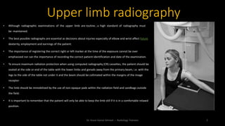
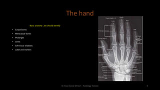
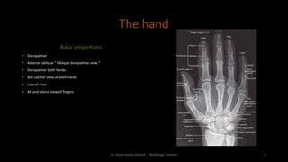
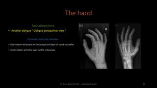
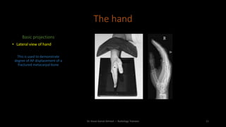
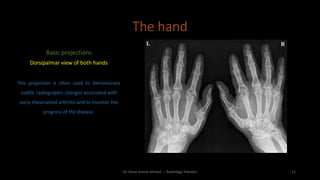
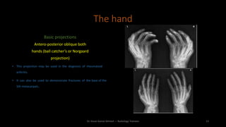
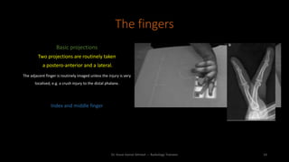
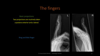
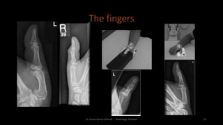
The document discusses guidelines for proper upper limb radiography techniques. It emphasizes maintaining high standards, ensuring correct patient identification and positioning to minimize radiation exposure. Proper hand positioning and identification of anatomical structures is important. Standard projections of the hand include dorsopalmar, oblique, lateral, and views of both hands. Finger projections typically include PA and lateral views. The thumb also requires PA and lateral views. Overall care is needed to obtain clear images while protecting patients.
2. • Although radiographic examinations of the upper limb are routine ; a high standard of radiography must
be maintained.
• The best possible radiographs are essential as decisions about injuries especially of elbow and wrist affect future
dexterity, employment and earnings of the patient.
• The importance of registering the correct right or left marker at the time of the exposure cannot be over
emphasized nor can the importance of recording the correct patient identification and date of the examination.
• To ensure maximum radiation protection when using computed radiography (CR) cassettes, the patient should be
seated at the side or end of the table with the lower limbs and gonads away from the primary beam, i.e. with the
legs to the side of the table not under it and the beam should be collimated within the margins of the image
receptor
• The limb should be immobilized by the use of non-opaque pads within the radiation field and sandbags outside
the field.
• It is important to remember that the patient will only be able to keep the limb still if it is in a comfortable relaxed
position.
Upper limb radiography
Dr. Kosar Kamal Ahmed --- Radiology Trainees 2
3. • Direct digital radiography (DDR) allows more flexibility for the examination as the
detector can be positioned:
• Similar to a CR procedure with the detector used like a table procedure.
• Erect, which allows the extremity to be positioned with the patient standing
and reduces examination times
• DDR detectors have, in the main, a large receptive field (normally 43 cm × 43 cm) and
the limb can be positioned any where within that field for upper-limb examinations.
Dr. Kosar Kamal Ahmed --- Radiology Trainees 3
4. The hand
Basic anatomy ; we should identify
• Carpal bones
• Metacarpal bones
• Phalanges
• Joints
• Soft tissue shadows
• Label and markers
Dr. Kosar Kamal Ahmed --- Radiology Trainees 4
5. The hand
Basic projections
• Dorsipalmar
• Anterior oblique “ Oblique dorsipalmar view “
• Dorsipalmar both hands
• Ball catcher view of both hands
• Lateral view
• AP and lateral view of fingers
Dr. Kosar Kamal Ahmed --- Radiology Trainees 5
6. The hand
Basic projections
• Dorsipalmar
Position of patient and image receptor
• The patient is seated alongside the table with the affected arm nearest to
the table.
• The forearm is pronated and placed on the table with the palmer
surface of the hand in contact with the image receptor.
• The fingers are separated and extended but relaxed to ensure that
they remain in contact with the image receptor.
• The wrist is adjusted so that the radial and ulna styloid processes are
equidistant from the image receptor.
• A sandbag is placed over the lower forearm for immobilisation
Dr. Kosar Kamal Ahmed --- Radiology Trainees 6
7. The hand
Basic projections
• Dorsipalmar
Essential image characteristics
• The image should demonstrate all the phalanges, including the soft tissue
of the fingertips, the carpal and metacarpal bones and the distal end of
the radius and ulna.
• The interphalangeal and metacarpo-phalangeal and carpometacarpal
joints should be demonstrated clearly.
• No rotation of the hand.
Dr. Kosar Kamal Ahmed --- Radiology Trainees 7
8. The hand
Basic projections
• Anterior oblique “ Oblique dorsipalmar view “
Position of patient and image receptor
• From the basic postero-anterior position, the hand is externally rotated 45° with the
fingers extended.
• The fingers should be separated slightly and the hand supported on a 45° non-
opaque pad.
• A sandbag is placed over the lower end of the forearm for immobilisation.
Dr. Kosar Kamal Ahmed --- Radiology Trainees 8
9. The hand
Basic projections
• Anterior oblique “ Oblique dorsipalmar view “
Essential image characteristics
• The image should demonstrate all the phalanges, including the soft tissue of the
fingertips, the carpal and metacarpal bones and the distal end of the radius and ulna.
• The correct degree of rotation has been achieved when the heads of the 1st and 2nd
metacarpals are seen separated whilst those of the 4th and 5th are just superimposed.
Dr. Kosar Kamal Ahmed --- Radiology Trainees 9
10. The hand
Basic projections
• Anterior oblique “ Oblique dorsipalmar view “
Common faults and remedies
• Over rotation will project the metacarpals and digits on top of each other .
• Under rotation will fail to open out the metacarpals
Dr. Kosar Kamal Ahmed --- Radiology Trainees 10
11. The hand
Basic projections
• Lateral view of hand
This is used to demonstrate
degree of AP displacement of a
fractured metacarpal bone
Dr. Kosar Kamal Ahmed --- Radiology Trainees 11
12. The hand
Basic projections
Dorsipalmar view of both hands
This projection is often used to demonstrate
subtle radiographic changes associated with
early rheumatoid arthritis and to monitor the
progress of the disease.
Dr. Kosar Kamal Ahmed --- Radiology Trainees 12
13. The hand
Basic projections
Antero-posterior oblique both
hands (ball catcher’s or Norgaard
projection)
• This projection may be used in the diagnosis of rheumatoid
arthritis.
• It can also be used to demonstrate fractures of the base of the
5th metacarpals..
Dr. Kosar Kamal Ahmed --- Radiology Trainees 13
14. The fingers
Basic projections
Two projections are routinely taken
a postero-anterior and a lateral.
The adjacent finger is routinely imaged unless the injury is very
localised, e.g. a crush injury to the distal phalanx.
Index and middle finger
Dr. Kosar Kamal Ahmed --- Radiology Trainees 14
15. The fingers
Basic projections
Two projections are routinely taken
a postero-anterior and a lateral.
Ring and little finger
Dr. Kosar Kamal Ahmed --- Radiology Trainees 15