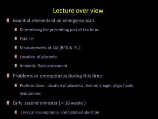
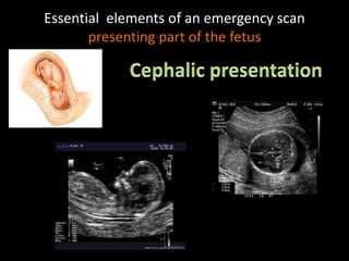
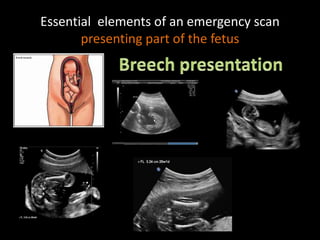
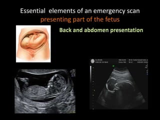
Second and third trimester
emergencies
Dr. Kosar Kamal Ahmed
H.D diagnostic radiology
Lecture 3
Lecture over view
Essential elements of an emergency scan
Determining the presenting part of the fetus
Fetal lie
Measurements of GA (BPD & FL )
Location of placenta
Amniotic fluid assessment
Problems or emergencies during this time
Preterm labor , location of placenta , haemorrhage , oligo / poly
hydramnios
Early second trimester ( < 16 weeks )
cervical incompitence and habitual abortion
Essential elements of an emergency scan
presenting part of the fetus
Essential elements of an emergency scan
presenting part of the fetus
Essential elements of an emergency scan
presenting part of the fetus
Essential elements of an emergency scan
The BPD is the maximum diameter of a transverse
section of the fetal skull at the level of the
.parietal eminences
Essential elements of an emergency scan
• A single optimal measurement of the BPD will predict the
• gestational age to within ± 5 days.
• It is more accurate at predicting the date of delivery than an
optimal menstrual history.
• It is more accurate than CRL ( Why ?)
• Recent data report that HC is more accurate than BPD
• Lateral ventricles view:
• rugby-football-shaped skull
• a long midline equidistant from the proximal
and distal skull echoes
• the cavum septum pellucidum bissecting the
midline one-third of the distance from the
synciput to the occiput
• the two anterior horns of the lateral
ventricles, symmetrically placed about the
Midline
• all or part of the posterior horns of the lateral
ventricles symmetrically placed about the
midline .
Thalami view :
● a rugby-football-shaped skull
● a short midline equidistant from the proximal
and distal skull echoes
● the cavum septum pellucidum bisecting the
midline one-third of the distance from the
synciput to the occiput
● the thalami
-This measurment is as accurate as BPD
in the prediction of GS
-The upper femur should be selected for
measurement
-To ensure that the section obtained is
not oblique ( soft tissue should be
visible beyond both ends of the
femur and the bone should not
appear to merge with the skin of the
thigh at any point
Essential elements of an emergency scan
• The placenta is best identified by scanning the uterus
longitudinally and is easily recognized by its more echogenic
pattern compared with that of the underlying myometrium
• Placenta can be in :
– Proper position
– Low lying
– Marginal previa
– Previa
Essential elements of an emergency scan
1. Subjective assessment :
With experience, it is possible to classify amniotic fluid volume into the broad categories
absent, low, normal, increased and excessive.
2. Single deepest pool :
The size of the deepest cord-free pool of amniotic fluid is assessed with the ultrasound
probe perpendicular to the maternal abdomen; The vertical depth of the largest pool is
measured A depth of 2–3 cm is normal
3. Amniotic fluid index :
This is a semiquantitativetechnique for assessing amniotic fluid volume,Using the maternal
umbilicus as a reference point, the abdomen is divided into four quarters.
With the ultrasound probe held in the longitudinal axis of the mother and perpendicular to
the floor the largest vertical pool depth in each quadrant is recorded.
Essential elements of an emergency scan
Subjective method
Essential elements of an emergency scan
Second and third trimester emergencies

Second and third trimester emergencies

  • 1.
    Second and thirdtrimester emergencies Dr. Kosar Kamal Ahmed H.D diagnostic radiology Lecture 3
  • 2.
    Lecture over view Essentialelements of an emergency scan Determining the presenting part of the fetus Fetal lie Measurements of GA (BPD & FL ) Location of placenta Amniotic fluid assessment Problems or emergencies during this time Preterm labor , location of placenta , haemorrhage , oligo / poly hydramnios Early second trimester ( < 16 weeks ) cervical incompitence and habitual abortion
  • 3.
    Essential elements ofan emergency scan presenting part of the fetus
  • 4.
    Essential elements ofan emergency scan presenting part of the fetus
  • 5.
    Essential elements ofan emergency scan presenting part of the fetus
  • 6.
    Essential elements ofan emergency scan The BPD is the maximum diameter of a transverse section of the fetal skull at the level of the .parietal eminences
  • 7.
    Essential elements ofan emergency scan • A single optimal measurement of the BPD will predict the • gestational age to within ± 5 days. • It is more accurate at predicting the date of delivery than an optimal menstrual history. • It is more accurate than CRL ( Why ?) • Recent data report that HC is more accurate than BPD
  • 8.
    • Lateral ventriclesview: • rugby-football-shaped skull • a long midline equidistant from the proximal and distal skull echoes • the cavum septum pellucidum bissecting the midline one-third of the distance from the synciput to the occiput • the two anterior horns of the lateral ventricles, symmetrically placed about the Midline • all or part of the posterior horns of the lateral ventricles symmetrically placed about the midline .
  • 9.
    Thalami view : ●a rugby-football-shaped skull ● a short midline equidistant from the proximal and distal skull echoes ● the cavum septum pellucidum bisecting the midline one-third of the distance from the synciput to the occiput ● the thalami
  • 11.
    -This measurment isas accurate as BPD in the prediction of GS -The upper femur should be selected for measurement -To ensure that the section obtained is not oblique ( soft tissue should be visible beyond both ends of the femur and the bone should not appear to merge with the skin of the thigh at any point
  • 12.
    Essential elements ofan emergency scan • The placenta is best identified by scanning the uterus longitudinally and is easily recognized by its more echogenic pattern compared with that of the underlying myometrium • Placenta can be in : – Proper position – Low lying – Marginal previa – Previa
  • 13.
    Essential elements ofan emergency scan
  • 14.
    Essential elements ofan emergency scan
  • 15.
    Essential elements ofan emergency scan
  • 16.
    Essential elements ofan emergency scan 1. Subjective assessment : With experience, it is possible to classify amniotic fluid volume into the broad categories absent, low, normal, increased and excessive. 2. Single deepest pool : The size of the deepest cord-free pool of amniotic fluid is assessed with the ultrasound probe perpendicular to the maternal abdomen; The vertical depth of the largest pool is measured A depth of 2–3 cm is normal 3. Amniotic fluid index : This is a semiquantitativetechnique for assessing amniotic fluid volume,Using the maternal umbilicus as a reference point, the abdomen is divided into four quarters. With the ultrasound probe held in the longitudinal axis of the mother and perpendicular to the floor the largest vertical pool depth in each quadrant is recorded.
  • 17.
    Essential elements ofan emergency scan Subjective method
  • 18.
    Essential elements ofan emergency scan
  • 19.
    Essential elements ofan emergency scan