Blood and hematopoietic system
•Download as PPTX, PDF•
13 likes•1,543 views

Blood is essential to life. Blood circulates through our body and delivers essential substances like oxygen and nutrients to the body’s cells. It also transports metabolic waste products away from those same cells. There is no substitute for blood. It cannot be made or manufactured. Generous blood donors are the only source of blood for patients in need of a blood transfusion.

Recommended
More Related Content
What's hot
What's hot (20)
Similar to Blood and hematopoietic system
Similar to Blood and hematopoietic system (20)
More from A M O L D E O R E
More from A M O L D E O R E (20)
Recently uploaded
Recently uploaded (20)
Blood and hematopoietic system
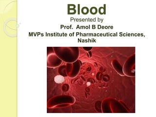
- 1. Blood Presented by Prof. Amol B Deore MVPs Institute of Pharmaceutical Sciences, Nashik
- 2. What is blood? Blood is a specialized fluid connective tissue. Blood is a suspension in which blood cells are distributed in fluid plasma. Blood consists of two components: the blood cells, and the fluid plasma. Blood makes up about 8% of body weight (Average volume: 5-6 liters in male and 4-5 liters in female).
- 3. Color of blood: reddish brown PH: slightly alkaline 7.35 to 7.45.
- 4. Composition of Blood Whole blood is composed of two portions: Plasma constitutes about 55% blood cells make up about 45% of the total blood volume.
- 6. PLASMA (55%) BLOOD CELLS (45%) 1) Water (98% of plasma) 2) Plasma proteins Albumin, globulins, glycoproteins & antibodies 3) Clotting factors Fibrinogen, prothrombin, thromboplastin 4) Electrolytes ions Na+ , K+ , Ca+2 , Mg, Cu, iodine, cobalt, bicarbonate, iron, phosphate, 5) Nutrients Carbohydrates, proteins, lipids, amino acids, fatty acids, vitamins 6) Gases Oxygen, carbon dioxide, nitrogen 7) Enzymes acid phosphatase 8) Hormones Growth hormone, thyroid hormone, insulin, testosterone, oxytocin, antidiuretic hormone 9) Waste products Urea, uric acid, creatinine, ammonia 1) Erythrocytes or Red blood cells (RBCs), 2) Leukocytes or White blood cells (WBCs), 3) Thrombocytes or Platelets
- 7. Functions of the blood 1. It transports oxygen from the lungs to the cells of the body. 2. It transports carbon dioxide from the cells to lungs for excretion. 3. It transports nutrients, ions, and water from the digestive tract to cells. 4. It transports waste products from cells to sweat glands and kidneys. 5. It transports hormones to target organs and enzymes to body cells.
- 8. 6. It regulates physiological pH and acid base electrolyte balance. 7. It helps to regulate normal body temperature (98.40F). 8. It helps to prevent blood loss from rupturing blood vessels through the blood clotting mechanism. 9. It protects against foreign microbes, virus, bacteria, parasites, worms, fungi and others due to immune defence system of leukocytes.
- 9. The process of blood cell formation takes place in bone marrow is known as hematopoiesis. Red bone marrow found in epiphysis of long and flat bones such as pelvic girdle, sternum, cranium, ribs, vertebrae, scapula, femur and humerus. Hematopoiesis occurs in red bone marrow, which is composed of pluripotent stem cells. Formation of the blood
- 10. All blood cells are produced from pluripotent stem cells and pass through several developmental stages before entering in the blood.
- 12. RED BLOOD CELLS
- 13. RBCs (or erythrocytes) are circular biconcave discs shaped. They are non-nucleated and simple in structure. They are composed of a network of protein called stroma, cytoplasm, and the oxygen carrying red pigment called hemoglobin which is responsible for the red color of whole blood.
- 14. Properties of Red Blood Cells Scientific name: Erythrocytes Color Reddish- brown (due to Hemoglobin pigment) Stroma Consist of antigens and Hemoglobin pigment Normal counts In male: 4.5-6 million RBCs/µL In female: 4-5 million RBCs/µL Higher values in male due to higher levels of testosterone and erythropoietin hormones Life span ~120 days (3-4 months) Production site Red bone marrow
- 15. Function : 1. It transports oxygen from the lungs to cells of the body 2. It transports carbon dioxide from the cells to the lungs for excretion Disorders: Anemia, polycythemia
- 16. Hemoglobin Red blood cells contain the oxygen- carrying protein hemoglobin, which is a pigment that gives its red color to the blood. Hemoglobin molecule consists of a polypeptide protein called globin and four non-protein pigments called hemes. Each heme contains four iron atoms (ferrous, Fe+2). Each iron atom can carry one molecule of oxygen; therefore one hemoglobin molecule can carry four molecules of oxygen.
- 18. Hemoglobin that is carrying oxygen is bright red in color, whereas hemoglobin not carrying oxygen is a darker red in color. Female shows less Hb count because maximum blood transported in to the fetus, during pregnancy and bleeding during menstruation cycle. Each Erythrocyte contains about 280 million molecules of hemoglobin.
- 19. Table 5.3: Properties of hemoglobin Molecular weight 68,000 units Normal count In male: 13-18 gm% of blood In female: 12-16 gm% of blood In infants: 13-20 gm% of blood Color Brownish red (due to ferrous pigment) Life span About 120 days Capacity 1 gram of Hb can carry 1.34 ml of oxygen molecules to body tissues.
- 20. Functions of Hemoglobin 1. Transport of oxygen In the lungs, molecular oxygen binds to Hb to form oxyhemoglobin (pure blood). Oxyhemoglobin is transported to body tissue to supply O2. O2 + Hb Oxyhemoglobin 2. Transport of carbon dioxide The CO2 formed in body tissues due to biochemical reactions. CO2 molecules bind with Hb to form carbaminohemoglobin (impure blood) to transport back to the lungs. CO2 + Hb carbaminohemoglobin
- 21. Erythropoiesis
- 22. Disorders of RBCS Polycythemia It is characterized by abnormally rise in RBC count more than 7 million/µL in blood. Blood viscosity and blood pressure become elevated. That may result in risk of internal injury to blood vessels, hemorrhage, blood clotting, thrombosis etc. Causes include- Over transfusion of blood, abnormally large no. of erythrocytes etc.
- 23. Anemia Anemia is characterized by reduction in oxygen carrying capacity of blood. It is caused by four factors: decrease in the normal number of erythrocytes; decrease of normal amounts of hemoglobin in the RBCs; deficiency of normal hemoglobin; or production of abnormal hemoglobin. Anemia reduces the amount of oxygen that RBCs can transport resulting in a lack of energy, shortness of breath on minor exertions, exhaustion, increased heart rate, anorexia, pale skin, and a general feeling of tiredness.
- 24. Sickle cell anemia Sickle cell anemia is a genetically inherited disease characterized by abnormal hemoglobin. The abnormal hemoglobin forms sickle shape RBC (S or C shape) when deoxygenated. Hence oxygen carrying capacity of blood becomes reduced. The life span of sickle RBCs become reduced due to early hemolysis. Sickle shaped RBCs do not move smoothly through small blood vessels like capillaries, veins.
- 25. Iron-deficiency anemia Iron-deficiency anemia results from nutritional deficiencies of iron (ferrous) from the body, resulting in lower RBC production. It is most commonly observe in pregnancy because of deficiency of iron in RBCs. The normal daily requirement of iron in women is 3 mg. Its causes include dietary deficiency of iron, calorie controlled diet, poorly planned food, malabsorption of iron in body, peptic ulcer, intestinal ulcer, menstruation, blood loss etc.
- 26. Megaloblastic anemia (pernicious anemia) It is characterized by deficiency of Vitamin B12 and folic acid which affect maturation of RBCs during erythropoiesis. Abnormally large sized RBCs found in the blood. Life span of RBCs becomes reduced up to 45-50 days. Its causes include deficiency of intrinsic factor which is needed for absorption of Vitamin B12, gastritis or peptic ulcer.
- 28. The two most important blood group systems are ABO and Rh factor. They determine some blood groups: A, B, AB, O and Rh positive; Rh negative The ABO blood group system is based on presence or absence of two major antigens on the RBC membrane (stroma): antigen A and antigen B. In addition, individuals make antibodies but not to their own type of antigen. Hence on incompatible blood transfusion, antibodies would be produced by immune system causing agglutination/ antigen-antibody reaction.
- 29. Classification of ABO blood groups with compatible blood transfusion Blood Group Antigen on RBC membrane Antibody in Plasma Can receive blood from? (compatible) Can donate blood to? (compatible) A A Anti-B A,O A,AB B B Anti-A B,O B,AB AB A and B -------- A,B,AB,O (Universal receiver) AB O -------- Anti-A & Anti-B O A,B,AB,O (Universal Donor)
- 31. Blood transfusion Match blood transfusion If individual donates blood to compatible blood group as shown in table, then immune system of recipient person accept it. This is called as match blood transfusion. Mismatch blood transfusion If individual donates blood to incompatible blood group, then immune system of recipient person starts producing antibodies that result in antigen: antibody complex reaction (clumping or agglutination). It causes hemolysis, loss of RBCs, headache, difficulty for breathing, jaundice, kidney failure etc. This is called as mismatch blood transfusion.
- 33. Universal Donor Individuals with blood group O do not have antigen on their RBC membrane. Hence if they donate blood to any blood groups A, B, AB, & O then recipient will not produce any antibody. It becomes compatible (match transfusion) and safe. Therefore blood group O is known as “Universal Donor”. Individuals with O Rh-negative blood may be given to people with any other blood type.
- 34. Universal Recipient Individuals with blood group AB contain both antigens A and B on their RBC membrane but neither produces anti-A nor Anti-B antibodies. Transfusion of A, B, AB, O blood groups in such individuals is safe and compatible, because there are no antibodies to react with antigen (match transfusion). Therefore blood group AB is known as “Universal Recipient”.
- 35. Rhesus System (Rh Factor) The Rh blood group was named after the Rhesus monkeys. Rhesus monkeys consist of Rh antigen on their RBC membrane. This Rh antigen (or antigen D) was later discovered in humans. Hence it is identified as Rhesus factor. If Rh-antigen found on the RBC membrane, then blood is Rh positive. About 85% people have Rh antigen on RBCs membranes, they are Rh- positive. They do not produce anti-Rh antibodies. Whereas 15% people have no Rh-antigen on RBCs membrane, they are Rh-negative. They can capable of producing anti-Rh antibodies.
- 36. Rh factor Antigen on RBC membrane Antibody in Plasma Rh +ve Rh --- Rh -ve ---- Anti-Rh antibody If Rh-negative person receives a blood from Rh- positive person then the Rh-negative person begins to produce anti-Rh antibodies against the foreign blood. This is mismatched blood transfusion. This initial mismatch has no immediate serious complications because it takes the body time to react and produce antibodies.
- 37. However, if same Rh-negative person again receives a blood transfusion of Rh-positive blood (mismatched) then the patient’s anti-Rh antibodies will undergo agglutination reaction. Agglutination leads to hemolysis, kidney failure, difficulty for breathing, jaundice etc. Hence, Rh negative person should not receive blood from Rh positive person.
- 38. Compatible blood transfusion chart
- 40. White blood cells or leukocytes have nucleus (polymorphonuclear). They are colorless because they do not contain hemoglobin. Their general function is to fight against inflammation and microbial infection. Leukocytes are categories into two major types: granulocytes and agranulocytes.
- 43. GRANULOCYTES Granulocytes have cytoplasmic granules. These granules consist of lysosomal enzymes, strong oxidants and several inflammatory mediators including cytokines, prostaglandins, interleukins, histamine and heparin. Granulocytes are polymorphonuclear i.e. having two or more lobes of nucleus. Granulocytes are summarized below.
- 44. Phagocytosis
- 45. TYPE FIGURE FUNCTIONING Neutrophils 40-75% Phagocytosis: It is the process of engulfment, digestion, killing & fragmentation of microbes. It is performed by lysosomal enzymes, defensins, and strong oxidants of cytoplasmic granules. WBCs are act as immune system of body against foreign pathogens. Basophils 1% Release histamine and prostaglandin from granules which leads to inflammation and allergic reactions. Basophils also release heparin to stop blood clotting.
- 46. Agranulocytes Agranulocytes do not have cytoplasmic granules. They also consist of cytoplasmic granules. Due to their small size they cannot be seen under microscope. Eosinophils 1-6% Phagocytosis of parasites, worms and microbes. Promote allergic inflammation, asthmatic allergy, skin allergy etc.
- 47. Monocytes 2-10% They circulate in the blood and promote Phagocytosis. Some monocytes migrate from blood into tissues then develop into macrophages to promote phagocytosis. Monocytes release interleukin & Prostaglandin which act on hypothalamus causing rise In body temperature (fever) due to microbial infection.
- 48. Lymphocytes 20-50% B-lymphocytes produce antibodies, showing antigen- antibody reaction. T-lymphocytes fight against viruses, bacteria, cancer cells, transplanted tissue cells etc.
- 49. Table 5.8: Properties of white blood Cells Scientific name Leukocytes Size and shape Circular, spherical shaped 10-20 µm Color Colorless (no pigment) Normal counts In male and In female: 4000-11,000 WBCs/µL Life span Few hours to few days After phagocytosis the microbes, WBC itself also get dies. Hence its life span become few hours to few days. Production site Red Bone marrow, Thymus gland Destruction site Spleen and liver, lymph node, tonsils Function Immune defense, Inflammatory response, Antibody production, Cellular immune response and Phagocytosis Disorders Leukopenia, Leukocytosis, Leukemia
- 50. Disorders of WBC Leukopenia Leukopenia is characterized low WBC count; less than 4000 WBCs/µL. It’s causes include chemotherapy, exposure of X-rays, alpha rays and other radioactive substances, Bone marrow failure, enlargement of spleen, aplastic anemia; certain infection like typhoid, dengue, AIDS etc. Symptoms include frequent headache, anemia, heavy bleeding and menstruation in woman; inflammation on chicks, lips, tongue and tonsils etc.
- 51. Leukocytosis It is characterized by high WBC count; more than 30,000 WBCs/µL. Its causes include acute infection, hay fever, hepatitis, cancer, tuberculosis, pneumonia, bronchial asthma, stress, excess exercise, arthritis, skin allergy, burning etc. Symptoms include- fever, weakness, tired, sick, feeling dizzy, sweaty, tingling in the arms, legs, abdomen; weight loss, loss of appetite etc.
- 52. Leukemia Leukemia is characterized by an overproduction of immature white blood cells (more than 200,000 WBCs/µL). These immature cells cannot perform their normal functions, and the person becomes very susceptible to infection. Its causes include exposure of radiations like X-rays, UV rays, gamma rays, Chemical exposures, Smoking, and stressful lifestyle.
- 53. Agranulocytosis It is a condition in which there is severe leukopenia characterized by fall in WBC count below 500 WBCs/µL.
- 54. Table 5.9: Difference between RBC and WBC
- 55. Sr. No. Points Red blood cells White blood cells 1. Scientific Name Erythrocytes Leukocytes 2. Shape Biconcave disc Spherical disc 3. Color Red brown Colorless 4. Hemoglobin pigment Present Absent 5. Normal Count In male: 4.5-6 million RBCs/µL In female: 4-5million RBCs/µL In male and In female: 4000-11,000 WBCs/µL 6. Life span 120 days Few hours to few days 7. Function O2 and CO2 transport Immune defense system, Phagocytosis 8. Types --------- Granulocytes, agranulocytes 9. Nucleus Absent Polymorphonuclear 10. Disorders Anemia, polycythemia Leukemia, Leukopenia, leukocytosis 11. Cytoplasmic granules Absent Present 12. Antigens Antigen A, Antigen B, & Rh factor Absent
- 56. PLATELETS
- 57. Table 5.10: Properties of Platelets Scientific name Thrombocytes Size and shape Disc shaped, 2-4 µm size Fig 5.6: Platelets Normal counts In male and In female: 250,000-400,000 platelets/µL Life span 9-11 days Production site Red bone marrow Destruction site Spleen and liver Cytoplasmic granules Alpha granules: clotting factors Dense granules: ADP, Ca2+ , thromboxane A2, prostaglandin, and serotonin Function Hemostasis: stoppage of bleeding Blood clotting, Platelets help to repair slightly damaged blood vessels Disorders Thrombocytopenia
- 58. Hemostasis Hemostasis refers to the stoppage of bleeding i.e. arrest of blood loss. If any small blood vessel like blood capillary is damaged or ruptured, then there are three basic mechanisms that promote hemostasis to arrest the bleeding. Platelets play vital role in hemostasis.
- 59. The series of three mechanisms of hemostasis include: 1) Vascular Spasm When platelets come in contact with a blood vessel damaged, they adhere on damaged wall. Platelets undergo degranulation and release serotonin and thromboxane A2, which cause contraction of blood vessel (vascular spasm). Thereby reducing blood loss from rupturing blood vessel.
- 60. 2. Platelet Plug Formation In this stage, platelets stick on damaged part of blood vessel. This process is called as platelet adhesion. Platelet adhesion promotes degranulation to release ADP and thromboxane-A2 in blood. These chemicals attract other platelets to stick on each other. This gathering of platelets is called as platelet aggregation. The aggregation of large number of platelets form a mass at damaged site is called as a platelet plug. Platelet plug acts as a temporary seal.
- 61. Table 5.11: Blood clotting factors Factor No. Description I Fibrinogen II Prothrombin III Thromboplastin (or tissue factor) IV Calcium ions V Labile factor VI Does not exist VII Stable factor VIII Antihemophilic factor A IX Antihemophilic factor B X Stuart factor XI Antihemophilic factor C XII Hagemans factor XIII Fibrin stabilizing factor
- 63. Blood Clotting (or coagulation) Blood clotting is a complex process of the formation of semisolid insoluble gel of blood & fibrin threads at the damaged wall of blood vessel. Blood clotting involves several enzymes and other chemicals known as clotting factors.
- 64. Stages of blood clotting are discussed below Stage 1: Formation of prothrombinase (prothrombin activator) Stage 2: Conversion of prothrombin into the thrombin Stage 3: Conversion of fibrinogen into fibrin
- 65. The clot is made up from fibrin. Fibrin is a thread-like protein. The blood clot contains RBCs and platelets. This blood clot creates a wall across the breaking site in the blood vessel. Once the clot has formed and bleeding has stopped, clot retraction and fibrinolysis occur.
- 70. Normal range of clotting time: 4-11 minutes Serum: Serum is a clear, transparent and sticky fluid coming out from shrinking of blood clot. Factors affecting blood coagulation Physiological factors: Clotting time is reduced during menstruation and parturition. Pathological factors: Clotting time becomes prolong in hemophilia, liver diseases, afibrinogenemia, diabetes, vitamin K deficiency, and Christmas disease
- 71. Disorders of Platelets Thrombocytopenia Thrombocytopenia is a disorder characterized by reduction in platelet count below 40,000 platelets/µL. It may be due to reduction in tare of platelet production or increase in rate of destruction from spleen and liver. Symptoms includes Frequent bleeding from gums, nose, skin, teeth etc.; Internal organ bleeding like brain hemorrhage, gastric ulcer; Bleeding gums, skin rash, red colored patch on skin etc.
- 72. Hemophilia Hemophilia is characterized by the inability of the blood to clot properly. Hemophilia is a genetic blood disorder involves deficiency of blood clotting factors including factor VIII, factor IX and factor XI. Without factor VIII, the first stage of chemical clotting cannot be completed, and prothrombinase is not formed. Blood clotting factors are needed to stop bleeding after a cut or injury and to prevent spontaneous bleeding.
- 73. Symptoms includes Prolonged spontaneous bleeding due to minor trauma (injury); Bleeding from the joints; Bleeding from muscles leads to severe pain; Blood in urine; Frequent nose and dental bleeding.
- 74. Thrombosis Clotting in such an unbroken blood vessel (usually a vein) is called thrombosis. Usually platelets get deposited on that to form a blood clot known as thrombus. It may dissolve spontaneously. Embolism If pieces of a blood clot or air bubble get transported into the bloodstream, it is called an embolus. When an embolus becomes blocked in a blood vessel and cuts off circulation, it is known as an embolism.
- 75. Thanking u