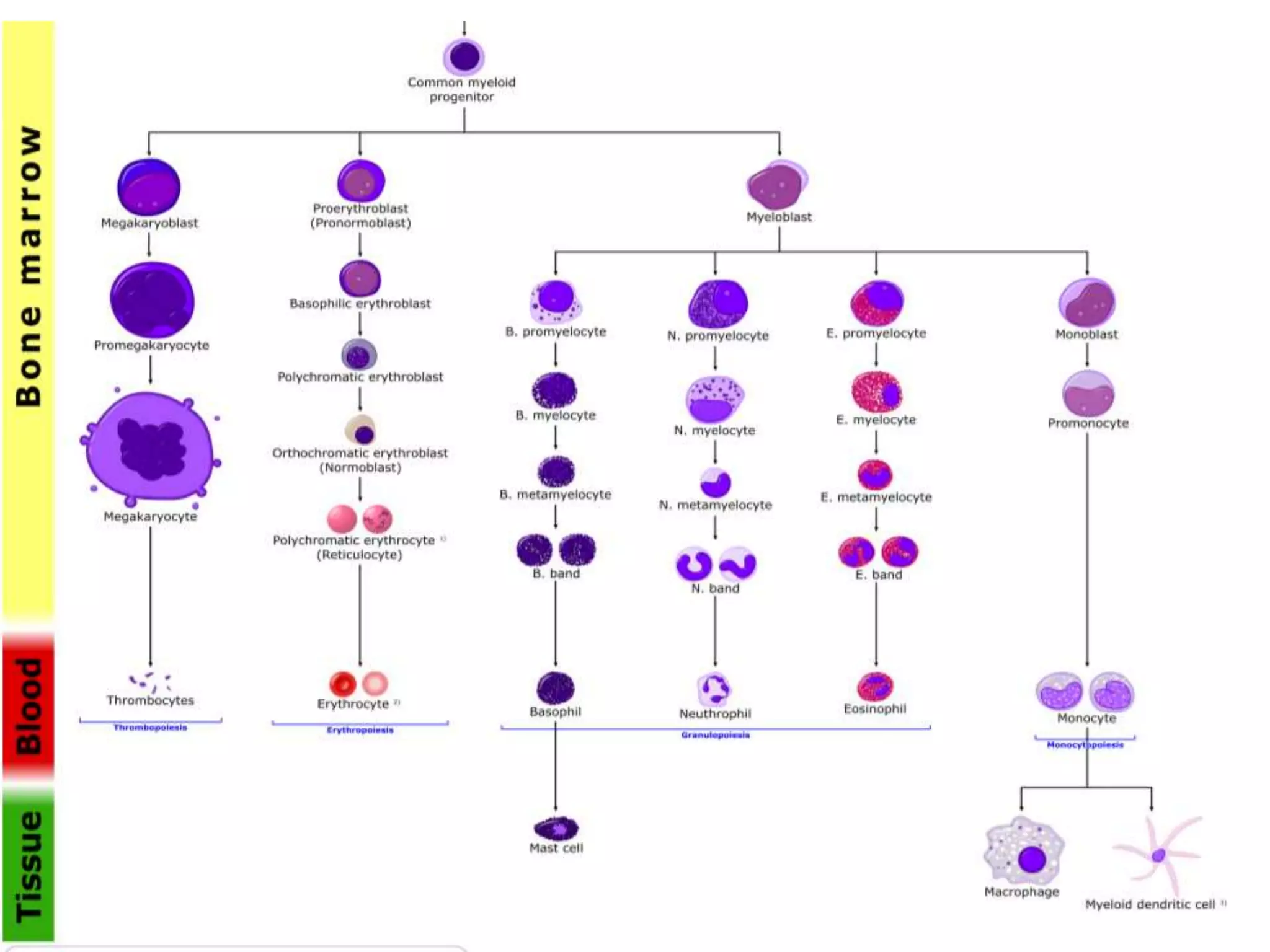
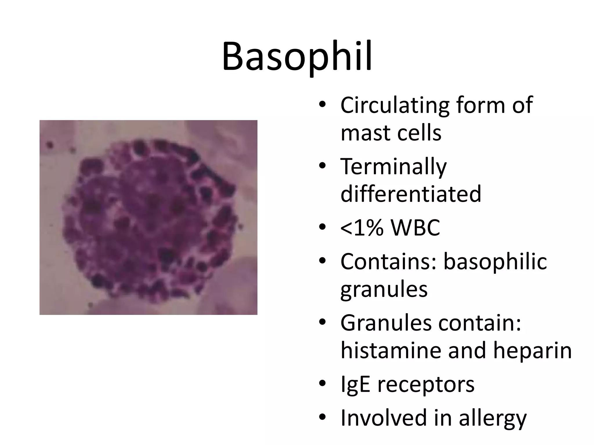
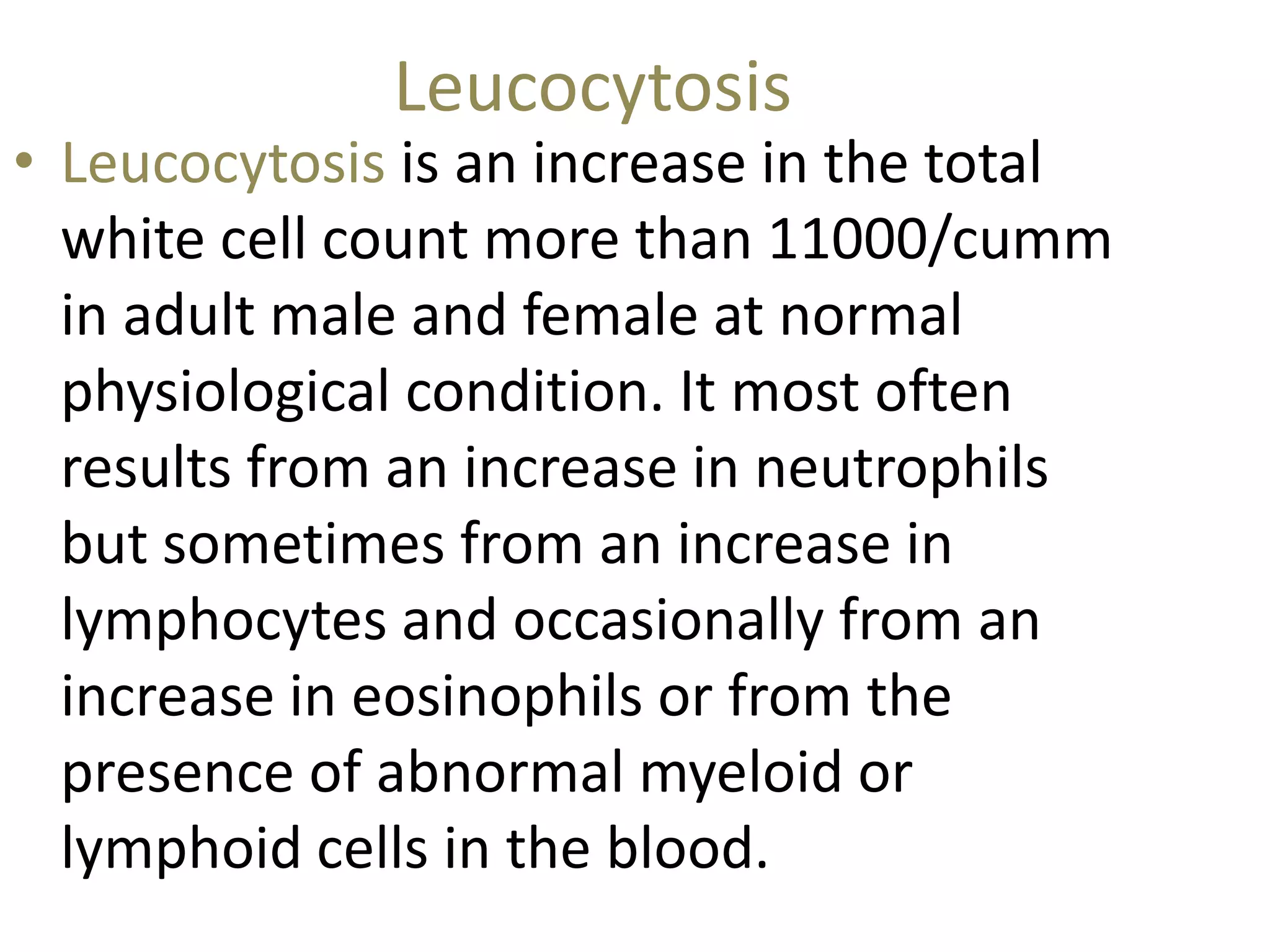
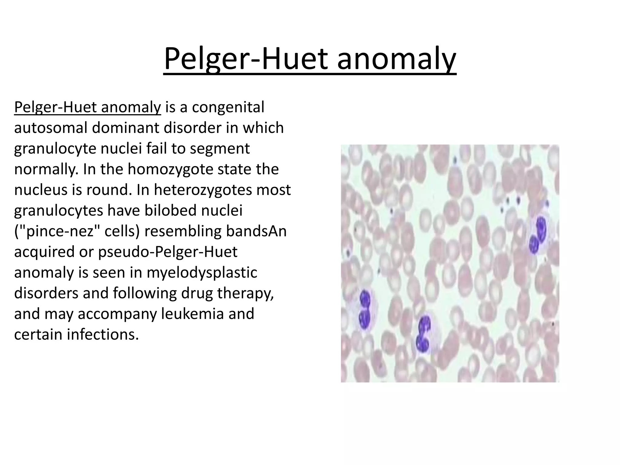
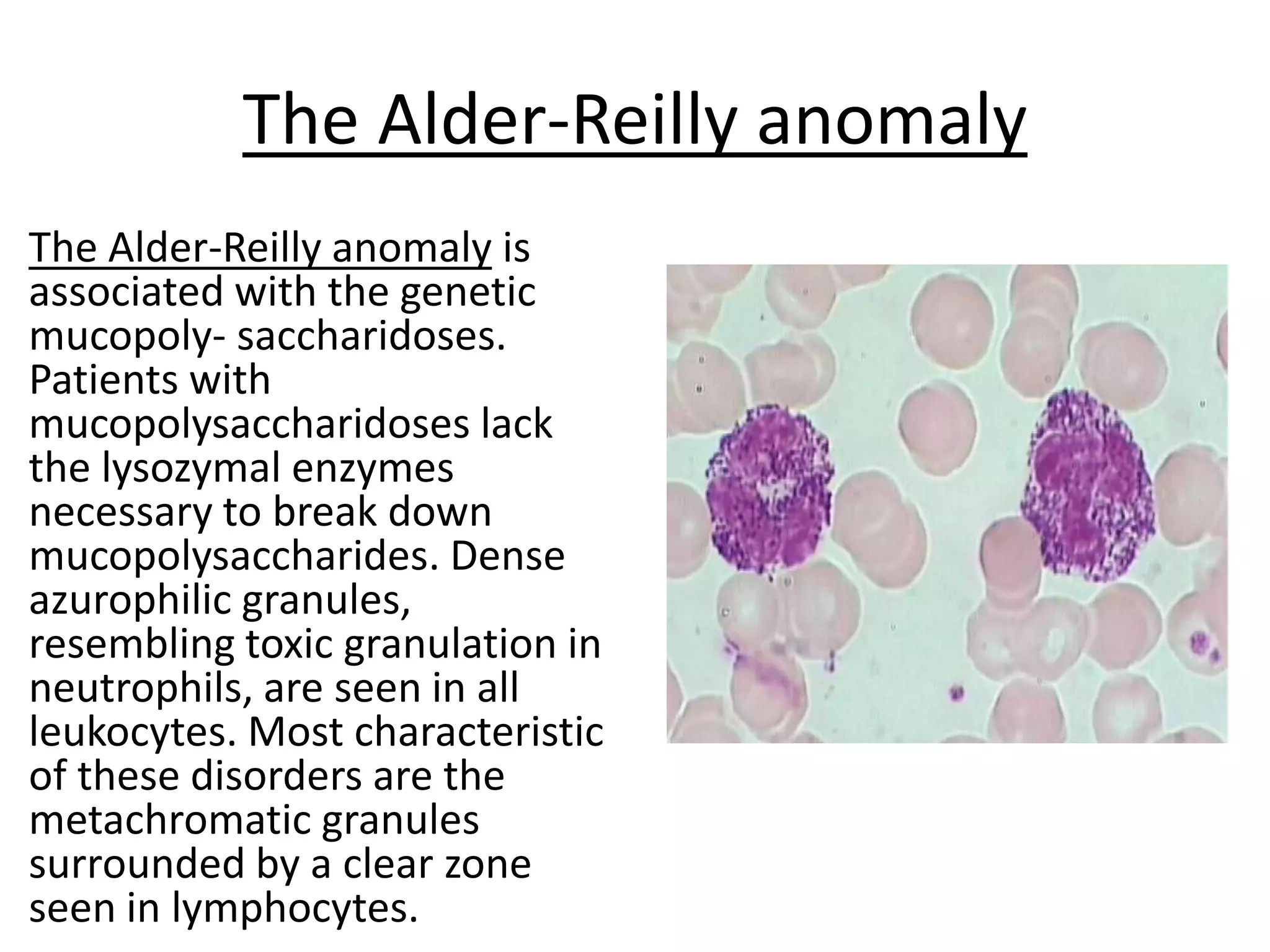
Morphology of Normal White
Blood Cells
Granulocytes
Neutrophil
• PMN-Polymorphonuclear
Leucocytes.
• 60-70% WBC
• Appearance: pink granules in
cytoplasm, nucleus has 3-4
lobes
• Function: Phagocytosis of
bacteria
• Azurophilic (1°) granules are
"lysosomes of PMNs", occur
in all leukocytes
Eosinophil (Eos)
• Bilobed nucleus
• 2-4% of WBC
• Recruited to sites of
inflammation
• Function: Involved in allergy,
parasitic infections
• Contains: eosinophilic
granules
• Granules contain: major basic
protein
• Terminally differentiated
Azurophilic granuels
Basophil
• Circulating form of
mast cells
• Terminally
differentiated
• <1% WBC
• Contains: basophilic
granules
• Granules contain:
histamine and heparin
• IgE receptors
• Involved in allergy
Monocyte/ Macrophage
Monocyte
• 3-8% WBC
• Circulating form (precursor) of
tissue macrophages
• Recruited to sites of inflammation
Macrophages
• Phagocytosis, bacterial killing,
antigen presentation
• Peritoneal cavity: peritoneal
macrophages
• Lung: alveolar macrophages
• Spleen: splenic macrophages
• Liver: Kupffer cells
lymphocyte
•Appearance: small (same size as
RBCs), little visible cytoplasm
•NO specific granules
• 20-25% of WBC
•T cells: CMI (for viral infections)
• B cells: humoral (antibody)
• Natural Killer Cells
Leucocytosis
• Leucocytosis is an increase in the total
white cell count more than 11000/cumm
in adult male and female at normal
physiological condition. It most often
results from an increase in neutrophils
but sometimes from an increase in
lymphocytes and occasionally from an
increase in eosinophils or from the
presence of abnormal myeloid or
lymphoid cells in the blood.
CAUSES OF LEUKOCYTOSIS
PHYSIOLOGICAL CAUSES:-
• EXERCISE
• STRESS
• Medications
• Idiopathic
• Pregnancy
• Trauma
CAUSES OF LEUKOCYTOSIS
• Pathological causes:-
• Infection
• Tissue death, burns, cancer
• Metabolic disorders, diabetes, overactive
thyroid
• Inflammation, Rheumatoid arthritis
• Bone marrow disease, leukemia
• Inflammation
• Tissue damage
Pathological causes:-
• Leukaemia
• Bacterial infection
• Fungal infection
• Arthritis
• Myocardial infarction
• Carcinoma
• Myeloproliferative disorders
• Connective tissue disease
• Dermatitis
NEUTROPHILIA
DEFINATION
• When absolute neutrophil count(ANC) is above
7500/µl or more than 70%neutrophils in the
differential WBC counts(DLC)
CAUSES:
Acute infectionse.g.pneumonia,cholecysitis,meningitis
• Other inflammation e.g.gout, collagen vascular disease
• Acute hemorrhage
• Acute hypoxia,Heat stress
Causes of Neutrophilia:
• Metabolic and endocrine disorders, e.g. diabetic
ketoacidosis, acute renal failure, Cushing’s syndrome,
thyrotoxic crisis
• Malignant disease
• Myeloproliferative and leukaemic disorders,,
• neutrophilic leukaemia, acute myeloid leukaemia
(not commonly), other rare leukaemias,
polycythaemia rubra vera,
• chemotherapy, treatment of megaloblastic anaemia
Neutropenia
Defination
When absolute neutrophil count(ANC) is less
than 1500/cumm.
Causes :-
• Neutropenia and immune deficiency
syndromes of
• various causes, e.g. cyclical neutropenia,
severe
• congenital neutropenia, chronic idiopathic
neutropenia,
• autoimmune neutropenia
Lymphocytosis
Defination:-
• When the differential count of lymphocytes is more
than 40% or absolute lymphocyte count is > than
4000/cumm.
Causes
• Acute infection e.g. pertussis, infectious
mononucleosis, viral hepatitis
• Chronic infection e.g. brucellosis, TB, syphilis
• Hematopoietic disorders e.g.lymphoid leukemia,
lymphosarcoma
• Relative lymphocytosis
Lymphocytopenia
Defination:-
• Absolute lymphocyte count below 1500/µm is
referred to as lymphocytopenia.
Common causes:-
• Acute infections
• Severe bone marrow failure
• Corticosteroid and immunosuppresive therapy
• Widespread irradiation
More common causes of
Eosinophilia
• Allergic diseases, e.g. atopic eczema, asthma,
allergic rhinitis (hay fever), acute urticaria, allergic
• Parasitic infection e.g. filariasis
• Skin diseases, e.g. pemphigus, bullous pemphigoid,
• Bronchopulmonary aspergillosis and other
• Bronchoallergic fungal infections
• Drug hypersensitivity (particularly to gold,
sulphonamides, penicillin, nitrofurantoin) including
Basophilia
• Chronic myeloid leukemia
• Polycythemia vera
• Myelosclerosis
• Myxoedema
• Ulcerative colitis
• Following splenectomy
• Hodgkin’s disease
• Urticaria pigmentosa
Common causes of Monocytosis
• Chronic infection including miliary tuberculosis
and
• congenital syphilis
• Chronic inflammatory conditions including
Crohn’s
disease, ulcerative colitis, rheumatoid arthritis and
• systemic lupus erythematosus
• Carcinoma
• Myeloproliferative and leukaemic conditions
including CMML, atypical CML, JMML, CGL*,
MDS, systemic mastocytosis, AML
Morphologic abnormalities of leukocytes
• Hypersegmentation
• Toxic granulation
• L Dohle bodies
• Pelger-Huet anomaly
• The Chediak-Higashi syndrome
• May-Hegglin anomaly
• The Alder-Reilly anomaly
Hypersegmentation,
• Hypersegmentation,
the presence of
abnormally
increased nuclear
lobulation, is one of
the first hematologic
abnormalities seen
in megaloblastic
anemia
Toxic granulation
Toxic granulation is found in
severe inflammatory states.
The toxic granules are
azurophilic, usually found in
the promyelocyte,
metamyelocyte, band, and
segmented stages. The toxic
granulation is thought to be
due to impaired cytoplasmic
maturation,in the effort to
rapidly generate large
numbers of granulocytes.
L Dohle bodies
• L Dohle bodies are single or
multiple blue cytoplasmic
inclusions. They represent
remnants of rough
endoplasmic reticulum from
earlier maturational stages.
They are associated with
myeloid "left shifts" and are
seen in conjunction with
toxic granulation.
Pelger-Huet anomaly
Pelger-Huet anomaly is a congenital
autosomal dominant disorder in which
granulocyte nuclei fail to segment
normally. In the homozygote state the
nucleus is round. In heterozygotes most
granulocytes have bilobed nuclei
("pince-nez" cells) resembling bandsAn
acquired or pseudo-Pelger-Huet
anomaly is seen in myelodysplastic
disorders and following drug therapy,
and may accompany leukemia and
certain infections.
The Chediak-Higashi syndrome
is a rare autosomal recessive
condition associated with
abnormally large leukocyte
granules resulting from fusion
of lysozymes. This disorder
may affect granulocytes,
leukocytes, and monocytes.
Chemotaxis and phagocytosis
is defective. Platelets lack
dense granules and platelet
function is abnormal. Giant
melanosomes in occular and
skin tissues result in
hypopigmentation.
May-Hegglin anomaly
May-Hegglin anomaly is a
rare autosomal dominant
abnormality characterized by
large pale basophilic
inclusions resembling Dohle
bodies and appear to be
altered RNA. Giant platelets,
and sometimes
thrombocytopenia are
associated with this. The
anomaly is usually benign
but may be associated with
bleeding.
The Alder-Reilly anomaly
The Alder-Reilly anomaly is
associated with the genetic
mucopoly- saccharidoses.
Patients with
mucopolysaccharidoses lack
the lysozymal enzymes
necessary to break down
mucopolysaccharides. Dense
azurophilic granules,
resembling toxic granulation in
neutrophils, are seen in all
leukocytes. Most characteristic
of these disorders are the
metachromatic granules
surrounded by a clear zone
seen in lymphocytes.
LEUKAEMOID REACTION
• LEUKAEMOID REACTION IS DEFINED AS A REACTIVE
EXCESSIVE LEUCOCYTOSIS IN THE PERIPHERAL
BLOOD RESEMBLING THAT OF LEUKAEMIA IN A
SUBJECT NOT HAVING LEUKAEMIA.
• THE USUAL FEATURES OF LEUKAEMIA SUCH AS
SPLEENOMEGALY, LYMPHADENOPATHY AND
HAEMORRHAGES ARE USUALY ABSENT.AND
FEATURES OF UNDERLYING DISEASE ARE USUALLY
PRESENT.
Feature Leukamoid
reaction
CML
Nature of D’se Reactive Stem cell disorder
Cause + nt - nt
Splenomegaly - nt + nt
TLC <50,000 1 to 5 lacs
Toxic granules in
neutrophil
+ nt - nt
Basophilia - nt + nt
Eosinophilia - nt + nt
NAP Score ↑ ↑ (150-350) ↓ ↓ (0-40)
Ph’chromosome -nt + nt
TYPES OF LEUKAEMOID REACTION
• 1. MYELOID LEUKAEMOID REACTION
• 2.LYMPHOID LEUKAEMOID REACTION
MYELOID LEUKAMOID REACTION
• CAUSES:
• 1.INFECTION Eg STAPH PNEUMONIA,
TB,MENINGITIS, DIPTHERIA, SEPSIS.
• 2.INTOXICATION: Eg ECLAMPSIA
• 3.MALIGNANY DISEASE Eg MULTIPLE
MYELOMA,HODGKINS etc.
• 4.SEVERE HEMORRHAGE AND SEVERE HEMOLYSIS
LYMPHOID LEUKAEMIA RECTION
• CAUSES:
• 1 INFECTION Eg. INFECTIUOS
MONONUCLEOSIS, CMV, PERTUSIS, CHICKEN
POX, TB, MEASELS.
• 2. MALIGNANT DISEASE RARELY.
Acute Leukemia
DEFINITION:
• HETEROGENEOUS GROUP OF MALIGNANT
DISORDERS WHICH IS CHARACTERIZED BY
UNCONTROLLED CLONAL PROLIFERATION AND
ACCUMULATION OF BLASTS CELLS IN THE
BONE MARROW AND BODY TISSUES
• SUDDEN ONSET
• IF LEFT UNTREATED IS FATAL WITHIN A FEW
WEEKS OR MONTHS
Comparison of acute and chronic leukemias
Acute Chronic
Age All ages Usually adults
Clinical onset Sudden Insidious
Course (untreated) 6 mo or less 2-6 years
Leukemic cells Immature
>30% blasts
More mature cells
Anemia Prominent Mild
Thrombocytopenia Prominent Mild
WBC count Variable Increased
Lymphadenopathy Mild Present; often
Splenomegaly Mild Present; often
CAUSES OF ACUTE LEUKEMIA
• PRE LEUKEMIA – Myelodysplastic or myeloproliferative syndromes
can evolve into AML.
• CHEMICAL EXPOSURE- Alkylating agents,benzene, aromatic organic
solvents.
• RADIATION - Atomic bombings of Hiroshima and Nagasaki,X rays ,
victims of Chernobyl nuclear reactor.
• GENETICS –Down syndrome,Ataxia telengectasia , Klinefelter’s
syndrome , Fanconi’s anaemia.
• Naturally occuring retroviruses and the human T – cell lymphotropic
viruses cause adult- ALL

WBC normal and abnormal final.pptx

  • 1.
    Morphology of NormalWhite Blood Cells
  • 4.
  • 5.
    Neutrophil • PMN-Polymorphonuclear Leucocytes. • 60-70%WBC • Appearance: pink granules in cytoplasm, nucleus has 3-4 lobes • Function: Phagocytosis of bacteria • Azurophilic (1°) granules are "lysosomes of PMNs", occur in all leukocytes
  • 6.
    Eosinophil (Eos) • Bilobednucleus • 2-4% of WBC • Recruited to sites of inflammation • Function: Involved in allergy, parasitic infections • Contains: eosinophilic granules • Granules contain: major basic protein • Terminally differentiated Azurophilic granuels
  • 7.
    Basophil • Circulating formof mast cells • Terminally differentiated • <1% WBC • Contains: basophilic granules • Granules contain: histamine and heparin • IgE receptors • Involved in allergy
  • 8.
    Monocyte/ Macrophage Monocyte • 3-8%WBC • Circulating form (precursor) of tissue macrophages • Recruited to sites of inflammation Macrophages • Phagocytosis, bacterial killing, antigen presentation • Peritoneal cavity: peritoneal macrophages • Lung: alveolar macrophages • Spleen: splenic macrophages • Liver: Kupffer cells
  • 9.
    lymphocyte •Appearance: small (samesize as RBCs), little visible cytoplasm •NO specific granules • 20-25% of WBC •T cells: CMI (for viral infections) • B cells: humoral (antibody) • Natural Killer Cells
  • 10.
    Leucocytosis • Leucocytosis isan increase in the total white cell count more than 11000/cumm in adult male and female at normal physiological condition. It most often results from an increase in neutrophils but sometimes from an increase in lymphocytes and occasionally from an increase in eosinophils or from the presence of abnormal myeloid or lymphoid cells in the blood.
  • 11.
    CAUSES OF LEUKOCYTOSIS PHYSIOLOGICALCAUSES:- • EXERCISE • STRESS • Medications • Idiopathic • Pregnancy • Trauma
  • 12.
    CAUSES OF LEUKOCYTOSIS •Pathological causes:- • Infection • Tissue death, burns, cancer • Metabolic disorders, diabetes, overactive thyroid • Inflammation, Rheumatoid arthritis • Bone marrow disease, leukemia • Inflammation • Tissue damage
  • 13.
    Pathological causes:- • Leukaemia •Bacterial infection • Fungal infection • Arthritis • Myocardial infarction • Carcinoma • Myeloproliferative disorders • Connective tissue disease • Dermatitis
  • 14.
    NEUTROPHILIA DEFINATION • When absoluteneutrophil count(ANC) is above 7500/µl or more than 70%neutrophils in the differential WBC counts(DLC) CAUSES: Acute infectionse.g.pneumonia,cholecysitis,meningitis • Other inflammation e.g.gout, collagen vascular disease • Acute hemorrhage • Acute hypoxia,Heat stress
  • 15.
    Causes of Neutrophilia: •Metabolic and endocrine disorders, e.g. diabetic ketoacidosis, acute renal failure, Cushing’s syndrome, thyrotoxic crisis • Malignant disease • Myeloproliferative and leukaemic disorders,, • neutrophilic leukaemia, acute myeloid leukaemia (not commonly), other rare leukaemias, polycythaemia rubra vera, • chemotherapy, treatment of megaloblastic anaemia
  • 16.
    Neutropenia Defination When absolute neutrophilcount(ANC) is less than 1500/cumm. Causes :- • Neutropenia and immune deficiency syndromes of • various causes, e.g. cyclical neutropenia, severe • congenital neutropenia, chronic idiopathic neutropenia, • autoimmune neutropenia
  • 17.
    Lymphocytosis Defination:- • When thedifferential count of lymphocytes is more than 40% or absolute lymphocyte count is > than 4000/cumm. Causes • Acute infection e.g. pertussis, infectious mononucleosis, viral hepatitis • Chronic infection e.g. brucellosis, TB, syphilis • Hematopoietic disorders e.g.lymphoid leukemia, lymphosarcoma • Relative lymphocytosis
  • 18.
    Lymphocytopenia Defination:- • Absolute lymphocytecount below 1500/µm is referred to as lymphocytopenia. Common causes:- • Acute infections • Severe bone marrow failure • Corticosteroid and immunosuppresive therapy • Widespread irradiation
  • 19.
    More common causesof Eosinophilia • Allergic diseases, e.g. atopic eczema, asthma, allergic rhinitis (hay fever), acute urticaria, allergic • Parasitic infection e.g. filariasis • Skin diseases, e.g. pemphigus, bullous pemphigoid, • Bronchopulmonary aspergillosis and other • Bronchoallergic fungal infections • Drug hypersensitivity (particularly to gold, sulphonamides, penicillin, nitrofurantoin) including
  • 20.
    Basophilia • Chronic myeloidleukemia • Polycythemia vera • Myelosclerosis • Myxoedema • Ulcerative colitis • Following splenectomy • Hodgkin’s disease • Urticaria pigmentosa
  • 21.
    Common causes ofMonocytosis • Chronic infection including miliary tuberculosis and • congenital syphilis • Chronic inflammatory conditions including Crohn’s disease, ulcerative colitis, rheumatoid arthritis and • systemic lupus erythematosus • Carcinoma • Myeloproliferative and leukaemic conditions including CMML, atypical CML, JMML, CGL*, MDS, systemic mastocytosis, AML
  • 22.
    Morphologic abnormalities ofleukocytes • Hypersegmentation • Toxic granulation • L Dohle bodies • Pelger-Huet anomaly • The Chediak-Higashi syndrome • May-Hegglin anomaly • The Alder-Reilly anomaly
  • 23.
    Hypersegmentation, • Hypersegmentation, the presenceof abnormally increased nuclear lobulation, is one of the first hematologic abnormalities seen in megaloblastic anemia
  • 24.
    Toxic granulation Toxic granulationis found in severe inflammatory states. The toxic granules are azurophilic, usually found in the promyelocyte, metamyelocyte, band, and segmented stages. The toxic granulation is thought to be due to impaired cytoplasmic maturation,in the effort to rapidly generate large numbers of granulocytes.
  • 25.
    L Dohle bodies •L Dohle bodies are single or multiple blue cytoplasmic inclusions. They represent remnants of rough endoplasmic reticulum from earlier maturational stages. They are associated with myeloid "left shifts" and are seen in conjunction with toxic granulation.
  • 26.
    Pelger-Huet anomaly Pelger-Huet anomalyis a congenital autosomal dominant disorder in which granulocyte nuclei fail to segment normally. In the homozygote state the nucleus is round. In heterozygotes most granulocytes have bilobed nuclei ("pince-nez" cells) resembling bandsAn acquired or pseudo-Pelger-Huet anomaly is seen in myelodysplastic disorders and following drug therapy, and may accompany leukemia and certain infections.
  • 27.
    The Chediak-Higashi syndrome TheChediak-Higashi syndrome is a rare autosomal recessive condition associated with abnormally large leukocyte granules resulting from fusion of lysozymes. This disorder may affect granulocytes, leukocytes, and monocytes. Chemotaxis and phagocytosis is defective. Platelets lack dense granules and platelet function is abnormal. Giant melanosomes in occular and skin tissues result in hypopigmentation.
  • 28.
    May-Hegglin anomaly May-Hegglin anomalyis a rare autosomal dominant abnormality characterized by large pale basophilic inclusions resembling Dohle bodies and appear to be altered RNA. Giant platelets, and sometimes thrombocytopenia are associated with this. The anomaly is usually benign but may be associated with bleeding.
  • 29.
    The Alder-Reilly anomaly TheAlder-Reilly anomaly is associated with the genetic mucopoly- saccharidoses. Patients with mucopolysaccharidoses lack the lysozymal enzymes necessary to break down mucopolysaccharides. Dense azurophilic granules, resembling toxic granulation in neutrophils, are seen in all leukocytes. Most characteristic of these disorders are the metachromatic granules surrounded by a clear zone seen in lymphocytes.
  • 30.
    LEUKAEMOID REACTION • LEUKAEMOIDREACTION IS DEFINED AS A REACTIVE EXCESSIVE LEUCOCYTOSIS IN THE PERIPHERAL BLOOD RESEMBLING THAT OF LEUKAEMIA IN A SUBJECT NOT HAVING LEUKAEMIA. • THE USUAL FEATURES OF LEUKAEMIA SUCH AS SPLEENOMEGALY, LYMPHADENOPATHY AND HAEMORRHAGES ARE USUALY ABSENT.AND FEATURES OF UNDERLYING DISEASE ARE USUALLY PRESENT.
  • 31.
    Feature Leukamoid reaction CML Nature ofD’se Reactive Stem cell disorder Cause + nt - nt Splenomegaly - nt + nt TLC <50,000 1 to 5 lacs Toxic granules in neutrophil + nt - nt Basophilia - nt + nt Eosinophilia - nt + nt NAP Score ↑ ↑ (150-350) ↓ ↓ (0-40) Ph’chromosome -nt + nt
  • 32.
    TYPES OF LEUKAEMOIDREACTION • 1. MYELOID LEUKAEMOID REACTION • 2.LYMPHOID LEUKAEMOID REACTION
  • 33.
    MYELOID LEUKAMOID REACTION •CAUSES: • 1.INFECTION Eg STAPH PNEUMONIA, TB,MENINGITIS, DIPTHERIA, SEPSIS. • 2.INTOXICATION: Eg ECLAMPSIA • 3.MALIGNANY DISEASE Eg MULTIPLE MYELOMA,HODGKINS etc. • 4.SEVERE HEMORRHAGE AND SEVERE HEMOLYSIS
  • 34.
    LYMPHOID LEUKAEMIA RECTION •CAUSES: • 1 INFECTION Eg. INFECTIUOS MONONUCLEOSIS, CMV, PERTUSIS, CHICKEN POX, TB, MEASELS. • 2. MALIGNANT DISEASE RARELY.
  • 35.
    Acute Leukemia DEFINITION: • HETEROGENEOUSGROUP OF MALIGNANT DISORDERS WHICH IS CHARACTERIZED BY UNCONTROLLED CLONAL PROLIFERATION AND ACCUMULATION OF BLASTS CELLS IN THE BONE MARROW AND BODY TISSUES • SUDDEN ONSET • IF LEFT UNTREATED IS FATAL WITHIN A FEW WEEKS OR MONTHS
  • 36.
    Comparison of acuteand chronic leukemias Acute Chronic Age All ages Usually adults Clinical onset Sudden Insidious Course (untreated) 6 mo or less 2-6 years Leukemic cells Immature >30% blasts More mature cells Anemia Prominent Mild Thrombocytopenia Prominent Mild WBC count Variable Increased Lymphadenopathy Mild Present; often Splenomegaly Mild Present; often
  • 37.
    CAUSES OF ACUTELEUKEMIA • PRE LEUKEMIA – Myelodysplastic or myeloproliferative syndromes can evolve into AML. • CHEMICAL EXPOSURE- Alkylating agents,benzene, aromatic organic solvents. • RADIATION - Atomic bombings of Hiroshima and Nagasaki,X rays , victims of Chernobyl nuclear reactor. • GENETICS –Down syndrome,Ataxia telengectasia , Klinefelter’s syndrome , Fanconi’s anaemia. • Naturally occuring retroviruses and the human T – cell lymphotropic viruses cause adult- ALL