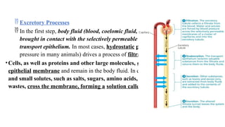
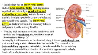
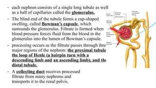
Urinary
structure
Whether an animal lives on land, in salt water, or in fresh water, water
balance depends on the regulation of solute movement between internal fluids
and the external environment. Much of this movement is handled by excretory
systems. These systems are central to homeostasis because they dispose of
metabolic wastes and control body fluid composition
 Excretory Processes
 In the first step, body fluid (blood, coelomic fluid, or hemolymph) is
brought in contact with the selectively permeable membrane of a
transport epithelium. In most cases, hydrostatic pressure (blood
pressure in many animals) drives a process of filtration.
•Cells, as well as proteins and other large molecules, cannot cross the
epithelial membrane and remain in the body fluid. In contrast, water
and small solutes, such as salts, sugars, amino acids, and nitrogenous
wastes, cross the membrane, forming a solution called the filtrate.
- The filtrate is converted to a waste fluid by the specific
transport of materials into or out of the filtrate.
 The process of selective reabsorption recovers useful molecules and
water from the filtrate and returns them to the body fluids.
- Valuable solutes—including glucose, certain salts, vitamins, hormones,
and amino acids—are reabsorbed by active transport.
 Nonessential solutes and wastes are left in the filtrate or are added to
it by selective secretion, which also occurs by active transport.
pumping of various solutes adjusts the osmotic movement of water into or out of
the filtrate.
 In the last step—excretion—the processed filtrate containing
nitrogenous wastes is released from the body as urine.
The major function of the urinary system
Its major function is to control the composition and volume of body fluids. The kidneys perform this
function through multiple processes:
1.Excretion. The kidneys are the major excretory organs of the body. They
remove waste products from the blood. Many waste products are toxic, but most are metabolic by-
products of cells and substances absorbed from the intestine.
The skin, liver, lungs, and intestines eliminate some of these waste products, but they cannot
compensate if the kidneys fail to function.
2.Regulation of blood volume and pressure. The kidneys play a major role in controlling the
extracellular fluid volume in the body. They can produce either a large volume of dilute urine or a small
volume of concentrated urine. Thereby, the kidneys regulate blood volume and blood pressure.
3.Regulation of the concentration of solutes in the blood. The kidneys help
regulate the concentration of the major molecules and ions, such as glucose,
Na+
, Cl−
, K+
, Ca2+
, HCO3
-
, and HPO4
2-
.
4.Regulation of extracellular fluid pH. The kidneys excrete variable amounts
of H+ to help regulate extracellular fluid pH.
5.Regulation of red blood cell synthesis. The kidneys secrete a hormone,
erythropoietin, which regulates the synthesis of red blood cells in bone
marrow.
6.Regulation of vitamin D synthesis. The kidneys play an important role in
controlling blood levels of Ca2+ regulating the synthesis of vitamin D.
- in humans, the excretory system consists of a
pair of kidneys, bean-shaped organs about 10
cm in length, as well as organs
•for transporting and storing urine.
•Urine produced by each kidney exits through a duct
called the ureter; the two ureters drain into a
common sac called the urinary bladder.
•During urination, urine is expelled from the bladder
through a tube called the urethra, which empties to
the outside near the vagina in females and through
the penis in males. Sphincter muscles near the
junction of the urethra and bladder regulate urination.
- Each kidney has an outer renal cortex
and an inner renal medulla. Both regions are
supplied with blood by a renal artery and
drained by a renal vein. Within the cortex and
medulla lie tightly packed excretory tubules and
associated blood vessels. The inner renal
pelvis collects urine from the excretory tubules
and passes it to the urinary bladder.
- Weaving back and forth across the renal cortex and
medulla are the nephrons, the functional units of
the vertebrate kidney. Of
the roughly 1 million nephrons in a human kidney, 85% are cortical nephrons,
which reach only a short distance into the medulla. The remainder, the
juxtamedullary nephrons, extend deep into the medulla. Juxtamedullary
nephrons are essential for production of urine that is hyperosmotic to body
fluids, a key adaptation for water conservation in mammals.
- each nephron consists of a single long tubule as well
as a ball of capillaries called the glomerulus.
- The blind end of the tubule forms a cup-shaped
swelling, called Bowman’s capsule, which
surrounds the glomerulus. Filtrate is formed when
blood pressure forces fluid from the blood in the
glomerulus into the lumen of Bowman’s capsule.
- processing occurs as the filtrate passes through three
major regions of the nephron: the proximal tubule,
the loop of Henle (a hairpin turn with a
descending limb and an ascending limb), and the
distal tubule.
- A collecting duct receives processed
filtrate from many nephrons and
transports it to the renal pelvis.
- each nephron is supplied with blood by an afferent
arteriole, an offshoot of the renal artery that
branches and forms the capillaries of the
glomerulus. The capillaries converge as they leave the
glomerulus, forming an efferent arteriole.
- Branches of this vessel form the peritubular capillaries,
which surround the proximal and distal tubules. Other
branches extend downward and form the vasa recta,
hairpin-shaped capillaries that serve the renal medulla,
including the long loop of Henle of juxtamedullary
nephrons.

Urinary structure.pptx jkbjhvghccjvhjkbiut

  • 1.
    Urinary structure Whether an animallives on land, in salt water, or in fresh water, water balance depends on the regulation of solute movement between internal fluids and the external environment. Much of this movement is handled by excretory systems. These systems are central to homeostasis because they dispose of metabolic wastes and control body fluid composition
  • 2.
     Excretory Processes In the first step, body fluid (blood, coelomic fluid, or hemolymph) is brought in contact with the selectively permeable membrane of a transport epithelium. In most cases, hydrostatic pressure (blood pressure in many animals) drives a process of filtration. •Cells, as well as proteins and other large molecules, cannot cross the epithelial membrane and remain in the body fluid. In contrast, water and small solutes, such as salts, sugars, amino acids, and nitrogenous wastes, cross the membrane, forming a solution called the filtrate.
  • 3.
    - The filtrateis converted to a waste fluid by the specific transport of materials into or out of the filtrate.  The process of selective reabsorption recovers useful molecules and water from the filtrate and returns them to the body fluids. - Valuable solutes—including glucose, certain salts, vitamins, hormones, and amino acids—are reabsorbed by active transport.  Nonessential solutes and wastes are left in the filtrate or are added to it by selective secretion, which also occurs by active transport. pumping of various solutes adjusts the osmotic movement of water into or out of the filtrate.  In the last step—excretion—the processed filtrate containing nitrogenous wastes is released from the body as urine.
  • 4.
    The major functionof the urinary system Its major function is to control the composition and volume of body fluids. The kidneys perform this function through multiple processes: 1.Excretion. The kidneys are the major excretory organs of the body. They remove waste products from the blood. Many waste products are toxic, but most are metabolic by- products of cells and substances absorbed from the intestine. The skin, liver, lungs, and intestines eliminate some of these waste products, but they cannot compensate if the kidneys fail to function. 2.Regulation of blood volume and pressure. The kidneys play a major role in controlling the extracellular fluid volume in the body. They can produce either a large volume of dilute urine or a small volume of concentrated urine. Thereby, the kidneys regulate blood volume and blood pressure.
  • 5.
    3.Regulation of theconcentration of solutes in the blood. The kidneys help regulate the concentration of the major molecules and ions, such as glucose, Na+ , Cl− , K+ , Ca2+ , HCO3 - , and HPO4 2- . 4.Regulation of extracellular fluid pH. The kidneys excrete variable amounts of H+ to help regulate extracellular fluid pH. 5.Regulation of red blood cell synthesis. The kidneys secrete a hormone, erythropoietin, which regulates the synthesis of red blood cells in bone marrow. 6.Regulation of vitamin D synthesis. The kidneys play an important role in controlling blood levels of Ca2+ regulating the synthesis of vitamin D.
  • 6.
    - in humans,the excretory system consists of a pair of kidneys, bean-shaped organs about 10 cm in length, as well as organs •for transporting and storing urine. •Urine produced by each kidney exits through a duct called the ureter; the two ureters drain into a common sac called the urinary bladder. •During urination, urine is expelled from the bladder through a tube called the urethra, which empties to the outside near the vagina in females and through the penis in males. Sphincter muscles near the junction of the urethra and bladder regulate urination.
  • 7.
    - Each kidneyhas an outer renal cortex and an inner renal medulla. Both regions are supplied with blood by a renal artery and drained by a renal vein. Within the cortex and medulla lie tightly packed excretory tubules and associated blood vessels. The inner renal pelvis collects urine from the excretory tubules and passes it to the urinary bladder. - Weaving back and forth across the renal cortex and medulla are the nephrons, the functional units of the vertebrate kidney. Of the roughly 1 million nephrons in a human kidney, 85% are cortical nephrons, which reach only a short distance into the medulla. The remainder, the juxtamedullary nephrons, extend deep into the medulla. Juxtamedullary nephrons are essential for production of urine that is hyperosmotic to body fluids, a key adaptation for water conservation in mammals.
  • 8.
    - each nephronconsists of a single long tubule as well as a ball of capillaries called the glomerulus. - The blind end of the tubule forms a cup-shaped swelling, called Bowman’s capsule, which surrounds the glomerulus. Filtrate is formed when blood pressure forces fluid from the blood in the glomerulus into the lumen of Bowman’s capsule. - processing occurs as the filtrate passes through three major regions of the nephron: the proximal tubule, the loop of Henle (a hairpin turn with a descending limb and an ascending limb), and the distal tubule. - A collecting duct receives processed filtrate from many nephrons and transports it to the renal pelvis.
  • 9.
    - each nephronis supplied with blood by an afferent arteriole, an offshoot of the renal artery that branches and forms the capillaries of the glomerulus. The capillaries converge as they leave the glomerulus, forming an efferent arteriole. - Branches of this vessel form the peritubular capillaries, which surround the proximal and distal tubules. Other branches extend downward and form the vasa recta, hairpin-shaped capillaries that serve the renal medulla, including the long loop of Henle of juxtamedullary nephrons.