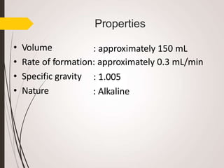
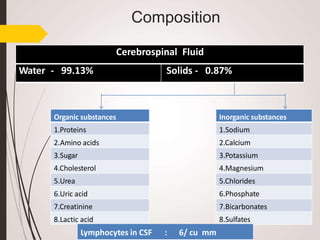
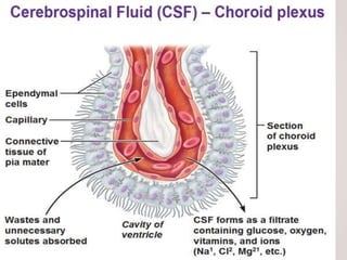
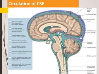
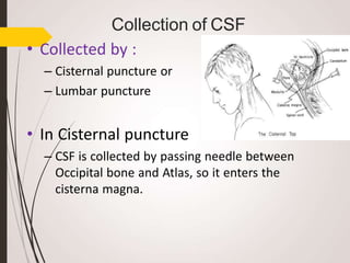
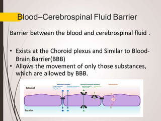
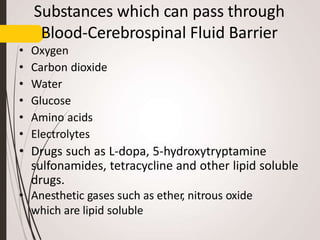
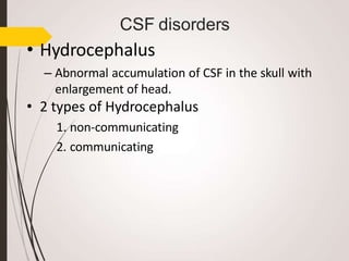
Introduction
• CSF is clear, colorless and
transparent fluid present in
the cerebral ventricles,
spinal canal, and
subarachnoid spaces.
• Circulates through cavity of
the:
– Brain
– Subarachnoid space
– Central canal of spinal cord
• Part of Extracellular fluid
(ECF)
Properties
• Volume : approximately 150 mL
• Rate of formation: approximately 0.3 mL/min
• Specific gravity
• Nature
: 1.005
: Alkaline
Composition
Inorganic substances
1.Sodium
2.Calcium
3.Potassium
4.Magnesium
5.Chlorides
6.Phosphate
7.Bicarbonates
8.Sulfates
Cerebrospinal Fluid
Water - 99.13% Solids - 0.87%
Organic substances
1.Proteins
2.Amino acids
3.Sugar
4.Cholesterol
5.Urea
6.Uric acid
7.Creatinine
8.Lactic acid
Lymphocytes in CSF : 6/ cu mm
Continued…
• As CSF is part of ECF therefore it contain
more amount of Sodium than Potassium.
• Contains some lymphocytes.
• CSF secreted by ventricles does not contain
any cell.
• The lymphocytes are added when it flows in
the spinal cord.
Formation of CSF
• Site of formation
– Formed by the choroid plexus
situated within the
ventricles.
– The choroid plexus are tuft of
capillary projections present
inside ventricles and covered
by:
• Pia matter
• Ependymal covering.
Mechanism of formation
• Formed by process of secretion.
• Uses energy.
• Active transport mechanism is involved in secretion.
Substances affecting the formation of CSF
•Pilocarpine, ether and extracts of pituitary gland
stimulates the secretion of CSF by stimulating
choroid plexus.
•Pilocarpine is a medication used to treat increased
pressure inside the eye and dry mouth
•Injection of isotonic saline also stimulates CSF
formation.
Continued…
• Injection of hypotonic saline
– Causes greater rise in capillary pressure and
intracranial pressure,
– Fall in osmotic pressure leading to increase in CSF
formation.
• Hypertonic saline
– Decreases CSF formation
– Decreases CSF pressure
Circulation of CSF
Absorption of CSF
• Mostly absorbed by the Arachnoid villi
into Dural sinuses and Spinal veins.
• Small amount is absorbed along the
perineural spaces into cervical lymphatics
and in to the perivascular spaces.
• Normally, 500 mL of CSF is formed
everyday and equal amount is absorbed.
• Absorption is done by filtration due to
pressure gradient between hydrostatic
pressure in the subarachnoid space fluid
and the pressure that exists in the Dural
sinus blood.
FUNCTIONS OF CSF
Protection
Nutrition
Removal of waste
Lubrication
Continued…
• Regulation of Cranial Content Volume
– Is very essential because
– When substances are absorbed into the venous sinuses,
intracranial pressure is raised,
– Intracranial pressure in turn interferes with the cerebral
circulation causing Asphyxia.
• It is prevented by greater absorption of CSF to give
space for increasing cranial contents.
• Medium of Exchange
• CSF is the medium through which substances like :
– Nutritive substances
– Waste materials
are exchanged between blood and brain tissues.
Collection of CSF
• Collected by :
– Cisternal puncture or
– Lumbar puncture
• In Cisternal puncture
– CSF is collected by passing needle between
Occipital bone and Atlas, so it enters the
cisterna magna.
Continued…
• In Lumbar puncture
– The Lumbar puncture needle is
introduced into the subarachnoid
space in the Lumbar region
, between the third and fourth Lumbar
spines.
– Uses of Lumbar puncture
• Collecting CSF for diagnostic purpose.
• Injecting drugs for spinal anesthesia,
analgesia and chemotherapy.
• Measuring pressure exerted by CSF.
Blood–Cerebrospinal Fluid Barrier
Barrier between the blood and cerebrospinal fluid .
• Exists at the Choroid plexus and Similar to Blood-
Brain Barrier(BBB)
• Allows the movement of only those substances,
which are allowed by BBB.
Substances which can pass through
Blood-Cerebrospinal Fluid Barrier
• Oxygen
• Carbon dioxide
• Water
• Glucose
• Amino acids
• Electrolytes
• Drugs such as L-dopa, 5-hydroxytryptamine
sulfonamides, tetracycline and other lipid soluble
drugs.
• Anesthetic gases such as ether, nitrous oxide
which are lipid soluble
Substanes which can’t pass through
Blood-Cerebrospinal Fluid Barrier
• Injurious chemical agents.
• Pathogens such as bacteria.
• Drugs such as Penicillin and the
Catecholamines.
• Dopamine also can’t pass therefore
Parkinsonism is treated with L-dopa instead
of dopamine.
Continued…
• Bile pigments
– However barrier is not well developed in infants.
– The bile pigments enter the brain tissues .
– During jaundice in infants, the bile pigments
enter the brain and causes damage of Basal
ganglia leading to Kernicterus.
CSF disorders
• Hydrocephalus
– Abnormal accumulation of CSF in the skull with
enlargement of head.
• 2 types of Hydrocephalus
1. non-communicating
2. communicating
1. Non-communicating Hydrocephalus or
Internal Hydrocephalus
Due to the obstruction of any of the foramen
through which CSF escapes results in dilation of
ventricular cavity.
2. Communicating Hydrocephalus or External
Hydrocephalus
3. Due to blockage of Arachnoid villi.
Symptoms of Hydrocephalus:
 Headache, Vomiting Atrophy of brain Mental weakness
Convulsions
INDICATIONS OF CSF
EXAMINATION
1. Infections: meningitis, encephalitis.
2. Inflammatory conditions: Sarcoidosis,
Neurosyphilis, SLE.
3. Infiltrative conditions: Leukemia, lymphoma
4. Administration of drugs in CSF (Therapeutic
aim): Antibiotics
5. Anticancer drugs
6. Anesthetic drugs
Cerebrospinal fluid ppt

Cerebrospinal fluid ppt

  • 2.
    Introduction • CSF isclear, colorless and transparent fluid present in the cerebral ventricles, spinal canal, and subarachnoid spaces. • Circulates through cavity of the: – Brain – Subarachnoid space – Central canal of spinal cord • Part of Extracellular fluid (ECF)
  • 3.
    Properties • Volume :approximately 150 mL • Rate of formation: approximately 0.3 mL/min • Specific gravity • Nature : 1.005 : Alkaline
  • 4.
    Composition Inorganic substances 1.Sodium 2.Calcium 3.Potassium 4.Magnesium 5.Chlorides 6.Phosphate 7.Bicarbonates 8.Sulfates Cerebrospinal Fluid Water- 99.13% Solids - 0.87% Organic substances 1.Proteins 2.Amino acids 3.Sugar 4.Cholesterol 5.Urea 6.Uric acid 7.Creatinine 8.Lactic acid Lymphocytes in CSF : 6/ cu mm
  • 5.
    Continued… • As CSFis part of ECF therefore it contain more amount of Sodium than Potassium. • Contains some lymphocytes. • CSF secreted by ventricles does not contain any cell. • The lymphocytes are added when it flows in the spinal cord.
  • 6.
    Formation of CSF •Site of formation – Formed by the choroid plexus situated within the ventricles. – The choroid plexus are tuft of capillary projections present inside ventricles and covered by: • Pia matter • Ependymal covering.
  • 8.
    Mechanism of formation •Formed by process of secretion. • Uses energy. • Active transport mechanism is involved in secretion.
  • 9.
    Substances affecting theformation of CSF •Pilocarpine, ether and extracts of pituitary gland stimulates the secretion of CSF by stimulating choroid plexus. •Pilocarpine is a medication used to treat increased pressure inside the eye and dry mouth •Injection of isotonic saline also stimulates CSF formation.
  • 10.
    Continued… • Injection ofhypotonic saline – Causes greater rise in capillary pressure and intracranial pressure, – Fall in osmotic pressure leading to increase in CSF formation. • Hypertonic saline – Decreases CSF formation – Decreases CSF pressure
  • 11.
  • 12.
    Absorption of CSF •Mostly absorbed by the Arachnoid villi into Dural sinuses and Spinal veins. • Small amount is absorbed along the perineural spaces into cervical lymphatics and in to the perivascular spaces. • Normally, 500 mL of CSF is formed everyday and equal amount is absorbed. • Absorption is done by filtration due to pressure gradient between hydrostatic pressure in the subarachnoid space fluid and the pressure that exists in the Dural sinus blood.
  • 13.
  • 14.
    Continued… • Regulation ofCranial Content Volume – Is very essential because – When substances are absorbed into the venous sinuses, intracranial pressure is raised, – Intracranial pressure in turn interferes with the cerebral circulation causing Asphyxia. • It is prevented by greater absorption of CSF to give space for increasing cranial contents. • Medium of Exchange • CSF is the medium through which substances like : – Nutritive substances – Waste materials are exchanged between blood and brain tissues.
  • 15.
    Collection of CSF •Collected by : – Cisternal puncture or – Lumbar puncture • In Cisternal puncture – CSF is collected by passing needle between Occipital bone and Atlas, so it enters the cisterna magna.
  • 16.
    Continued… • In Lumbarpuncture – The Lumbar puncture needle is introduced into the subarachnoid space in the Lumbar region , between the third and fourth Lumbar spines. – Uses of Lumbar puncture • Collecting CSF for diagnostic purpose. • Injecting drugs for spinal anesthesia, analgesia and chemotherapy. • Measuring pressure exerted by CSF.
  • 17.
    Blood–Cerebrospinal Fluid Barrier Barrierbetween the blood and cerebrospinal fluid . • Exists at the Choroid plexus and Similar to Blood- Brain Barrier(BBB) • Allows the movement of only those substances, which are allowed by BBB.
  • 18.
    Substances which canpass through Blood-Cerebrospinal Fluid Barrier • Oxygen • Carbon dioxide • Water • Glucose • Amino acids • Electrolytes • Drugs such as L-dopa, 5-hydroxytryptamine sulfonamides, tetracycline and other lipid soluble drugs. • Anesthetic gases such as ether, nitrous oxide which are lipid soluble
  • 19.
    Substanes which can’tpass through Blood-Cerebrospinal Fluid Barrier • Injurious chemical agents. • Pathogens such as bacteria. • Drugs such as Penicillin and the Catecholamines. • Dopamine also can’t pass therefore Parkinsonism is treated with L-dopa instead of dopamine.
  • 20.
    Continued… • Bile pigments –However barrier is not well developed in infants. – The bile pigments enter the brain tissues . – During jaundice in infants, the bile pigments enter the brain and causes damage of Basal ganglia leading to Kernicterus.
  • 21.
    CSF disorders • Hydrocephalus –Abnormal accumulation of CSF in the skull with enlargement of head. • 2 types of Hydrocephalus 1. non-communicating 2. communicating
  • 22.
    1. Non-communicating Hydrocephalusor Internal Hydrocephalus Due to the obstruction of any of the foramen through which CSF escapes results in dilation of ventricular cavity. 2. Communicating Hydrocephalus or External Hydrocephalus 3. Due to blockage of Arachnoid villi. Symptoms of Hydrocephalus:  Headache, Vomiting Atrophy of brain Mental weakness Convulsions
  • 23.
    INDICATIONS OF CSF EXAMINATION 1.Infections: meningitis, encephalitis. 2. Inflammatory conditions: Sarcoidosis, Neurosyphilis, SLE. 3. Infiltrative conditions: Leukemia, lymphoma 4. Administration of drugs in CSF (Therapeutic aim): Antibiotics 5. Anticancer drugs 6. Anesthetic drugs