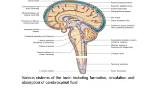
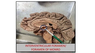
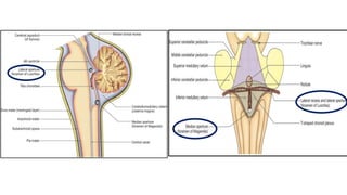
Cerebrospinal Fluid
Dr. Amaan
23rd Jan 2023/ Monday
Various cisterns of the brain including formation, circulation and
absorption of cerebrospinal fluid
Cerebrospinal Fluid
 The CSF is a modified tissue fluid
• CSF is clear, colorless liquid secreted by choroid plexus
• Present outside the brain in the subarachnoid space and spinal cord.
• Within the brain in its ventricles
 The brain and CSF have approximately the same specific gravity, so
that the brain simply floats in the fluid
• CSF replaces lymph in the CNS
FORMATION
• The bulk of the CSF is formed by the choroid plexuses of the
lateral ventricles
• The total quantity is about 150 ml
• Formed at the rate of about 200 ml per hour or 5000 ml per day
• The normal pressure of CSF is 60 to 100 mm of water
CSF CIRCULATION
Each lateral ventricle to the third ventricle through the interventricular
foramen of Monro.
Third ventricle, it passes to the fourth ventricle through the cerebral
aqueduct.
From the fourth ventricle, the CSF passes to the subarachnoid spaces of
the cerebrum and the vertebral canal through the median and lateral
apertures of the fourth ventricle
Some of it passes down the central canal of spinal cord.
LATERAL VENTRICLES
THIRD VENTRICLES
(in between hypothalamus of both side )
INTERVENTRICULAR FORAMEN/
FORAMEN OF MONRO
CEREBRAL AQUEDUCT
FOURTH VENTRICLE
(communicating with the central canal of Spinal cord)
CSF ABSORPTION
• Chiefly through the arachnoid villi and granulations,
and is thus drained into the cranial venous sinuses
• Veins related to spinal nerves
FUNCTIONS OF CSF
1. CSF decreases the sudden pressure or
forces on delicate nervous tissue
2. CSF nourishes nervous tissue
• CSF comes in contact with neurons
• It provides nourishment and returns products of metabolism to the
venous sinuses
3. Neurons cannot live without glucose and oxygen for more than 3–5
minutes. These are constantly provided by CSF.
3. Pineal gland secretions reach pituitary gland via CSF
4. Cushions the brain within its solid vault
5. There is no CSF brain barrier,
• so drugs can reach the neurons through CSF
6. There is blood CSF barrier
CLINICAL ANATOMY
• Hydrocephalus:
It is the dilatation of the ventricular system and occurs due to
obstruction of CSF circulation
a. Communicating,
b. Non-communicating:
a.Communicating hydrocephalus/ external hydrocephalus
• If the obstruction is outside the ventricular system,
• Usually in the subarachnoid space or arachnoid granulations
• This occurs due to fibrosis following meningitis
Clinical features are:
• Head size is rather large
• Tense anterior fontanelle
• Dilated veins over thin scalp
b. Non-communicating hydrocephalus/ internal hydrocephalus
• If the obstruction is within the ventricular system
• Usually caused by a tumour or inflammation
• A shunt procedure is employed to divert the CSF from the ventricular
system into the peritoneal cavity
• Drainage of CSF at regular intervals is of therapeutic value in
meningitis.
• Headaches of unknown aetiology are also known to have been cured
by a mere lumbar puncture with drainage of CSF.
• Obstruction in the vertebral canal produces Froin’s syndrome or
loculation syndrome.
This is characterised by yellowish discolouration of CSF
(xanthochromia) below the level of obstruction, and its spontaneous
coagulation after withdrawal due to a high protein content.

CSF.pptx

  • 1.
  • 2.
    Various cisterns ofthe brain including formation, circulation and absorption of cerebrospinal fluid
  • 3.
    Cerebrospinal Fluid  TheCSF is a modified tissue fluid • CSF is clear, colorless liquid secreted by choroid plexus • Present outside the brain in the subarachnoid space and spinal cord. • Within the brain in its ventricles  The brain and CSF have approximately the same specific gravity, so that the brain simply floats in the fluid • CSF replaces lymph in the CNS
  • 4.
    FORMATION • The bulkof the CSF is formed by the choroid plexuses of the lateral ventricles • The total quantity is about 150 ml • Formed at the rate of about 200 ml per hour or 5000 ml per day • The normal pressure of CSF is 60 to 100 mm of water
  • 5.
    CSF CIRCULATION Each lateralventricle to the third ventricle through the interventricular foramen of Monro. Third ventricle, it passes to the fourth ventricle through the cerebral aqueduct. From the fourth ventricle, the CSF passes to the subarachnoid spaces of the cerebrum and the vertebral canal through the median and lateral apertures of the fourth ventricle Some of it passes down the central canal of spinal cord.
  • 7.
  • 8.
    THIRD VENTRICLES (in betweenhypothalamus of both side )
  • 9.
  • 10.
    CEREBRAL AQUEDUCT FOURTH VENTRICLE (communicatingwith the central canal of Spinal cord)
  • 12.
    CSF ABSORPTION • Chieflythrough the arachnoid villi and granulations, and is thus drained into the cranial venous sinuses • Veins related to spinal nerves
  • 13.
    FUNCTIONS OF CSF 1.CSF decreases the sudden pressure or forces on delicate nervous tissue 2. CSF nourishes nervous tissue • CSF comes in contact with neurons • It provides nourishment and returns products of metabolism to the venous sinuses 3. Neurons cannot live without glucose and oxygen for more than 3–5 minutes. These are constantly provided by CSF.
  • 14.
    3. Pineal glandsecretions reach pituitary gland via CSF 4. Cushions the brain within its solid vault 5. There is no CSF brain barrier, • so drugs can reach the neurons through CSF 6. There is blood CSF barrier
  • 15.
    CLINICAL ANATOMY • Hydrocephalus: Itis the dilatation of the ventricular system and occurs due to obstruction of CSF circulation a. Communicating, b. Non-communicating:
  • 16.
    a.Communicating hydrocephalus/ externalhydrocephalus • If the obstruction is outside the ventricular system, • Usually in the subarachnoid space or arachnoid granulations • This occurs due to fibrosis following meningitis Clinical features are: • Head size is rather large • Tense anterior fontanelle • Dilated veins over thin scalp
  • 17.
    b. Non-communicating hydrocephalus/internal hydrocephalus • If the obstruction is within the ventricular system • Usually caused by a tumour or inflammation • A shunt procedure is employed to divert the CSF from the ventricular system into the peritoneal cavity
  • 18.
    • Drainage ofCSF at regular intervals is of therapeutic value in meningitis. • Headaches of unknown aetiology are also known to have been cured by a mere lumbar puncture with drainage of CSF. • Obstruction in the vertebral canal produces Froin’s syndrome or loculation syndrome. This is characterised by yellowish discolouration of CSF (xanthochromia) below the level of obstruction, and its spontaneous coagulation after withdrawal due to a high protein content.