Downloaded 10 times
![XTT Assay
It is based on the reduction of a yellow tetrazolium salt (sodium 3´-[1- (phenylaminocarbonyl)- 3,4-
tetrazolium]-bis (4-methoxy6-nitro) benzene sulfonic acid hydrate or XTT) to an orange formazan
dye by metabolically active cells
XTT is simply for measuring proliferation and is therefore an excellent solution for quantitating cells
and determining their viability.
The XTT assay is used to measure cellular metabolic activity as an indicator of cell viability,
proliferation and cytotoxicity.
It is used for the measurement of cell proliferation in response to growth factors, cytokines and
nutrients
https://www.sigmaaldrich.com/technical-documents/protocols/biology/roche/cell-proliferation-kit-xtt-assay.html](https://image.slidesharecdn.com/unit1cytotoxicitylt3-210718092600/85/3-Cellular-cytotoxicity-7-320.jpg)
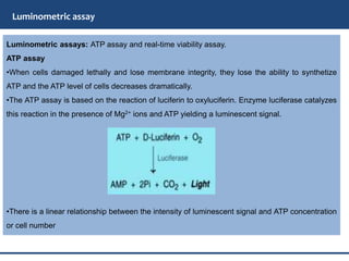
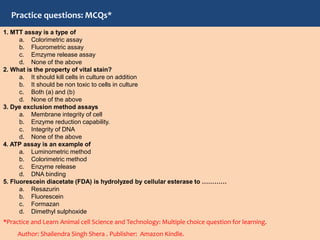

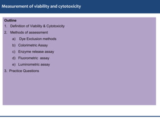
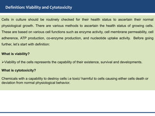
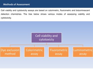
The document discusses the measurement of cell viability and cytotoxicity, defining both terms and outlining various assessment methods including dye exclusion, colorimetric, enzyme release, fluorometric, and luminometric assays. It explains how these methods work, their principles, and specific examples such as the MTT and XTT assays for colorimetric analysis, and the ATP assay for luminometric analysis. Additionally, it provides practice questions for learning about the principles and applications of these assays.


