DR. AMBEDKER COLLEGE,DEEKSHABHOOMI, NAGPUR
DEPARTMENT OF BIOTECHNOLOGY AND BIOCHEMISTRY
SEMINAR ON
2D-Electrophoresis
Presented by
PIYUSH GHOSHE
M.Sc. Biotechnology
Semester-1st
Introduction
• Introduction by O'Farrell and Klose in 1975
• Separation of mixed proteins
Principle
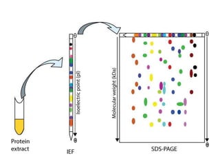
• In 2D Gel Electrophoresis proteins are
separated as per isoelectric point(pI) and
protein mass.
• It is an Combination of Iso-electric Focusing
(pI) and SDS-PAGE.
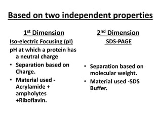
Based on two independent properties
1st Dimension
Iso-electric Focusing (pI)
pH at which a protein has
a neutral charge
• Separation based on
Charge.
• Material used -
Acrylamide +
ampholytes
+Riboflavin.
2nd Dimension
SDS-PAGE
• Separation based on
molecular weight.
• Material used -SDS
Buffer.
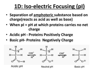
1D: Iso-electric Focusing (pI)
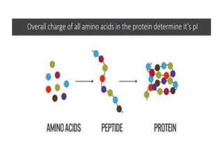
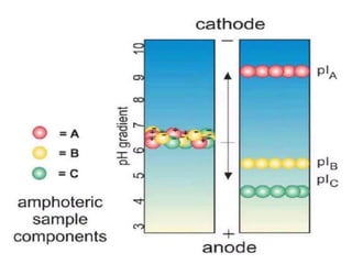
• Separation of amphoteric substance based on
charge(reacts as acid as well as base)
• When pI = pH at which proteins carries no net
charge
• Acidic pH - Proteins Positively Charge
• Basic pH- Proteins Negatively Charge
Acidic pH Neutral pH Basic pH
• IEF Gels are made up of Ampholytes-complex
mixture synthetic polyamino-polycarboxylic
acid.
• 1-2 mm thick IEF gel
• Expensive
• 0.5mm thin layer IEF
• Ampholytes + Riboflavin in Acrylamide is used.
• Separation is achieved by applying a potential
difference across a gel that contain a pH
gradient.
Chemicals used during IEF
• Ampholytes
• Riboflavin
• Acrylamide
• Spacer ( insulation tape)
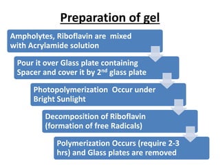
Preparation of gel
Ampholytes, Riboflavin are mixed
with Acrylamide solution
Pour it over Glass plate containing
Spacer and cover it by 2nd glass plate
Photopolymerization Occur under
Bright Sunlight
Decomposition of Riboflavin
(formation of free Radicals)
Polymerization Occurs (require 2-3
hrs) and Glass plates are removed
2D: SDS-PAGE
(Sodium Dodecyl Sulphate PolyAcrylamide Gel Electrophoresis)
Role of 2-Mercaptoethanol and SDS
• Separation of protein based on Molecular weight
Buffer and reagent for electrophoresis
1. TetraMethylEthyleneDiamine (TEMED)
2. Ammonium persulfate (APS)
3. Tris-HCl
4. Glycine
5. Bromophenol blue
6. Coomassie brilliant blue R250
7. Sodium dodecyl sulphate
8. Acrylamide
9. Bis-acrylamide
10. 2-Mercaptoethanol
11. Butanol  isopropanol
Preparation of Resolving Gel
Acrylamide solution 12%
(acrylamide & bisacrylamide)
Mixed with TEMED and APS
Add Tris HCl and Poured in
between the glass plate fitted
into the gel cassette
Allow them to Solidify and A
thin layer of organic solvent i.e.
butanol or isopropanol Is Added
Preparation of Stacking Gel
Acrylamide solution 4%
(acrylamide & bis-acrylamide)
Mixed with TEMED and APS
Add Tris HCl and Poured in
between the glass plate Above
the Resolving gel.
Allow them to Solidify
Then insert a IEF strip over it
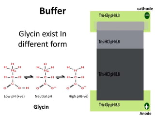
STACKING & RESOLVING GELS
STACKING GEL
• 4% of acrylamide
• Ordering/arrangin
g and conc. the
macromolecule
• High pore size
• pH 6.8
RESOLVING GEL
• 15% of acrylamide
• The actual zone of
separation
• Less pore size
• pH 8.8
Resolving Gel
Stacking Gel
Glycin exist In
different form
Low pH (+ve) Neutral pH High pH(-ve)
Glycin
Buffer cathode
Anode
Detection Of Proteins
1. Using chemical stains like
• Silver Nitrate
• Coomassie Brilliant Blue
• Western Blot
2. 2D gel analysis software
• BioNumerics 2D, Delta2D,
• ImageMaster
• PDQuest, Progenesis
Applications
• Analyze proteins
• Separation of proteins
• Determination of molecular mass
• To determine amino acid sequence
• Separation of DNA
REFERENCE
• O'Farrell, PH (1975); High resolution two-dimensional
electrophoresis of proteins; The Journal of Biologocal
Chemistry;Vol.250 NO.10; issue of May 25; 4007–
4021pg
• NPTEL – Biotechnology – Bioanalytical Techniques and
Bioinformatics (Module 4 Electrophoretic techniques)
2012,5-9pg
• Edited by Sameh Magdeldin;Gel Electrophoresis –
Principles and Basics; Published by InTech Janeza
Trdine 9, 51000 Rijeka, Croatia; 2012;62-65pg
• http://www.uvm.edu/~vgn/outreach/documents/Garfi
n_IEF_webArticle9-07.pdf
• http://en.m.wikipedia.org/wiki/Isoelectric_point
• http://m.youtube.com/watch?v=YV4gR4vucwY