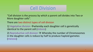
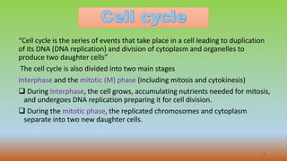
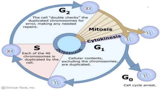
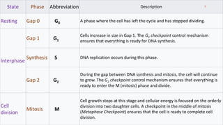
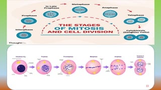
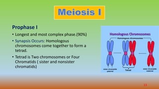
1
Presented To
Cell Division
“Cell division is the process by which a parent cell divides into Two or
More daughter cells”.
There are two distinct types of cell division
1) Vegetative division whereby each daughter cell is genetically
identical to the parent cell (mitosis)
2) Reproductive cell division  Whereby the number of Chromosomes
in the daughter cells is reduce by half to produce haploid gametes
(meiosis)
3
4
“Cell cycle is the series of events that take place in a cell leading to duplication
of its DNA (DNA replication) and division of cytoplasm and organelles to
produce two daughter cells”
The cell cycle is also divided into two main stages
Interphase and the mitotic (M) phase (including mitosis and cytokinesis)
 During Interphase, the cell grows, accumulating nutrients needed for mitosis,
and undergoes DNA replication preparing it for cell division.
 During the mitotic phase, the replicated chromosomes and cytoplasm
separate into two new daughter cells.
5
6
State Phase Abbreviation Description
Resting Gap 0 G0 A phase where the cell has left the cycle and has stopped dividing.
Interphase
Gap 1 G1
Cells increase in size in Gap 1. The G1 checkpoint control mechanism
ensures that everything is ready for DNA synthesis.
Synthesis S DNA replication occurs during this phase.
Gap 2 G2
During the gap between DNA synthesis and mitosis, the cell will continue
to grow. The G2 checkpoint control mechanism ensures that everything is
ready to enter the M (mitosis) phase and divide.
Cell
division
Mitosis M
Cell growth stops at this stage and cellular energy is focused on the orderly
division into two daughter cells. A checkpoint in the middle of mitosis
(Metaphase Checkpoint) ensures that the cell is ready to complete cell
division.
7
“Mitosis is the process whereby one cell divides, giving rise to two daughter cells
that are genetically identical to the parent cell”
Before a cell enters mitosis, each Chromosome replicates its DNA.
1) Prophase
• Condensing its chromosomes and initiating mitotic spindle formation.
• the genetic material in the nucleus consists of loosely packed chromatin.
• The two chromatids are joined at the centromere.
8
2) Prometaphase
phosphorylation of nuclear lamins causes the nuclear envelope to
disintegrate into small membrane vesicles.
3) Metaphase
The two centrosomes begin pulling the chromosomes towards opposite
ends of the cell.
4) Anaphase
• The cohesions that bind sister chromatids together are cleaved, forming two
identical daughter chromosomes.
• Shortening of the kinetochore microtubules pulls the newly formed daughter
chromosomes to opposite ends of the cell.
9
5) Telophase (the Greek word meaning "end“)
• reversal of prophase and prometaphase events.
• A new nuclear envelope forms using the membrane vesicles of the
parent cell's old nuclear envelope.
• Mitosis is complete. Each daughter nucleus has an identical set of
chromosomes
Cytokinesis
Cytokinesis is not a phase of mitosis but rather a separate process,
necessary for completing cell division.
10
11
“Meiosis is the cell division that takes place in the
germ cells. To generate male and female gametes.
(sperm and egg cells)”
These goals are accomplished in meiosis using a two-
step division process.
Step I  Homologue pairs separate during a first
round of cell division, called meiosis I.
Step II  Sister chromatids separate during a second
round, called meiosis II.
12
Prophase I
• Longest and most complex phase.(90%)
• Synapsis Occurs: Homologous
chromosomes come together to form a
tetrad.
• Tetrad is Two chromosomes or Four
Chromatids ( sister and nonsister
chromatids)
13
•Segments of nonsister
Chromatids break and
reattach to the other
Chromatid.
•The Chiasmata (chiasma)
are the sites of crossing
over.
14
• After crossing over, the spindle begins to capture chromosomes and move
them towards the center of the cell.
• This may seem familiar from mitosis, but there is a twist.
• Each chromosome attaches to microtubules from just one pole of the spindle.
15
Metaphase I
• Homologue pairs—not individual chromosomes—line up at the
metaphase plate for separation.
• When the homologous pairs line up at the metaphase plate.
• The orientation of each pair is Random.
Anaphase I
• The homologues are pulled apart and move apart to opposite ends of
the cell.
16
Telophase I
• The chromosomes arrive at opposite poles of the cell.
• The nuclear membrane re-forms and the chromosomes decondense.
• Cytokinesis  Forming two Haploid daughter cells.
• Cells will soon go through another round of division, meiosis II
17
Meiosis II
• Cells move from meiosis I to meiosis II without copying their DNA.
• Shorter and simpler process than meiosis I.
• Chromatids separate, making Haploid cells with non-duplicated chromosomes
18
prophase II
• Chromosomes condense and the nuclear envelope breaks down.
• Spindle formation.
• The two sister chromatids of each chromosome are captured by
microtubules from opposite spindle poles.
Metaphase II
• The chromosomes line up individually along the metaphase plate.
Anaphase II
• The sister chromatids separate and are pulled towards opposite poles of the
cell.
telophase II
• Nuclear membranes form , chromosomes decondense ,
• Cytokinesis splits the chromosome Four Haploid cells in which each
chromosome has just One chromatid.
19
20
“Process by which diploid or haploid precursor cells undergo cell
division and differentiation to form mature haploid gametes”
• The male and female germ cells are called Gametes.
• The gametes in human Males are produced by the testes.(sperm)
the process is known as Spermatogenesis.
• Gametes in human Females are produced by the ovaries. (Egg or
Ova) the process is known is Oogenesis.
21
• Gametes are derived from Primordial germ cells (PGCs).
• That are formed during 2nd Week (epiblast).
• During gastrulation they move through primitive streak and migrate to the
wall of the yolk sac.
• During the 4th week , the cell begin to migrate from the yolk sac toward the
developing gonads.
• Mitotic divisions increase their number during their migration and also when
they are arrive in the gonads.
• In preparation for fertilization, germ cells undergo gametogenesis, which
include meiosis to reduce the number of chromosomes and
Cytodifferentiation to complete their maturation.
22
23
Spermatogenesis
“Is the process by which haploid spermatozoa
develop from germ cells in the seminiferous
tubules of the testis”
•Starts with the division of spermatogonia and ends
with the formation of spermatozoa Begins at
puberty and then goes on continuously.
24
Steps of Spermatogenesis
• Spermatogenesis occurs in Seminiferous tubules.
• Stimulation of Anterior pituitary gonadotropin hormones.
• Spermatogenesis begins at an Average 13 years and continues.
• Sertoli cell are large , with overflowing cytoplasmic envelopes that surround
the developing spermatogonia all the way to the central lumen of the tubule.
• Meiosis.
Spermatogonia become progressively modified and enlarged to form
primary spermatocytes.
25
• Each primary Spermatocytes undergoes meiotic division to form Two secondary
Spermatocytes.
• After few days, Secondary spermatocytes divide to form Spermatids 
Spermatozoa (sperm).
• During the change from the spermatocyte to Spermatid Stage.
• 46 C (23 pair of C) of the Spermatocyte are Divide.
• 23 C goes  One Spermatid
• 23C goes  Second Spermatid.
• Gene are also divided.
• One half of genetic characteristics of the eventual Fetus are provided by the
father, with the other half being derived from the oocytes provided by the mother.
• Spermatogenesis period (from Spermatogonia to Spermatozoa) takes about 74
days.
26
27
Sex chromosomes
• In each Spermatogonium one of the 23 pairs of Chromosomes carries the
genetic information that determines the sex of each eventual offspring.
• X Chromosome is called Female chromosome.
• Y chromosome the male chromosome.
28
Formation of Sperm
• Each spermatozoon is composed of Head and a Tail.
• The Head comprises the condensed nucleus of the cell, with thin cytoplasmic
and cell membrane layer around its surface.
• Outside of anterior two third of the head is a thick cap called Acrosome. (
formed mainly from Golgi apparatus).
• Acrosome contain enzymes  Hyaluronidase and proteolytic enzymes.
• There enzymes role  allowing the sperm to enter the ovum and fertilize it.
29
• Tail of sperm called Flagellum. Three major
components;
1) A central Skeletal of 11 microtubules,
collectively called Axoneme.
2) A thin cell membrane covering the
Axoneme.
3) Proximal portion of tail where collection of
the mitochondria called Body of the tail.
• Back-and –forth movement of the tail
(flagellar movement)
• Energy is supplied in the form of ATP, which
is synthesized by mitochondria.
• Normal sperm move in fluid medium at
velocity of 1 to 4 mm⁄min.
30
Testosterone secreted from Leyding cells (intestitium of the testes) is essential for
growth and division.
Luteinizing hormone secreted from APG. Stimulates the leyding cells to secrete
Testosterone.
Follicle stimulating hormones Secreted from APG.Stimulates the Sertoli cells to
convert Spermatids to sperm ( ).
Estrogens formed from testosterone by the Sertoli cells, Also essential for
spermiogenesis.
Growth Hormone is necessary for controlling background metabolic function of the
testes.
31
Maturation of sperm in the Epididymis
• After formation in the seminiferous tubules,
• The sperm require several days to pass through the 6 meter long tubule of
the Epididymis.
• Removed sperm from seminiferous tubules are nonmotile and cannot
fertilize an ovum.
• After sperm in Epididymis for 18 to 24 hours , they develop the capability
of motility,
• Two testes of human adult form up to 120 million sperm each day.
• Most of these are stored in Epididymis, small quantity is stored in the vas
deference.
• The life expectancy of ejaculated sperm in the female genital tract is only 1
to 2 days.
32
33
Oogenesis
“The developing egg(oocytes) differentiates into a mature egg (ovum)
through a series of steps called oogenesis”
• During embryonic development , primordial germ cells form the dorsal
endoderm of the yolk sac migrate along the mesentery of the hind gut to
the outer surface of the ovary.
• Which covered by germinal epithelium.
• Once these primordial germ cells reach the germinal epithelium, they
migrate into the substance of the ovarian cortex and become oogonia or
primordial ova.
34
35
• Each primordial ovum collects around layer ,epithelioid characteristics are
called Granulosa cells.
• The ovum surrounded by single layer of granulosa cells is called primordial
follicle. It this stage the ovum is still immature and called primary oocyte.
• The oogonia in embryonic ovary complete mitotic replication and the first
stage of meiosis by the fifth month of fetal development.
• At birth the ovary contains about 1 to 2 million primary oocytes.
36
• The first meiotic division of the oocytes occur after puberty.
• Each oocyte divides in to Two cells.
• 1) large ovum (secondary oocyte)
• 2) A small polar body.
• Each of these cells contains 23 duplicated chromosomes.
• The ovum undergoes Second meiotic division.
• Sister chromatids separate, there are pause in meiosis.
• If the ovum is fertilized , the final step in meiosis occurs and the sister
chromatids in the ovum go to separate cells.
• After ovulation if the ovum is fertilized, final meiosis occurs.
37
• At puberty, only about 300,000 oocytes remain in the ovaries.
• Between about 13 and 46 years of age ,
• Only 400 to 500 of the primordial follicles develop, one each month.
• Remainder degenerate ( become atretic).
• After Menopause only a few primordial follicles remain in ovaries and
even these follicles degenerate soon.
38
39
 A Hypothalamic releasing hormones, called Gonadotropin releasing
hormones (GnRH)
From Anterior pituitary, FSH and LH.
The ovarian hormones, Estrogen and Progesterone.
These various hormones are secreted at drastically differing rates during
different parts of the female monthly sexual cycle.
40
41
42

Cell division and gametogenesis

  • 1.
  • 2.
  • 3.
    Cell Division “Cell divisionis the process by which a parent cell divides into Two or More daughter cells”. There are two distinct types of cell division 1) Vegetative division whereby each daughter cell is genetically identical to the parent cell (mitosis) 2) Reproductive cell division  Whereby the number of Chromosomes in the daughter cells is reduce by half to produce haploid gametes (meiosis) 3
  • 4.
  • 5.
    “Cell cycle isthe series of events that take place in a cell leading to duplication of its DNA (DNA replication) and division of cytoplasm and organelles to produce two daughter cells” The cell cycle is also divided into two main stages Interphase and the mitotic (M) phase (including mitosis and cytokinesis)  During Interphase, the cell grows, accumulating nutrients needed for mitosis, and undergoes DNA replication preparing it for cell division.  During the mitotic phase, the replicated chromosomes and cytoplasm separate into two new daughter cells. 5
  • 6.
  • 7.
    State Phase AbbreviationDescription Resting Gap 0 G0 A phase where the cell has left the cycle and has stopped dividing. Interphase Gap 1 G1 Cells increase in size in Gap 1. The G1 checkpoint control mechanism ensures that everything is ready for DNA synthesis. Synthesis S DNA replication occurs during this phase. Gap 2 G2 During the gap between DNA synthesis and mitosis, the cell will continue to grow. The G2 checkpoint control mechanism ensures that everything is ready to enter the M (mitosis) phase and divide. Cell division Mitosis M Cell growth stops at this stage and cellular energy is focused on the orderly division into two daughter cells. A checkpoint in the middle of mitosis (Metaphase Checkpoint) ensures that the cell is ready to complete cell division. 7
  • 8.
    “Mitosis is theprocess whereby one cell divides, giving rise to two daughter cells that are genetically identical to the parent cell” Before a cell enters mitosis, each Chromosome replicates its DNA. 1) Prophase • Condensing its chromosomes and initiating mitotic spindle formation. • the genetic material in the nucleus consists of loosely packed chromatin. • The two chromatids are joined at the centromere. 8
  • 9.
    2) Prometaphase phosphorylation ofnuclear lamins causes the nuclear envelope to disintegrate into small membrane vesicles. 3) Metaphase The two centrosomes begin pulling the chromosomes towards opposite ends of the cell. 4) Anaphase • The cohesions that bind sister chromatids together are cleaved, forming two identical daughter chromosomes. • Shortening of the kinetochore microtubules pulls the newly formed daughter chromosomes to opposite ends of the cell. 9
  • 10.
    5) Telophase (theGreek word meaning "end“) • reversal of prophase and prometaphase events. • A new nuclear envelope forms using the membrane vesicles of the parent cell's old nuclear envelope. • Mitosis is complete. Each daughter nucleus has an identical set of chromosomes Cytokinesis Cytokinesis is not a phase of mitosis but rather a separate process, necessary for completing cell division. 10
  • 11.
  • 12.
    “Meiosis is thecell division that takes place in the germ cells. To generate male and female gametes. (sperm and egg cells)” These goals are accomplished in meiosis using a two- step division process. Step I  Homologue pairs separate during a first round of cell division, called meiosis I. Step II  Sister chromatids separate during a second round, called meiosis II. 12
  • 13.
    Prophase I • Longestand most complex phase.(90%) • Synapsis Occurs: Homologous chromosomes come together to form a tetrad. • Tetrad is Two chromosomes or Four Chromatids ( sister and nonsister chromatids) 13
  • 14.
    •Segments of nonsister Chromatidsbreak and reattach to the other Chromatid. •The Chiasmata (chiasma) are the sites of crossing over. 14
  • 15.
    • After crossingover, the spindle begins to capture chromosomes and move them towards the center of the cell. • This may seem familiar from mitosis, but there is a twist. • Each chromosome attaches to microtubules from just one pole of the spindle. 15
  • 16.
    Metaphase I • Homologuepairs—not individual chromosomes—line up at the metaphase plate for separation. • When the homologous pairs line up at the metaphase plate. • The orientation of each pair is Random. Anaphase I • The homologues are pulled apart and move apart to opposite ends of the cell. 16
  • 17.
    Telophase I • Thechromosomes arrive at opposite poles of the cell. • The nuclear membrane re-forms and the chromosomes decondense. • Cytokinesis  Forming two Haploid daughter cells. • Cells will soon go through another round of division, meiosis II 17
  • 18.
    Meiosis II • Cellsmove from meiosis I to meiosis II without copying their DNA. • Shorter and simpler process than meiosis I. • Chromatids separate, making Haploid cells with non-duplicated chromosomes 18
  • 19.
    prophase II • Chromosomescondense and the nuclear envelope breaks down. • Spindle formation. • The two sister chromatids of each chromosome are captured by microtubules from opposite spindle poles. Metaphase II • The chromosomes line up individually along the metaphase plate. Anaphase II • The sister chromatids separate and are pulled towards opposite poles of the cell. telophase II • Nuclear membranes form , chromosomes decondense , • Cytokinesis splits the chromosome Four Haploid cells in which each chromosome has just One chromatid. 19
  • 20.
  • 21.
    “Process by whichdiploid or haploid precursor cells undergo cell division and differentiation to form mature haploid gametes” • The male and female germ cells are called Gametes. • The gametes in human Males are produced by the testes.(sperm) the process is known as Spermatogenesis. • Gametes in human Females are produced by the ovaries. (Egg or Ova) the process is known is Oogenesis. 21
  • 22.
    • Gametes arederived from Primordial germ cells (PGCs). • That are formed during 2nd Week (epiblast). • During gastrulation they move through primitive streak and migrate to the wall of the yolk sac. • During the 4th week , the cell begin to migrate from the yolk sac toward the developing gonads. • Mitotic divisions increase their number during their migration and also when they are arrive in the gonads. • In preparation for fertilization, germ cells undergo gametogenesis, which include meiosis to reduce the number of chromosomes and Cytodifferentiation to complete their maturation. 22
  • 23.
  • 24.
    Spermatogenesis “Is the processby which haploid spermatozoa develop from germ cells in the seminiferous tubules of the testis” •Starts with the division of spermatogonia and ends with the formation of spermatozoa Begins at puberty and then goes on continuously. 24
  • 25.
    Steps of Spermatogenesis •Spermatogenesis occurs in Seminiferous tubules. • Stimulation of Anterior pituitary gonadotropin hormones. • Spermatogenesis begins at an Average 13 years and continues. • Sertoli cell are large , with overflowing cytoplasmic envelopes that surround the developing spermatogonia all the way to the central lumen of the tubule. • Meiosis. Spermatogonia become progressively modified and enlarged to form primary spermatocytes. 25
  • 26.
    • Each primarySpermatocytes undergoes meiotic division to form Two secondary Spermatocytes. • After few days, Secondary spermatocytes divide to form Spermatids  Spermatozoa (sperm). • During the change from the spermatocyte to Spermatid Stage. • 46 C (23 pair of C) of the Spermatocyte are Divide. • 23 C goes  One Spermatid • 23C goes  Second Spermatid. • Gene are also divided. • One half of genetic characteristics of the eventual Fetus are provided by the father, with the other half being derived from the oocytes provided by the mother. • Spermatogenesis period (from Spermatogonia to Spermatozoa) takes about 74 days. 26
  • 27.
  • 28.
    Sex chromosomes • Ineach Spermatogonium one of the 23 pairs of Chromosomes carries the genetic information that determines the sex of each eventual offspring. • X Chromosome is called Female chromosome. • Y chromosome the male chromosome. 28
  • 29.
    Formation of Sperm •Each spermatozoon is composed of Head and a Tail. • The Head comprises the condensed nucleus of the cell, with thin cytoplasmic and cell membrane layer around its surface. • Outside of anterior two third of the head is a thick cap called Acrosome. ( formed mainly from Golgi apparatus). • Acrosome contain enzymes  Hyaluronidase and proteolytic enzymes. • There enzymes role  allowing the sperm to enter the ovum and fertilize it. 29
  • 30.
    • Tail ofsperm called Flagellum. Three major components; 1) A central Skeletal of 11 microtubules, collectively called Axoneme. 2) A thin cell membrane covering the Axoneme. 3) Proximal portion of tail where collection of the mitochondria called Body of the tail. • Back-and –forth movement of the tail (flagellar movement) • Energy is supplied in the form of ATP, which is synthesized by mitochondria. • Normal sperm move in fluid medium at velocity of 1 to 4 mm⁄min. 30
  • 31.
    Testosterone secreted fromLeyding cells (intestitium of the testes) is essential for growth and division. Luteinizing hormone secreted from APG. Stimulates the leyding cells to secrete Testosterone. Follicle stimulating hormones Secreted from APG.Stimulates the Sertoli cells to convert Spermatids to sperm ( ). Estrogens formed from testosterone by the Sertoli cells, Also essential for spermiogenesis. Growth Hormone is necessary for controlling background metabolic function of the testes. 31
  • 32.
    Maturation of spermin the Epididymis • After formation in the seminiferous tubules, • The sperm require several days to pass through the 6 meter long tubule of the Epididymis. • Removed sperm from seminiferous tubules are nonmotile and cannot fertilize an ovum. • After sperm in Epididymis for 18 to 24 hours , they develop the capability of motility, • Two testes of human adult form up to 120 million sperm each day. • Most of these are stored in Epididymis, small quantity is stored in the vas deference. • The life expectancy of ejaculated sperm in the female genital tract is only 1 to 2 days. 32
  • 33.
  • 34.
    Oogenesis “The developing egg(oocytes)differentiates into a mature egg (ovum) through a series of steps called oogenesis” • During embryonic development , primordial germ cells form the dorsal endoderm of the yolk sac migrate along the mesentery of the hind gut to the outer surface of the ovary. • Which covered by germinal epithelium. • Once these primordial germ cells reach the germinal epithelium, they migrate into the substance of the ovarian cortex and become oogonia or primordial ova. 34
  • 35.
  • 36.
    • Each primordialovum collects around layer ,epithelioid characteristics are called Granulosa cells. • The ovum surrounded by single layer of granulosa cells is called primordial follicle. It this stage the ovum is still immature and called primary oocyte. • The oogonia in embryonic ovary complete mitotic replication and the first stage of meiosis by the fifth month of fetal development. • At birth the ovary contains about 1 to 2 million primary oocytes. 36
  • 37.
    • The firstmeiotic division of the oocytes occur after puberty. • Each oocyte divides in to Two cells. • 1) large ovum (secondary oocyte) • 2) A small polar body. • Each of these cells contains 23 duplicated chromosomes. • The ovum undergoes Second meiotic division. • Sister chromatids separate, there are pause in meiosis. • If the ovum is fertilized , the final step in meiosis occurs and the sister chromatids in the ovum go to separate cells. • After ovulation if the ovum is fertilized, final meiosis occurs. 37
  • 38.
    • At puberty,only about 300,000 oocytes remain in the ovaries. • Between about 13 and 46 years of age , • Only 400 to 500 of the primordial follicles develop, one each month. • Remainder degenerate ( become atretic). • After Menopause only a few primordial follicles remain in ovaries and even these follicles degenerate soon. 38
  • 39.
  • 40.
     A Hypothalamicreleasing hormones, called Gonadotropin releasing hormones (GnRH) From Anterior pituitary, FSH and LH. The ovarian hormones, Estrogen and Progesterone. These various hormones are secreted at drastically differing rates during different parts of the female monthly sexual cycle. 40
  • 41.
  • 42.