Downloaded 1,750 times

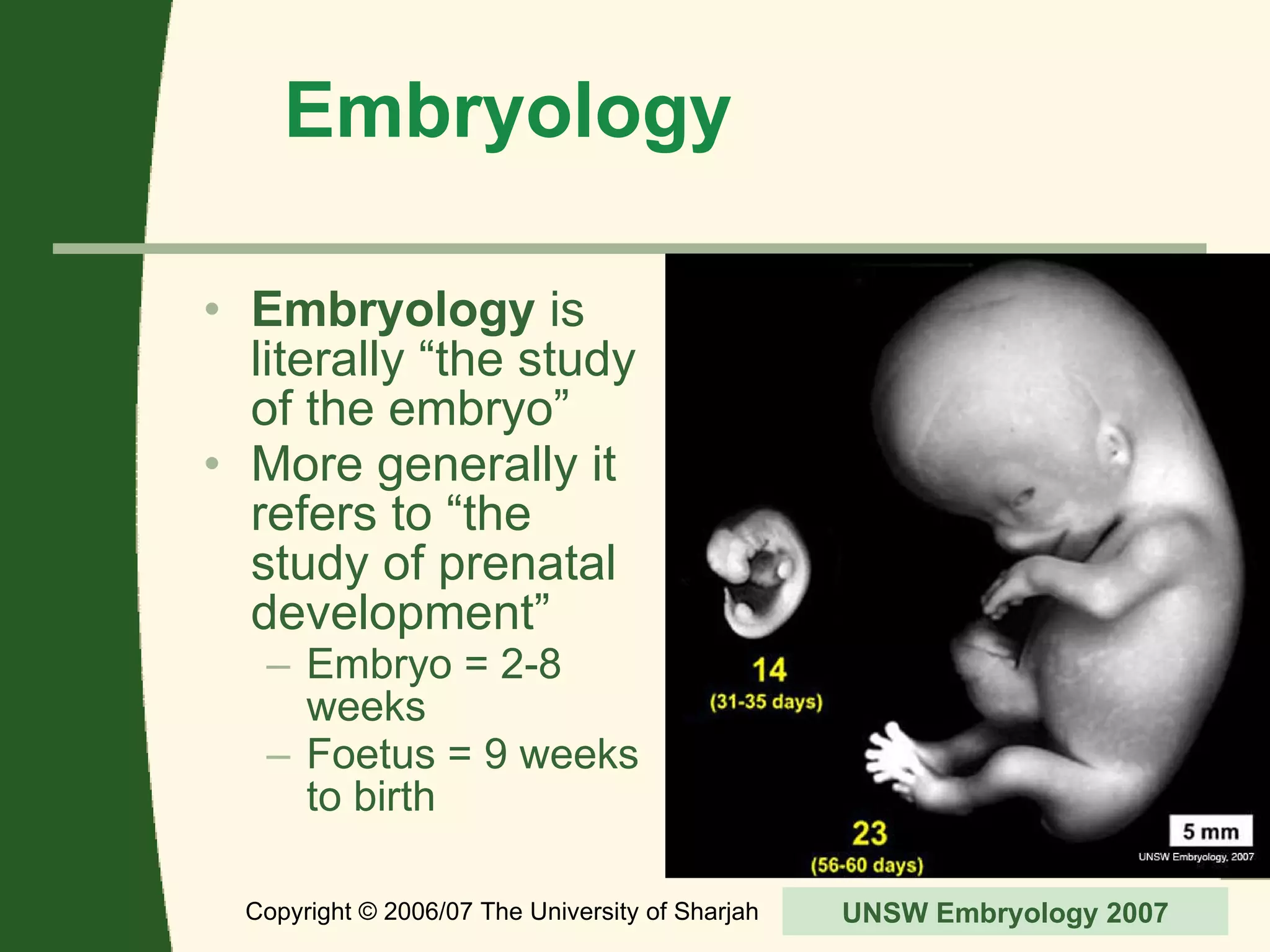


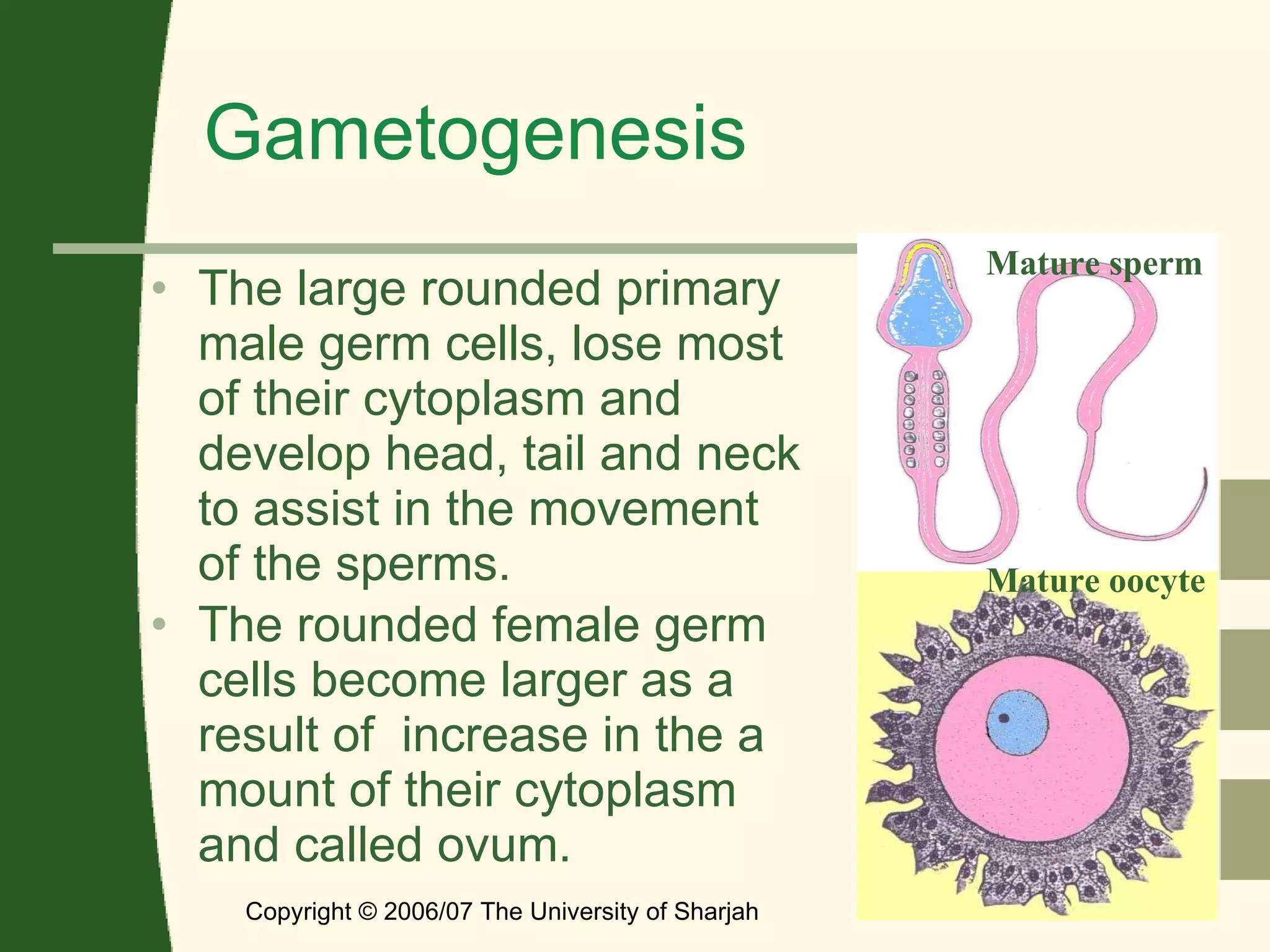
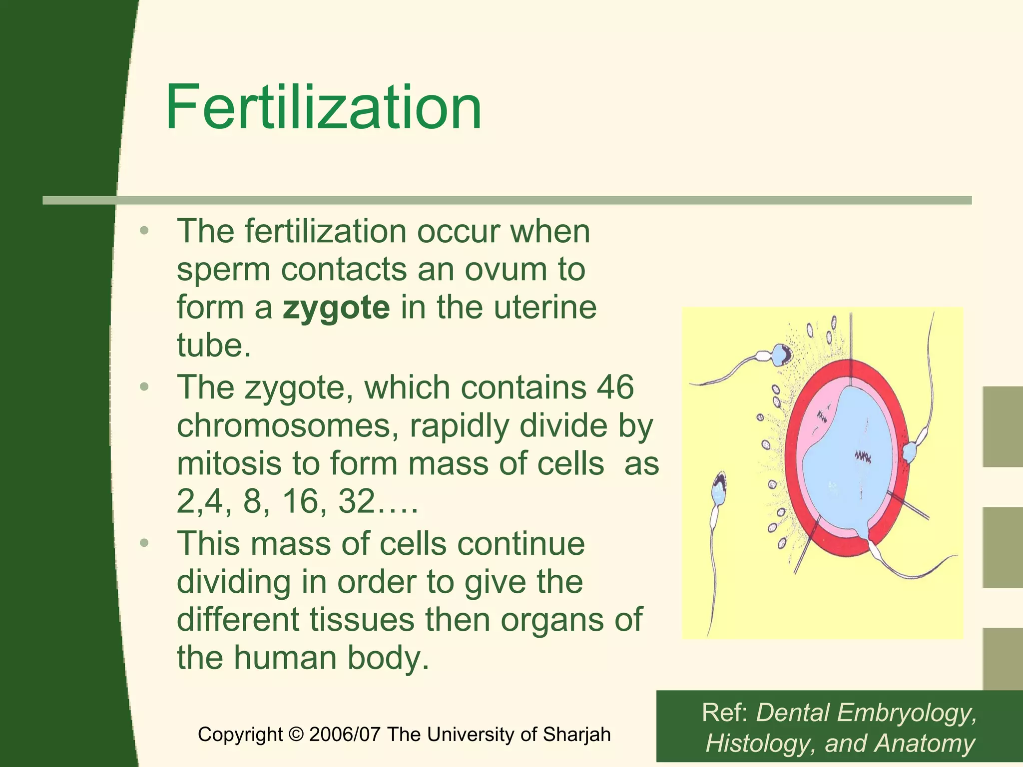


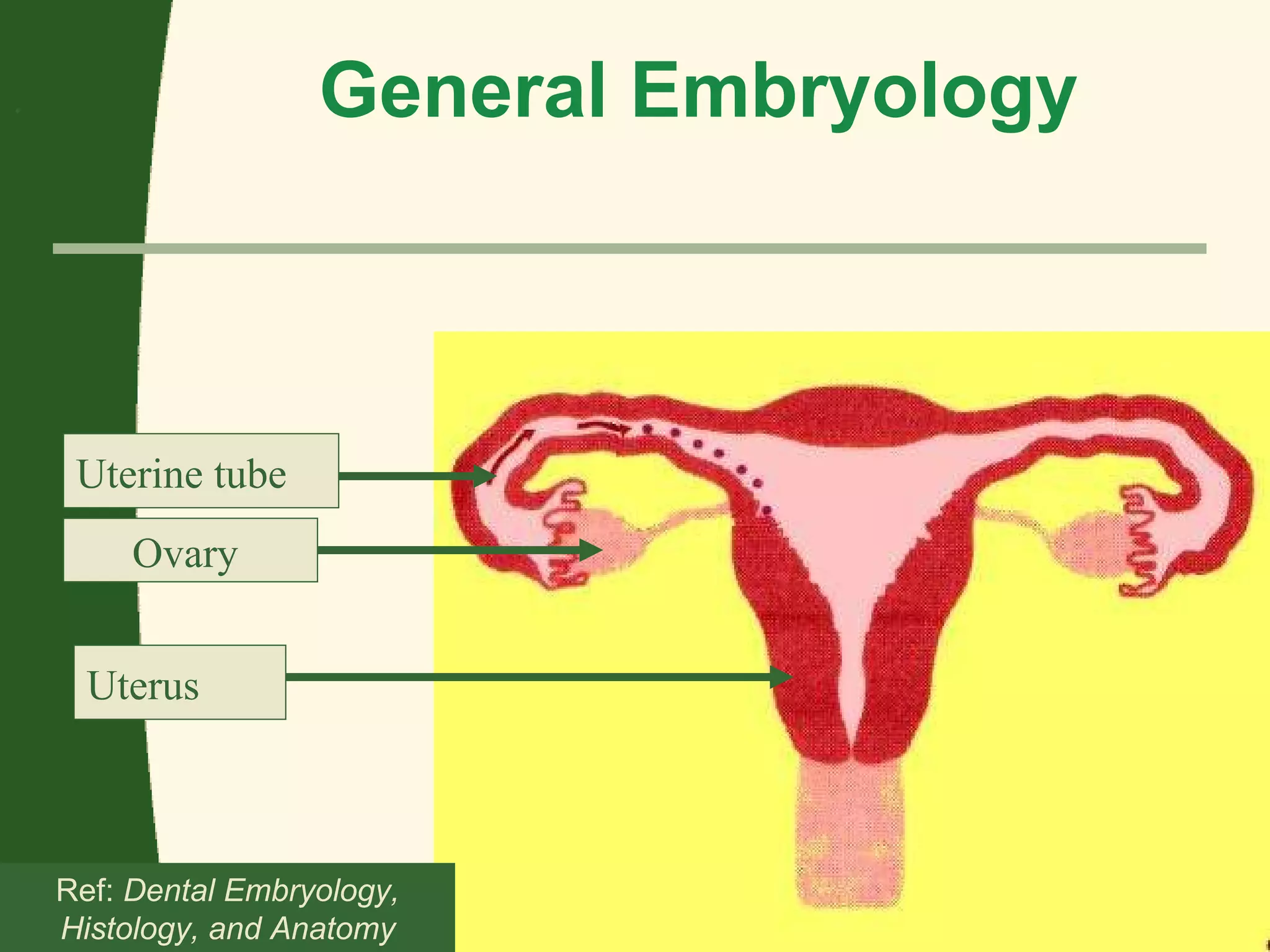


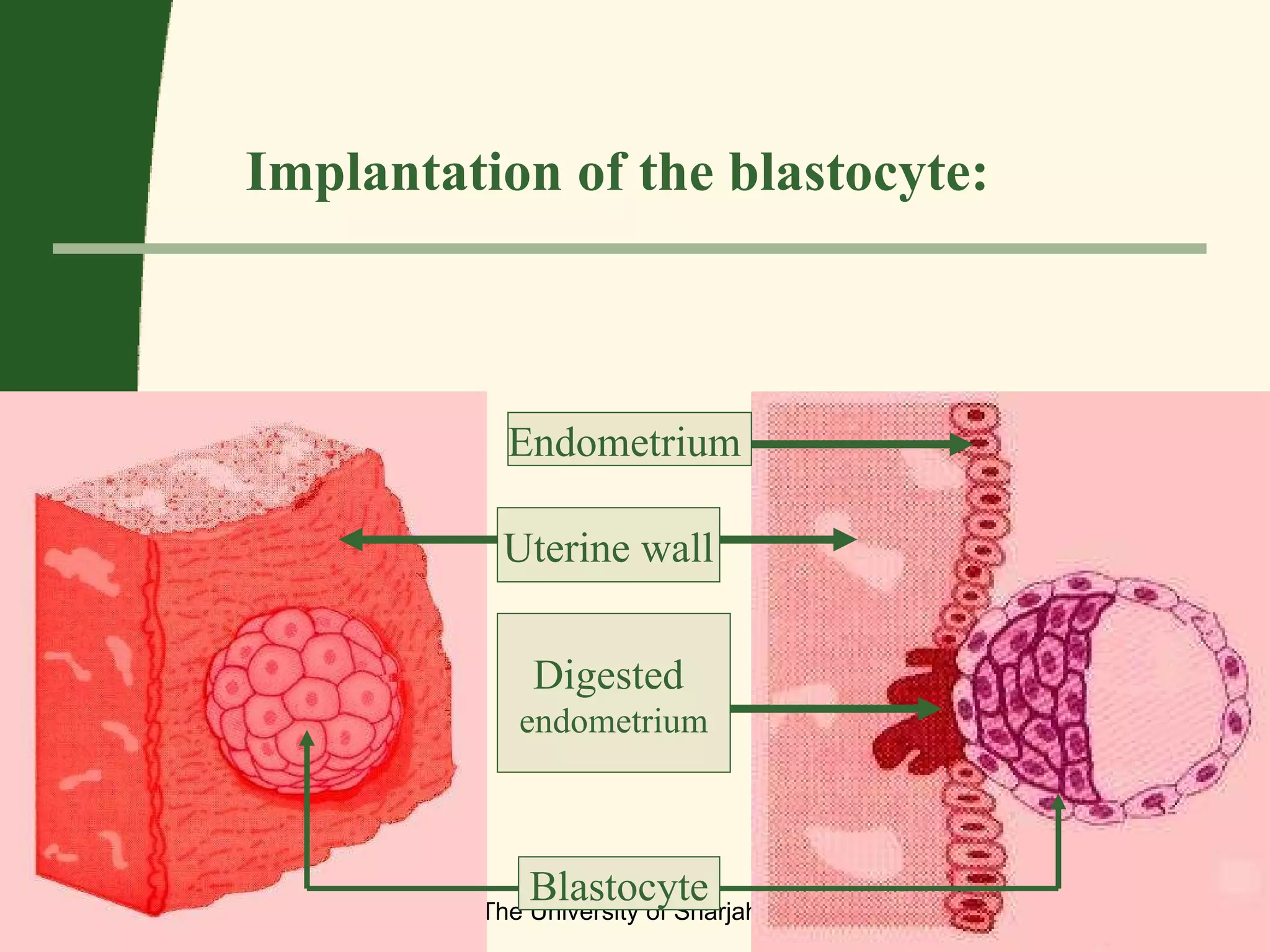

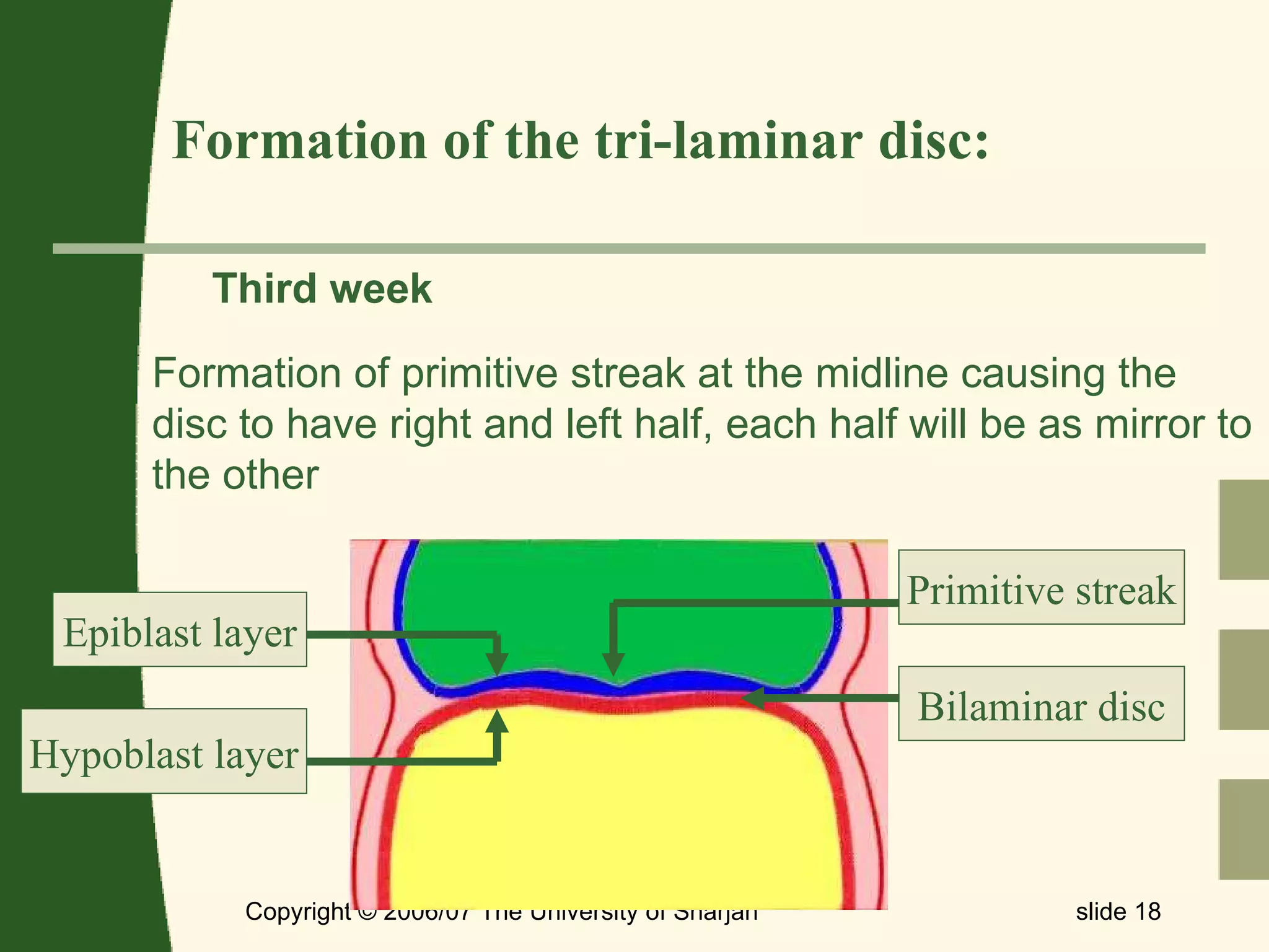


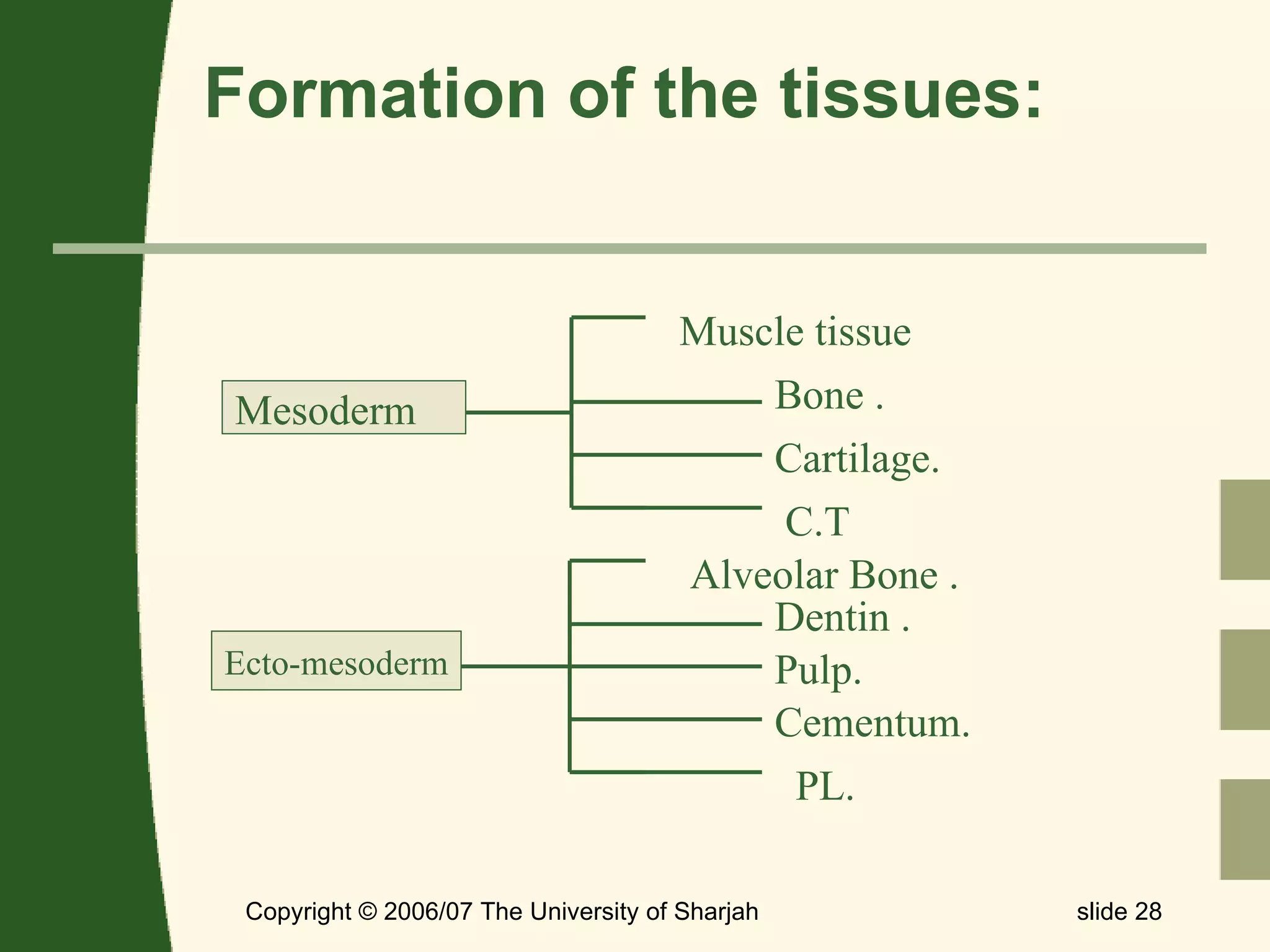


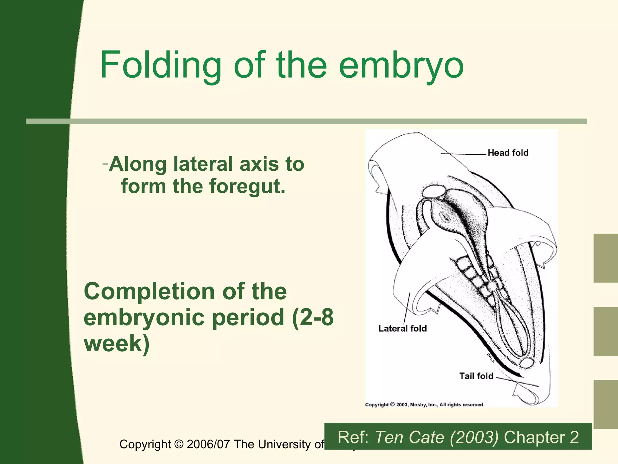


1. The document provides a general overview of human embryological processes including germ cell formation and fertilization, prenatal development through the formation of three layers, induction and differentiation, formation of the neural crest, and folding of the embryo. 2. Key stages discussed include gametogenesis, fertilization of the ovum by sperm to form a zygote, formation of the morula and blastocyst, implantation in the uterus, formation of the bilaminar and trilaminar germ discs, and development of the three germ layers and tissues from each layer. 3. Neural crest formation involves the development of the neural plate and tube from ectoderm and the migration of neural crest cells to


![Coded Agents – with UiPath SDK + LangGraph [Virtual Hands-on Workshop]](https://cdn.slidesharecdn.com/ss_thumbnails/codedagentsdeck-251215155422-5497c599-thumbnail.jpg?width=640&height=640&fit=bounds)
