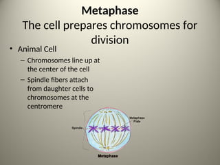
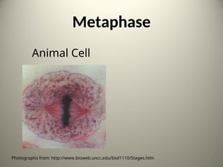
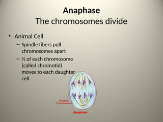
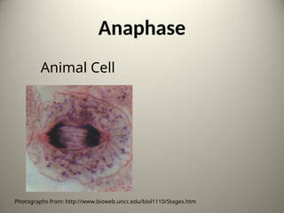
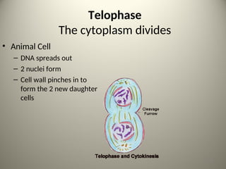
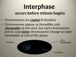
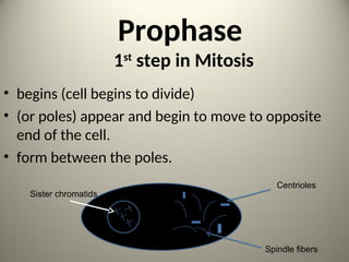
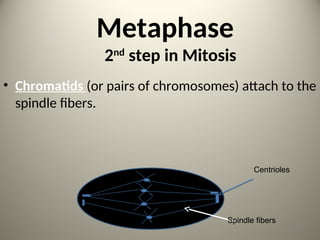
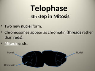
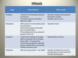
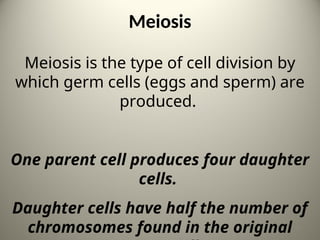
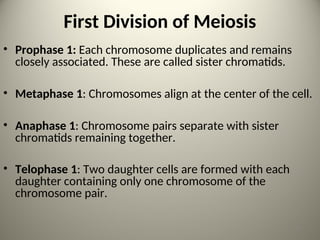
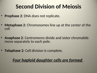
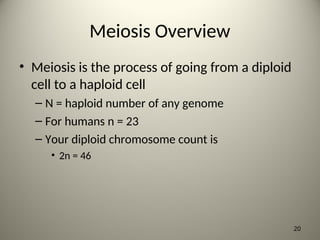
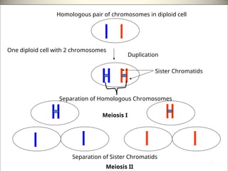
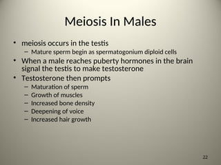
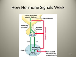
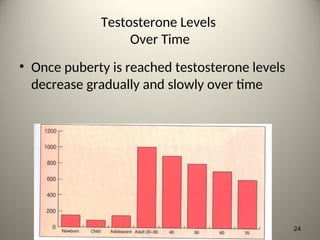
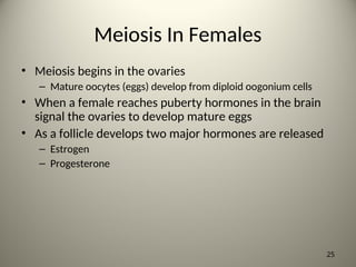
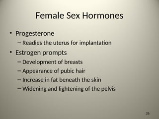
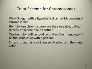
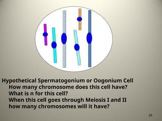
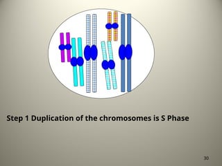
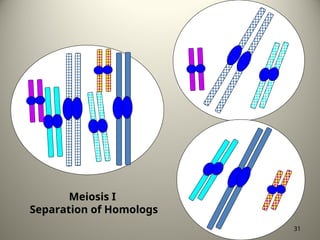
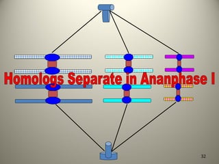
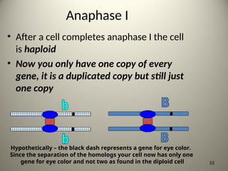
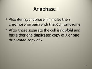
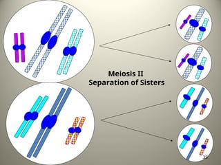
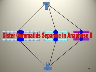
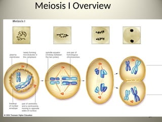
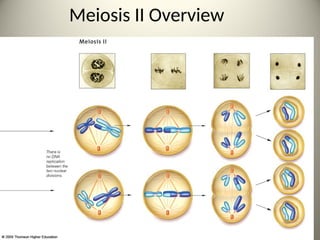
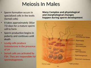
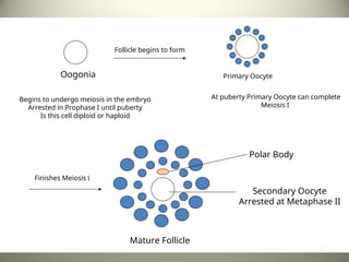
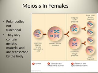
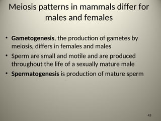
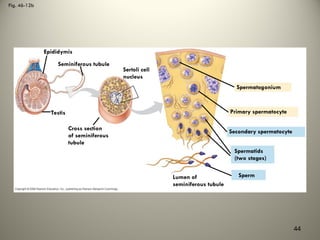
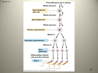
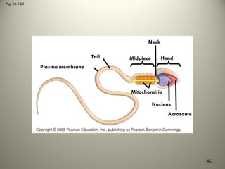
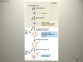
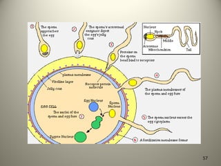
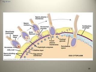
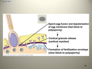
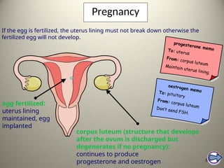
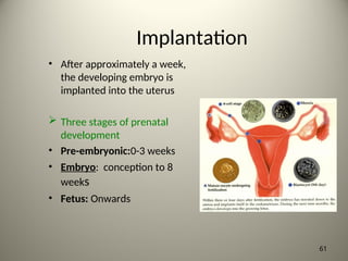
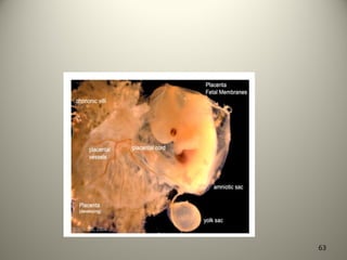
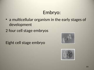
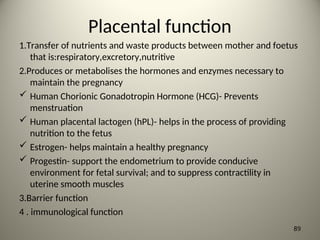
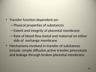
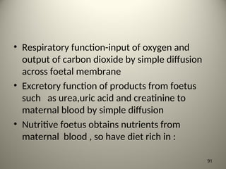
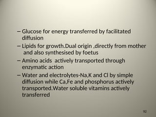
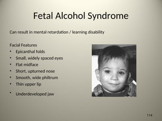
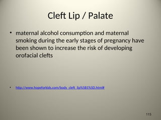
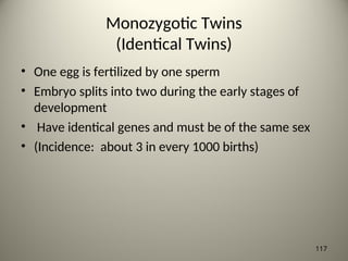
Fertilization-early Embryonic
Development
PRESENTED BY: SIAMANI BRIAN
(CURTESY: Prof. Bellington Vwalika
Consultant Obstetrician/Gynaecologist –UTH,
LUSAKA) 1
Mitosis
The process of cell division which
results in the production of two
daughter cells from a single parent
cell.
The daughter cells are identical to
one another and to the original
parent cell. 2
Mitosis can be divided into stages
4 Phases:-
Prophase
Metaphase
Anaphase
Telophase
3
Prophase
The cell prepares for nuclear division
• Animal Cell
– Packages DNA into
chromosomes
4
Metaphase
The cell prepares chromosomes for
division
• Animal Cell
– Chromosomes line up at
the center of the cell
– Spindle fibers attach
from daughter cells to
chromosomes at the
centromere
5
Metaphase
Animal Cell
Photographs from: http://www.bioweb.uncc.edu/biol1110/Stages.htm 6
Anaphase
The chromosomes divide
• Animal Cell
– Spindle fibers pull
chromosomes apart
– ½ of each chromosome
(called chromotid)
moves to each daughter
cell
7
Anaphase
Animal Cell
Photographs from: http://www.bioweb.uncc.edu/biol1110/Stages.htm 8
Telophase
The cytoplasm divides
• Animal Cell
– DNA spreads out
– 2 nuclei form
– Cell wall pinches in to
form the 2 new daughter
cells
9
Interphase
occurs before mitosis begins
• Chromosomes are copied (# doubles)
• Chromosomes appear as threadlike coils
(chromatin) at the start, but each chromosome
and its copy(sister chromosome) change to sister
chromatids at end of this phase
CELL
MEMBRANE
Nucleus
Cytoplasm
10
Prophase
1st
step in Mitosis
• begins (cell begins to divide)
• (or poles) appear and begin to move to opposite
end of the cell.
• form between the poles.
Centrioles
Sister chromatids
Spindle fibers 11
Metaphase
2nd
step in Mitosis
• Chromatids (or pairs of chromosomes) attach to the
spindle fibers.
Centrioles
Spindle fibers
12
Telophase
4th step in Mitosis
• Two new nuclei form.
• Chromosomes appear as chromatin (threads rather
than rods).
• Mitosis ends.
Nuclei
Nuclei
Chromatin
13
Mitosis
Stage Chromosomes Other events
Prophase •Consist of 2 chromatids joined by
centromere
•Condense (shorter and fatter)
•Become visible
•Nuclear envelope disintergrates
•Centrioles at poles
•Spindle starts to form
Metaphase •Still consist of 2 chromatids joined
by centromere
•Centromeres attach chromatid
pairs to spindle fibres
•Chromatid pairs line up along the
equator of the cell
•Spindle formed
Anaphase •Centromere holding chromatids
splits to separate the chromatids
(when split = chromosomes)
•Chromosomes pulled to opposite
poles by spindle fibres contracting
•Pulled centromere first
•Spindle fibres contract
Telophase •Become long and thin = not visible •Nuclear envelop forms around
chromosomes at each end of cell
•Spindle disintergrates
14
Meiosis
Meiosis is the type of cell division by
which germ cells (eggs and sperm) are
produced.
One parent cell produces four daughter
cells.
Daughter cells have half the number of
chromosomes found in the original 15
Meiosis
During meiosis, DNA replicates
once, but the nucleus divides
twice.
16
Meiosis
Four stages can be described for
each division of the nucleus.
17
First Division of Meiosis
• Prophase 1: Each chromosome duplicates and remains
closely associated. These are called sister chromatids.
• Metaphase 1: Chromosomes align at the center of the cell.
• Anaphase 1: Chromosome pairs separate with sister
chromatids remaining together.
• Telophase 1: Two daughter cells are formed with each
daughter containing only one chromosome of the
chromosome pair.
18
Second Division of Meiosis
• Prophase 2: DNA does not replicate.
• Metaphase 2: Chromosomes line up at the center of the
cell
• Anaphase 2: Centromeres divide and sister chromatids
move separately to each pole.
• Telophase 2: Cell division is complete.
Four haploid daughter cells are formed.
19
Meiosis Overview
• Meiosis is the process of going from a diploid
cell to a haploid cell
– N = haploid number of any genome
– For humans n = 23
– Your diploid chromosome count is
• 2n = 46
20
Homologous pair of chromosomes in diploid cell
Duplication
Sister Chromatids
Separation of Homologous Chromosomes
Meiosis I
Separation of Sister Chromatids
Meiosis II
One diploid cell with 2 chromosomes
21
Meiosis In Males
• meiosis occurs in the testis
– Mature sperm begin as spermatogonium diploid cells
• When a male reaches puberty hormones in the brain
signal the testis to make testosterone
• Testosterone then prompts
– Maturation of sperm
– Growth of muscles
– Increased bone density
– Deepening of voice
– Increased hair growth
22
How Hormone Signals Work
23
Testosterone Levels
Over Time
• Once puberty is reached testosterone levels
decrease gradually and slowly over time
24
Meiosis In Females
• Meiosis begins in the ovaries
– Mature oocytes (eggs) develop from diploid oogonium cells
• When a female reaches puberty hormones in the brain
signal the ovaries to develop mature eggs
• As a follicle develops two major hormones are released
– Estrogen
– Progesterone
25
Female Sex Hormones
• Progesterone
– Readies the uterus for implantation
• Estrogen prompts
– Development of breasts
– Appearance of pubic hair
– Increase in fat beneath the skin
– Widening and lightening of the pelvis
26
Part II
Mechanisms of Meiosis
27
Color Scheme for Chromosomes
• We will begin with a hypothetical cell which contains 6
chromosomes
• Homologous chromosomes are the same size, but not
directly attached to one another
• One homolog will be solid color the other homolog will
be the same color with a pattern
• Sister chromatids are of course attached and the same
color
28
Hypothetical Spermatogonium or Oogonium Cell
How many chromosome does this cell have?
What is n for this cell?
When this cell goes through Meiosis I and II
how many chromosomes will it have?
29
Step 1 Duplication of the chromosomes is S Phase
30
Meiosis I
Separation of Homologs
31
32
Anaphase I
• After a cell completes anaphase I the cell
is haploid
• Now you only have one copy of every
gene, it is a duplicated copy but still just
one copy
33
Hypothetically – the black dash represents a gene for eye color.
Since the separation of the homologs your cell now has only one
gene for eye color and not two as found in the diploid cell
Anaphase I
• Also during anaphase I in males the Y
chromosome pairs with the X chromosome
• After these separate the cell is haploid and
has either one duplicated copy of X or one
duplicated copy of Y
34
Meiosis II
Separation of Sisters
35
36
Meiosis I Overview
37
Meiosis II Overview
38
Meiosis In Males
• Sperm formation occurs in
specialized cells in the testis
(Sertoli cells)
• It takes approximately 30(or
72) days for a mature sperm
cell to form
• Sperm production begins in
puberty and continues until
death
• Leydig cells produce
testosterone in the presence
of LH
• Sertoli cells are activated by
FSH. They are responsible for
spermatogenesis.
39
Many Complex and physiological
and morphological changes
happen during sperm development
Meiosis in Females
• Females produce eggs from puberty until
menopause
• Female oogonia undergo meiosis but do not
complete meiosis II unless the egg becomes
fertilized
– Arrested in metaphase II
40
Begins to undergo meiosis in the embryo
Arrested in Prophase I until puberty
Is this cell diploid or haploid
Oogonia
Follicle begins to form
Primary Oocyte
Finishes Meiosis I
At puberty Primary Oocyte can complete
Meiosis I
Polar Body
Secondary Oocyte
Arrested at Metaphase II
Mature Follicle
41
Meiosis In Females
• Polar bodies are
not
functional
• They only
contain
genetic
material and
are reabsorbed
by the body
42
Meiosis patterns in mammals differ for
males and females
• Gametogenesis, the production of gametes by
meiosis, differs in females and males
• Sperm are small and motile and are produced
throughout the life of a sexually mature male
• Spermatogenesis is production of mature sperm
43
Fig. 46-12b
Epididymis
Seminiferous tubule
Sertoli cell
nucleus
Testis
Cross section
of seminiferous
tubule
Spermatogonium
Primary spermatocyte
Secondary spermatocyte
Spermatids
(two stages)
Sperm
Lumen of
seminiferous tubule
44
Fig. 46-12c
Primordial germ cell in embryo
Mitotic divisions
Spermatogonial
stem cell
Mitotic divisions
Spermatogonium
Mitotic divisions
Primary spermatocyte
Meiosis I
Secondary spermatocyte
Meiosis II
Early
spermatid
Differentiation (Sertoli
cells provide nutrients)
Sperm
2n
2n
2n
n n
n n n n
n n n n
45
Fig. 46-12d
46
• Eggs contain stored nutrients and are much larger
• Oogenesis is development of mature oocytes
(eggs) and can take many years
47
Fig. 46-12g
Primordial germ cell
Mitotic divisions
Oogonium
Mitotic divisions
Primary oocyte
(present at birth), arrested
in prophase of meiosis I
Completion of meiosis I
and onset of meiosis II
Secondary oocyte,
arrested at metaphase of
meiosis II
First
polar
body
Ovulation, sperm entry
Completion of meiosis II
Second
polar
body
Fertilized egg
2n
2n
n
n
n
n
In embryo
48
• Spermatogenesis differs from oogenesis:
– In oogenesis, one egg forms from each cycle of
meiosis; in spermatogenesis four sperm form from
each cycle of meiosis
– Oogenesis ceases later in life in females;
spermatogenesis continues throughout the adult life
of males
– Oogenesis has long interruptions; spermatogenesis
produces sperm from precursor cells in a continuous
sequence
49
The interplay of tropic and sex
hormones regulates mammalian
reproduction
• Human reproduction is coordinated by hormones
from the hypothalamus, anterior pituitary, and
gonads
• Gonadotropin-releasing hormone (GnRH) is
secreted by the hypothalamus and directs the
release of FSH and LH from the anterior pituitary
• FSH and LH regulate processes in the gonads and
the production of sex hormones
50
• The sex hormones are androgens, estrogens, and
progesterone
• Sex hormones regulate:
– The development of primary sex characteristics
during embryogenesis
– The development of secondary sex characteristics at
puberty
– Sexual behavior and sex drive
51
Fertilization
52
Fertilization:
the fusion of the sperm cell nucleus
with the egg cell nucleus to produce
a zygote (fertilized egg)
53
Fertilization:
• External
• Occurs outside of the body of the female
• Increased number of eggs produced to insure
the survival of the species
• Ex) fish and amphibians
54
Fertilization:
• Internal
• Occurs inside the body of the female
• Fewer number of eggs are produced
• Increased parental care insures species
survival
• Ex) mammals, reptiles, birds
55
Fertilization:
• fertilization in
mammals occurs
in the oviduct
• The ova is viable
for
approximately
24 hours after
ovulation
56
57
Fig. 47-3-5
Basal body
(centriole)
Sperm
head
Sperm-binding
receptors
Acrosome
Jelly coat Vitelline layer
Egg plasma
membrane
Hydrolytic enzymes
Acrosomal
process
Actin
filament
Sperm
nucleus
Sperm plasma
membrane
Fused
plasma
membranes
Fertilization
envelope
Cortical
granule
Perivitelline
space
EGG CYTOPLASM
58
Fig. 47-UN1
Sperm-egg fusion and depolarization
of egg membrane (fast block to
polyspermy)
Cortical granule release
(cortical reaction)
Formation of fertilization envelope
(slow block to polyspermy)
59
Pregnancy
60
If the egg is fertilized, the uterus lining must not break down otherwise the
fertilized egg will not develop.
corpus luteum (structure that develops
after the ovum is discharged but
degenerates if no pregnancy):
continues to produce
progesterone and oestrogen
egg fertilized:
uterus lining
maintained, egg
implanted
progesterone memo
To: uterus
From: corpus luteum
Maintain uterus lining.
oestrogen memo
To: pituitary
From: corpus luteum
Don’t send FSH.
Implantation
• After approximately a week,
the developing embryo is
implanted into the uterus
 Three stages of prenatal
development
• Pre-embryonic:0-3 weeks
• Embryo: conception to 8
weeks
• Fetus: Onwards
61
Embryonic Development
62
63
Embryo:
• a multicellular organism in the early stages of
development
2 four cell stage embryos
Eight cell stage embryo
64
Fig. 47-6
(a) Fertilized egg (b) Four-cell stage (c) Early blastula (d) Later blastula
65
Embryo:
The beginning developmental processes are
always the same in all animals:
1) cleavage
2) growth
3) differentiation
66
Embryo:
• after fertilization
the diploid
ZYGOTE
undergoes
cleavage divisions
in the oviduct
67
Cleavage
the first series of cell divisions by mitosis
after fertilization
Cell division is rapid, new cells do not take
time for the growth phase G1
cell growth does not occur so cells
decrease in size with each cleavage
division
68
Cleavage divisions
69
• Morula forms (solid ball of cells)
• Blastula forms (hollow ball of cells)
• Cells begin to grow before dividing
70
71
Differentation
• Gastrulation: one side
of the blastula
invaginates (indents)
forming a gastrula
• Three cell layers form
72
Differentiation
73
Fig. 47-16-5
Yolk sac
Mesoderm
Amnion
Chorion
Ectoderm
Extraembryonic
mesoderm
Trophoblast
Endoderm
Hypoblast
Expanding
region of
trophoblast
Epiblast
Maternal
blood
vessel
Allantois
Trophoblast
Hypoblast
Endometrial
epithelium
(uterine lining)
Inner cell mass
Blastocoel
Uterus
Epiblast
Amniotic
cavity
Expanding
region of
trophoblast
Yolk sac (from
hypoblast)
Chorion (from
trophoblast)
Extraembryonic
mesoderm cells
(from epiblast)
74
Differentiation
• The changing of unspecialized embryonic cells
into the specialized cells, tissues and organs of
a multicellular animal
75
Germ Layers
• Ectoderm Outer layer
• Nervous system including brain, spinal
cord and nerves
• Lining of the mouth, nostrils, and anus
• Epidermis of skin, sweat glands, hair, nails
76
Germ Layers
• Mesoderm Middle Layer
• Bones and muscles
• Blood and blood vessels
• Reproductive and excretory systems
• Inner layer (dermis) of skin
77
Germ Layers
• Endoderm Inner Layer
• Lining of digestive tract
• Lining of trachea, bronchi, and lungs
• Liver, pancreas
• Thyroid, parathyroid, thymus, urinary bladder
78
Placenta
• organ that
forms from the
embryo and
the uterus
79
Placenta
• contains blood vessels from the
mother and the developing baby
80
placenta
• Fully formed at 12 weeks
– 1st
wave of trophoblastic invasion at 12 weeks
– Second phase at 16 weeks
81
Placenta
• Oxygen & nutrients diffuse from the
mother’s blood vessels into the baby’s
blood vessels
• Wastes diffuse from the baby’s blood
vessels into the mother’s blood vessels
82
The placenta and fetal membranes
• Human placenta described as haemochorial
because of direct contact of chorion with
maternal blood
• It is deciduate because some maternal tissue
is shed at parturition
• Attached to uterine wall and link of mother
and fetus via umbilical cord
83
• Develops from 2 sources
• Principal component is foetal which develops from
chorion frondosum and maternal component consist
of decidual basalis
• Process begins at 6th
week and is completed by the
12th
week
• Until endof the 16th
week ,the placenta grows both in
thickness and circumference due to growth of
chorionic villi with accompanying expansion of
intervillous space
84
Placenta at term
• Gross anatomy
– Circular disc 15-20 diameter 2.5cm thick at center
– Weighs 500gm ,proportion to weight of fetus of
1:6
– Has fetal and materna surfaces
– Fetal surface
• Covered by smooth amnion with cord attached near
centre
85
• Maternal surface
– Is rough and spongy
– Mapped out into 15-20 lobes or cotyledons
• From chorionic plate arise the stem villi and extend
to the basal plate.These are the functional units of
the placenta
• Each villi is made up of outer syncytiotrophoblast(for
transfer),cytotrophoblast(for synthesis),central
stroma containing fetal capillaries etc and basement
membrane
86
Placental circulation
• 2-uteroplacental and foeto-placental
circulation
• Utero-placental concerned with circulaton of
maternal blood through intervillous space-
replaced every 3 to 4 times per minute
– Villi depend on maternal blood for nutrition ,thus
it is possible for chorionic villi to survive for a
varying period even after the foetus is dead
87
– 120-200 spiral arteries open into intervillous
space by piercing the basal plate
– There is cytotrophoblastic invasion into spiral
arteries upto the intramyometrial segment and is
completed by 16 weeks
• The umbilical cord has 2 arteries and one
vein.The arteries carry impure blood and veins
carry pure blood
88
Placental function
1.Transfer of nutrients and waste products between mother and foetus
that is:respiratory,excretory,nutritive
2.Produces or metabolises the hormones and enzymes necessary to
maintain the pregnancy
 Human Chorionic Gonadotropin Hormone (HCG)- Prevents
menstruation
 Human placental lactogen (hPL)- helps in the process of providing
nutrition to the fetus
 Estrogen- helps maintain a healthy pregnancy
 Progestin- support the endometrium to provide conducive
environment for fetal survival; and to suppress contractility in
uterine smooth muscles
3.Barrier function
4 . immunological function
89
• Transfer function dependent on:
– Physical properties of substances
– Extent and integrity of placental membrane
– Rate of blood flow,foetal and maternal on either
side of exchange membrane
• Mechanisms involved in transfer of substances
include: simple diffusion,active transfer,pinocytosis
and leakage through broken placental membrane
90
• Respiratory function-input of oxygen and
output of carbon dioxide by simple diffusion
across foetal membrane
• Excretory function of products from foetus
such as urea,uric acid and creatinine to
maternal blood by simple diffusion
• Nutritive foetus obtains nutrients from
maternal blood , so have diet rich in :
91
– Glucose for energy transferred by facilitated
diffusion
– Lipids for growth.Dual origin ,directly from mother
and also synthesised by foetus
– Amino acids actively transported through
enzymatic action
– Water and electrolytes-Na,K and Cl by simple
diffusion while Ca,Fe and phosphorus actively
transported.Water soluble vitamins actively
transferred
92
• Enzymatic function-numerous enzymes
elaborated in the placenta such as
oxytocinase and phospholipase A2
• Barrier function – fetal membrane protects
fetus against noxious agents .In general
substances of high molecular weight of more
than 500 daltons are held up but there are
exceptions
93
The foetal membranes
• Consist of 2 layers –outer chorion and inner
amnion
• Chorion ends at edge of placenta-has no
vessels or nerves
• Amnion inner and in contact with liqour amnii
– Functions
• Contribute to the formation of liquor amnii
• Intact membranes prevent ascending uterine infection
94
• Facilitate dilatation of the cervix during labour
• Has got enzymatic activity for steroid
hormone metabolism
• Rich source of glycerophospholipids
containing arachidonic acid –precursor of
prostaglandin E2 and F2alpha
95
Amniotic fluid
• Origin- precise origin is unknown.Theories include:
– As a transudate from maternal serum across
foetal membranes or from maternal circulation in
the placenta
– As a transudate across the umbilical cord or from
foetal circulation in the placenta or secretion from
amniotic epithelium
– Contribution from foetal urine-the fetus drinks
about 400ml of liqour everyday at term and
passes equal amount in urine
96
– Secretion from tracheobronchial tree and across
the foetal skin before the skin becomes keratinised
at the 20th
week
• Measures 30ml at 10 weeks and 300ml at 20
weeks reaching max of 1 litre at 36-38 weeks
• Physical features
– Colourless but near term becomes pale straw
coloured due lanugo and fetal squames.May be
turbid due to vernix caseosa
97
• Abnormal colour
– Meconium stained (green) due to foetal distress
– Golden colour in RH incompatibility due to
excessive hemolysis of feta RBC and hence
haemoglobin formation
– Greenish yellow in post maturity
– Dark coloured in concealed accidental
hemorrhage due to contamination of blood
– Dark brown(tobacco juice) amniotic fluid is found
in IUFD.Dark colour is due to frequent presence of
old HbA
98
• Functions
– Acts as shock absorber ,protecting the fetus from
possible extragenous injury
– Maintains an even pressure
– Distends amniotic sac and thereby allows for growth
and free movement of the fetus and prevents
adhesion between the foetal parts and amniotic sac
– Its nutritive value is negligible because of small
amount of protein and salt,however water supply to
foetus is quite adequate
99
• During labour
– Amnion and chorion are combined to form a
hydrostatic wedge which helps in dilatation of
the cervix
– During contration prevents marked interference
with placental circulation so long membranes
remain intact
– Flushes birth canal after first stage of labour and
by its aseptic and bactericidal action protects the
foetus and prevents ascending infection
100
• Clinical importance
– Study of it provides useful information about
status of foetus
– Intra amniotic instillation of chemicals used as a
method for induction of abortion
– Excess or less gives rise to hydramnios and
oligohydramnios
– ARM with drainage is helpful in induction of
labour
101
Umbilical Cord
• two arteries and a
vein Connects the
fetus to the
placenta
102
Amniotic Sac
• Contains fluid
(amniotic fluid) that
protects fetus by
giving it a stable
environment and
absorbing shock
103
• By the end of the 8th
week of pregnancy
the embryo is called a
fetus and all of the
major structures are
present
104
Later Stages of Fetal Development
105
• After blastocyst formation, the embryo implants
into the endometrium
• The embryo releases human chorionic
gonadotropin (hCG), which prevents menstruation
• Pregnancy, or gestation, is the condition of
carrying one or more embryos in the uterus
• Duration of pregnancy in other species correlates
with body size and maturity of the young at birth
106
First Trimester
Human gestation can be divided into three trimesters of
about three months each
The first trimester is the time of most radical change for
both the mother and the embryo
During implantation, the endometrium grows over the
blastocyst
During its first 2 to 4 weeks, the embryo obtains nutrients
directly from the endometrium
Meanwhile, the outer layer of the blastocyst, called the
trophoblast, mingles with the endometrium and
eventually forms the placenta
Blood from the embryo travels to the placenta through
arteries of the umbilical cord and returns via the umbilical
vein 107
Fig. 46-16
Placenta
Uterus
Umbilical cord
Chorionic villus,
containing fetal
capillaries
Maternal blood
pools
Maternal
arteries
Maternal
veins
Maternal
portion
of placenta
Fetal arteriole
Fetal venule
Umbilical cord
Fetal
portion of
placenta
(chorion)
Umbilical
arteries
Umbilical
vein
108
Splitting of the embryo during the first month of
development results in genetically identical twins
Release and fertilization of two eggs results in fraternal
and genetically distinct twins
The first trimester is the main period of organogenesis,
development of the body organs
All the major structures are present by 8 weeks, and
the embryo is called a fetus
• Changes occur in the mother
Growth of the placenta
Cessation of ovulation and the menstrual cycle
Breast enlargement
Nausea is also very common
109
Human gestation
• the period
between
fertilization and
birth
• approximately 38-
40 weeks
110
Teratogens
• Substances that may harm the
developing fetus and result in the
formation of birth defects
– Pre-embryonic (0-3 weeks) phase follow ‘all or
none principle’
– Embryonic phase(4-8weeks)- fetus is affected
– Fetal stage >8 weeks till delivery
111
Teratogens include:
• Alcohol, certain drugs/medications,
infections, and certain chemicals
112
113
Fetal Alcohol Syndrome
Can result in mental retardation / learning disability
Facial Features
• Epicanthal folds
• Small, widely spaced eyes
• Flat midface
• Short, upturned nose
• Smooth, wide philtrum
• Thin upper lip
• Underdeveloped jaw
114
Cleft Lip / Palate
• maternal alcohol consumption and maternal
smoking during the early stages of pregnancy have
been shown to increase the risk of developing
orofacial clefts
• http://www.hopeforkids.com/body_cleft_lip%5B1%5D.html#
115
How do twins form???
116
Monozygotic Twins
(Identical Twins)
• One egg is fertilized by one sperm
• Embryo splits into two during the early stages of
development
• Have identical genes and must be of the same sex
• (Incidence: about 3 in every 1000 births)
117
118
Dizygotic Twins
(Fraternal Twins)
• Two eggs are ovulated and each is fertilized by a
sperm cell
• No more genetically similar than any other sibling in
the family (can be same/different sexes)
• Maternal age, use of assisted reproductive
technologies are factors
• Incidence (6.7/1000 births in Japan to 40/1000 births
in Nigeria)
119
Thank you for
your attention !
120
Any questions?
121

5. Fertilization-Early Embryo Development.ppt

  • 1.
    Fertilization-early Embryonic Development PRESENTED BY:SIAMANI BRIAN (CURTESY: Prof. Bellington Vwalika Consultant Obstetrician/Gynaecologist –UTH, LUSAKA) 1
  • 2.
    Mitosis The process ofcell division which results in the production of two daughter cells from a single parent cell. The daughter cells are identical to one another and to the original parent cell. 2
  • 3.
    Mitosis can bedivided into stages 4 Phases:- Prophase Metaphase Anaphase Telophase 3
  • 4.
    Prophase The cell preparesfor nuclear division • Animal Cell – Packages DNA into chromosomes 4
  • 5.
    Metaphase The cell prepareschromosomes for division • Animal Cell – Chromosomes line up at the center of the cell – Spindle fibers attach from daughter cells to chromosomes at the centromere 5
  • 6.
    Metaphase Animal Cell Photographs from:http://www.bioweb.uncc.edu/biol1110/Stages.htm 6
  • 7.
    Anaphase The chromosomes divide •Animal Cell – Spindle fibers pull chromosomes apart – ½ of each chromosome (called chromotid) moves to each daughter cell 7
  • 8.
    Anaphase Animal Cell Photographs from:http://www.bioweb.uncc.edu/biol1110/Stages.htm 8
  • 9.
    Telophase The cytoplasm divides •Animal Cell – DNA spreads out – 2 nuclei form – Cell wall pinches in to form the 2 new daughter cells 9
  • 10.
    Interphase occurs before mitosisbegins • Chromosomes are copied (# doubles) • Chromosomes appear as threadlike coils (chromatin) at the start, but each chromosome and its copy(sister chromosome) change to sister chromatids at end of this phase CELL MEMBRANE Nucleus Cytoplasm 10
  • 11.
    Prophase 1st step in Mitosis •begins (cell begins to divide) • (or poles) appear and begin to move to opposite end of the cell. • form between the poles. Centrioles Sister chromatids Spindle fibers 11
  • 12.
    Metaphase 2nd step in Mitosis •Chromatids (or pairs of chromosomes) attach to the spindle fibers. Centrioles Spindle fibers 12
  • 13.
    Telophase 4th step inMitosis • Two new nuclei form. • Chromosomes appear as chromatin (threads rather than rods). • Mitosis ends. Nuclei Nuclei Chromatin 13
  • 14.
    Mitosis Stage Chromosomes Otherevents Prophase •Consist of 2 chromatids joined by centromere •Condense (shorter and fatter) •Become visible •Nuclear envelope disintergrates •Centrioles at poles •Spindle starts to form Metaphase •Still consist of 2 chromatids joined by centromere •Centromeres attach chromatid pairs to spindle fibres •Chromatid pairs line up along the equator of the cell •Spindle formed Anaphase •Centromere holding chromatids splits to separate the chromatids (when split = chromosomes) •Chromosomes pulled to opposite poles by spindle fibres contracting •Pulled centromere first •Spindle fibres contract Telophase •Become long and thin = not visible •Nuclear envelop forms around chromosomes at each end of cell •Spindle disintergrates 14
  • 15.
    Meiosis Meiosis is thetype of cell division by which germ cells (eggs and sperm) are produced. One parent cell produces four daughter cells. Daughter cells have half the number of chromosomes found in the original 15
  • 16.
    Meiosis During meiosis, DNAreplicates once, but the nucleus divides twice. 16
  • 17.
    Meiosis Four stages canbe described for each division of the nucleus. 17
  • 18.
    First Division ofMeiosis • Prophase 1: Each chromosome duplicates and remains closely associated. These are called sister chromatids. • Metaphase 1: Chromosomes align at the center of the cell. • Anaphase 1: Chromosome pairs separate with sister chromatids remaining together. • Telophase 1: Two daughter cells are formed with each daughter containing only one chromosome of the chromosome pair. 18
  • 19.
    Second Division ofMeiosis • Prophase 2: DNA does not replicate. • Metaphase 2: Chromosomes line up at the center of the cell • Anaphase 2: Centromeres divide and sister chromatids move separately to each pole. • Telophase 2: Cell division is complete. Four haploid daughter cells are formed. 19
  • 20.
    Meiosis Overview • Meiosisis the process of going from a diploid cell to a haploid cell – N = haploid number of any genome – For humans n = 23 – Your diploid chromosome count is • 2n = 46 20
  • 21.
    Homologous pair ofchromosomes in diploid cell Duplication Sister Chromatids Separation of Homologous Chromosomes Meiosis I Separation of Sister Chromatids Meiosis II One diploid cell with 2 chromosomes 21
  • 22.
    Meiosis In Males •meiosis occurs in the testis – Mature sperm begin as spermatogonium diploid cells • When a male reaches puberty hormones in the brain signal the testis to make testosterone • Testosterone then prompts – Maturation of sperm – Growth of muscles – Increased bone density – Deepening of voice – Increased hair growth 22
  • 23.
  • 24.
    Testosterone Levels Over Time •Once puberty is reached testosterone levels decrease gradually and slowly over time 24
  • 25.
    Meiosis In Females •Meiosis begins in the ovaries – Mature oocytes (eggs) develop from diploid oogonium cells • When a female reaches puberty hormones in the brain signal the ovaries to develop mature eggs • As a follicle develops two major hormones are released – Estrogen – Progesterone 25
  • 26.
    Female Sex Hormones •Progesterone – Readies the uterus for implantation • Estrogen prompts – Development of breasts – Appearance of pubic hair – Increase in fat beneath the skin – Widening and lightening of the pelvis 26
  • 27.
  • 28.
    Color Scheme forChromosomes • We will begin with a hypothetical cell which contains 6 chromosomes • Homologous chromosomes are the same size, but not directly attached to one another • One homolog will be solid color the other homolog will be the same color with a pattern • Sister chromatids are of course attached and the same color 28
  • 29.
    Hypothetical Spermatogonium orOogonium Cell How many chromosome does this cell have? What is n for this cell? When this cell goes through Meiosis I and II how many chromosomes will it have? 29
  • 30.
    Step 1 Duplicationof the chromosomes is S Phase 30
  • 31.
  • 32.
  • 33.
    Anaphase I • Aftera cell completes anaphase I the cell is haploid • Now you only have one copy of every gene, it is a duplicated copy but still just one copy 33 Hypothetically – the black dash represents a gene for eye color. Since the separation of the homologs your cell now has only one gene for eye color and not two as found in the diploid cell
  • 34.
    Anaphase I • Alsoduring anaphase I in males the Y chromosome pairs with the X chromosome • After these separate the cell is haploid and has either one duplicated copy of X or one duplicated copy of Y 34
  • 35.
  • 36.
  • 37.
  • 38.
  • 39.
    Meiosis In Males •Sperm formation occurs in specialized cells in the testis (Sertoli cells) • It takes approximately 30(or 72) days for a mature sperm cell to form • Sperm production begins in puberty and continues until death • Leydig cells produce testosterone in the presence of LH • Sertoli cells are activated by FSH. They are responsible for spermatogenesis. 39 Many Complex and physiological and morphological changes happen during sperm development
  • 40.
    Meiosis in Females •Females produce eggs from puberty until menopause • Female oogonia undergo meiosis but do not complete meiosis II unless the egg becomes fertilized – Arrested in metaphase II 40
  • 41.
    Begins to undergomeiosis in the embryo Arrested in Prophase I until puberty Is this cell diploid or haploid Oogonia Follicle begins to form Primary Oocyte Finishes Meiosis I At puberty Primary Oocyte can complete Meiosis I Polar Body Secondary Oocyte Arrested at Metaphase II Mature Follicle 41
  • 42.
    Meiosis In Females •Polar bodies are not functional • They only contain genetic material and are reabsorbed by the body 42
  • 43.
    Meiosis patterns inmammals differ for males and females • Gametogenesis, the production of gametes by meiosis, differs in females and males • Sperm are small and motile and are produced throughout the life of a sexually mature male • Spermatogenesis is production of mature sperm 43
  • 44.
    Fig. 46-12b Epididymis Seminiferous tubule Sertolicell nucleus Testis Cross section of seminiferous tubule Spermatogonium Primary spermatocyte Secondary spermatocyte Spermatids (two stages) Sperm Lumen of seminiferous tubule 44
  • 45.
    Fig. 46-12c Primordial germcell in embryo Mitotic divisions Spermatogonial stem cell Mitotic divisions Spermatogonium Mitotic divisions Primary spermatocyte Meiosis I Secondary spermatocyte Meiosis II Early spermatid Differentiation (Sertoli cells provide nutrients) Sperm 2n 2n 2n n n n n n n n n n n 45
  • 46.
  • 47.
    • Eggs containstored nutrients and are much larger • Oogenesis is development of mature oocytes (eggs) and can take many years 47
  • 48.
    Fig. 46-12g Primordial germcell Mitotic divisions Oogonium Mitotic divisions Primary oocyte (present at birth), arrested in prophase of meiosis I Completion of meiosis I and onset of meiosis II Secondary oocyte, arrested at metaphase of meiosis II First polar body Ovulation, sperm entry Completion of meiosis II Second polar body Fertilized egg 2n 2n n n n n In embryo 48
  • 49.
    • Spermatogenesis differsfrom oogenesis: – In oogenesis, one egg forms from each cycle of meiosis; in spermatogenesis four sperm form from each cycle of meiosis – Oogenesis ceases later in life in females; spermatogenesis continues throughout the adult life of males – Oogenesis has long interruptions; spermatogenesis produces sperm from precursor cells in a continuous sequence 49
  • 50.
    The interplay oftropic and sex hormones regulates mammalian reproduction • Human reproduction is coordinated by hormones from the hypothalamus, anterior pituitary, and gonads • Gonadotropin-releasing hormone (GnRH) is secreted by the hypothalamus and directs the release of FSH and LH from the anterior pituitary • FSH and LH regulate processes in the gonads and the production of sex hormones 50
  • 51.
    • The sexhormones are androgens, estrogens, and progesterone • Sex hormones regulate: – The development of primary sex characteristics during embryogenesis – The development of secondary sex characteristics at puberty – Sexual behavior and sex drive 51
  • 52.
  • 53.
    Fertilization: the fusion ofthe sperm cell nucleus with the egg cell nucleus to produce a zygote (fertilized egg) 53
  • 54.
    Fertilization: • External • Occursoutside of the body of the female • Increased number of eggs produced to insure the survival of the species • Ex) fish and amphibians 54
  • 55.
    Fertilization: • Internal • Occursinside the body of the female • Fewer number of eggs are produced • Increased parental care insures species survival • Ex) mammals, reptiles, birds 55
  • 56.
    Fertilization: • fertilization in mammalsoccurs in the oviduct • The ova is viable for approximately 24 hours after ovulation 56
  • 57.
  • 58.
    Fig. 47-3-5 Basal body (centriole) Sperm head Sperm-binding receptors Acrosome Jellycoat Vitelline layer Egg plasma membrane Hydrolytic enzymes Acrosomal process Actin filament Sperm nucleus Sperm plasma membrane Fused plasma membranes Fertilization envelope Cortical granule Perivitelline space EGG CYTOPLASM 58
  • 59.
    Fig. 47-UN1 Sperm-egg fusionand depolarization of egg membrane (fast block to polyspermy) Cortical granule release (cortical reaction) Formation of fertilization envelope (slow block to polyspermy) 59
  • 60.
    Pregnancy 60 If the eggis fertilized, the uterus lining must not break down otherwise the fertilized egg will not develop. corpus luteum (structure that develops after the ovum is discharged but degenerates if no pregnancy): continues to produce progesterone and oestrogen egg fertilized: uterus lining maintained, egg implanted progesterone memo To: uterus From: corpus luteum Maintain uterus lining. oestrogen memo To: pituitary From: corpus luteum Don’t send FSH.
  • 61.
    Implantation • After approximatelya week, the developing embryo is implanted into the uterus  Three stages of prenatal development • Pre-embryonic:0-3 weeks • Embryo: conception to 8 weeks • Fetus: Onwards 61
  • 62.
  • 63.
  • 64.
    Embryo: • a multicellularorganism in the early stages of development 2 four cell stage embryos Eight cell stage embryo 64
  • 65.
    Fig. 47-6 (a) Fertilizedegg (b) Four-cell stage (c) Early blastula (d) Later blastula 65
  • 66.
    Embryo: The beginning developmentalprocesses are always the same in all animals: 1) cleavage 2) growth 3) differentiation 66
  • 67.
    Embryo: • after fertilization thediploid ZYGOTE undergoes cleavage divisions in the oviduct 67
  • 68.
    Cleavage the first seriesof cell divisions by mitosis after fertilization Cell division is rapid, new cells do not take time for the growth phase G1 cell growth does not occur so cells decrease in size with each cleavage division 68
  • 69.
  • 70.
    • Morula forms(solid ball of cells) • Blastula forms (hollow ball of cells) • Cells begin to grow before dividing 70
  • 71.
  • 72.
    Differentation • Gastrulation: oneside of the blastula invaginates (indents) forming a gastrula • Three cell layers form 72
  • 73.
  • 74.
    Fig. 47-16-5 Yolk sac Mesoderm Amnion Chorion Ectoderm Extraembryonic mesoderm Trophoblast Endoderm Hypoblast Expanding regionof trophoblast Epiblast Maternal blood vessel Allantois Trophoblast Hypoblast Endometrial epithelium (uterine lining) Inner cell mass Blastocoel Uterus Epiblast Amniotic cavity Expanding region of trophoblast Yolk sac (from hypoblast) Chorion (from trophoblast) Extraembryonic mesoderm cells (from epiblast) 74
  • 75.
    Differentiation • The changingof unspecialized embryonic cells into the specialized cells, tissues and organs of a multicellular animal 75
  • 76.
    Germ Layers • EctodermOuter layer • Nervous system including brain, spinal cord and nerves • Lining of the mouth, nostrils, and anus • Epidermis of skin, sweat glands, hair, nails 76
  • 77.
    Germ Layers • MesodermMiddle Layer • Bones and muscles • Blood and blood vessels • Reproductive and excretory systems • Inner layer (dermis) of skin 77
  • 78.
    Germ Layers • EndodermInner Layer • Lining of digestive tract • Lining of trachea, bronchi, and lungs • Liver, pancreas • Thyroid, parathyroid, thymus, urinary bladder 78
  • 79.
    Placenta • organ that formsfrom the embryo and the uterus 79
  • 80.
    Placenta • contains bloodvessels from the mother and the developing baby 80
  • 81.
    placenta • Fully formedat 12 weeks – 1st wave of trophoblastic invasion at 12 weeks – Second phase at 16 weeks 81
  • 82.
    Placenta • Oxygen &nutrients diffuse from the mother’s blood vessels into the baby’s blood vessels • Wastes diffuse from the baby’s blood vessels into the mother’s blood vessels 82
  • 83.
    The placenta andfetal membranes • Human placenta described as haemochorial because of direct contact of chorion with maternal blood • It is deciduate because some maternal tissue is shed at parturition • Attached to uterine wall and link of mother and fetus via umbilical cord 83
  • 84.
    • Develops from2 sources • Principal component is foetal which develops from chorion frondosum and maternal component consist of decidual basalis • Process begins at 6th week and is completed by the 12th week • Until endof the 16th week ,the placenta grows both in thickness and circumference due to growth of chorionic villi with accompanying expansion of intervillous space 84
  • 85.
    Placenta at term •Gross anatomy – Circular disc 15-20 diameter 2.5cm thick at center – Weighs 500gm ,proportion to weight of fetus of 1:6 – Has fetal and materna surfaces – Fetal surface • Covered by smooth amnion with cord attached near centre 85
  • 86.
    • Maternal surface –Is rough and spongy – Mapped out into 15-20 lobes or cotyledons • From chorionic plate arise the stem villi and extend to the basal plate.These are the functional units of the placenta • Each villi is made up of outer syncytiotrophoblast(for transfer),cytotrophoblast(for synthesis),central stroma containing fetal capillaries etc and basement membrane 86
  • 87.
    Placental circulation • 2-uteroplacentaland foeto-placental circulation • Utero-placental concerned with circulaton of maternal blood through intervillous space- replaced every 3 to 4 times per minute – Villi depend on maternal blood for nutrition ,thus it is possible for chorionic villi to survive for a varying period even after the foetus is dead 87
  • 88.
    – 120-200 spiralarteries open into intervillous space by piercing the basal plate – There is cytotrophoblastic invasion into spiral arteries upto the intramyometrial segment and is completed by 16 weeks • The umbilical cord has 2 arteries and one vein.The arteries carry impure blood and veins carry pure blood 88
  • 89.
    Placental function 1.Transfer ofnutrients and waste products between mother and foetus that is:respiratory,excretory,nutritive 2.Produces or metabolises the hormones and enzymes necessary to maintain the pregnancy  Human Chorionic Gonadotropin Hormone (HCG)- Prevents menstruation  Human placental lactogen (hPL)- helps in the process of providing nutrition to the fetus  Estrogen- helps maintain a healthy pregnancy  Progestin- support the endometrium to provide conducive environment for fetal survival; and to suppress contractility in uterine smooth muscles 3.Barrier function 4 . immunological function 89
  • 90.
    • Transfer functiondependent on: – Physical properties of substances – Extent and integrity of placental membrane – Rate of blood flow,foetal and maternal on either side of exchange membrane • Mechanisms involved in transfer of substances include: simple diffusion,active transfer,pinocytosis and leakage through broken placental membrane 90
  • 91.
    • Respiratory function-inputof oxygen and output of carbon dioxide by simple diffusion across foetal membrane • Excretory function of products from foetus such as urea,uric acid and creatinine to maternal blood by simple diffusion • Nutritive foetus obtains nutrients from maternal blood , so have diet rich in : 91
  • 92.
    – Glucose forenergy transferred by facilitated diffusion – Lipids for growth.Dual origin ,directly from mother and also synthesised by foetus – Amino acids actively transported through enzymatic action – Water and electrolytes-Na,K and Cl by simple diffusion while Ca,Fe and phosphorus actively transported.Water soluble vitamins actively transferred 92
  • 93.
    • Enzymatic function-numerousenzymes elaborated in the placenta such as oxytocinase and phospholipase A2 • Barrier function – fetal membrane protects fetus against noxious agents .In general substances of high molecular weight of more than 500 daltons are held up but there are exceptions 93
  • 94.
    The foetal membranes •Consist of 2 layers –outer chorion and inner amnion • Chorion ends at edge of placenta-has no vessels or nerves • Amnion inner and in contact with liqour amnii – Functions • Contribute to the formation of liquor amnii • Intact membranes prevent ascending uterine infection 94
  • 95.
    • Facilitate dilatationof the cervix during labour • Has got enzymatic activity for steroid hormone metabolism • Rich source of glycerophospholipids containing arachidonic acid –precursor of prostaglandin E2 and F2alpha 95
  • 96.
    Amniotic fluid • Origin-precise origin is unknown.Theories include: – As a transudate from maternal serum across foetal membranes or from maternal circulation in the placenta – As a transudate across the umbilical cord or from foetal circulation in the placenta or secretion from amniotic epithelium – Contribution from foetal urine-the fetus drinks about 400ml of liqour everyday at term and passes equal amount in urine 96
  • 97.
    – Secretion fromtracheobronchial tree and across the foetal skin before the skin becomes keratinised at the 20th week • Measures 30ml at 10 weeks and 300ml at 20 weeks reaching max of 1 litre at 36-38 weeks • Physical features – Colourless but near term becomes pale straw coloured due lanugo and fetal squames.May be turbid due to vernix caseosa 97
  • 98.
    • Abnormal colour –Meconium stained (green) due to foetal distress – Golden colour in RH incompatibility due to excessive hemolysis of feta RBC and hence haemoglobin formation – Greenish yellow in post maturity – Dark coloured in concealed accidental hemorrhage due to contamination of blood – Dark brown(tobacco juice) amniotic fluid is found in IUFD.Dark colour is due to frequent presence of old HbA 98
  • 99.
    • Functions – Actsas shock absorber ,protecting the fetus from possible extragenous injury – Maintains an even pressure – Distends amniotic sac and thereby allows for growth and free movement of the fetus and prevents adhesion between the foetal parts and amniotic sac – Its nutritive value is negligible because of small amount of protein and salt,however water supply to foetus is quite adequate 99
  • 100.
    • During labour –Amnion and chorion are combined to form a hydrostatic wedge which helps in dilatation of the cervix – During contration prevents marked interference with placental circulation so long membranes remain intact – Flushes birth canal after first stage of labour and by its aseptic and bactericidal action protects the foetus and prevents ascending infection 100
  • 101.
    • Clinical importance –Study of it provides useful information about status of foetus – Intra amniotic instillation of chemicals used as a method for induction of abortion – Excess or less gives rise to hydramnios and oligohydramnios – ARM with drainage is helpful in induction of labour 101
  • 102.
    Umbilical Cord • twoarteries and a vein Connects the fetus to the placenta 102
  • 103.
    Amniotic Sac • Containsfluid (amniotic fluid) that protects fetus by giving it a stable environment and absorbing shock 103
  • 104.
    • By theend of the 8th week of pregnancy the embryo is called a fetus and all of the major structures are present 104
  • 105.
    Later Stages ofFetal Development 105
  • 106.
    • After blastocystformation, the embryo implants into the endometrium • The embryo releases human chorionic gonadotropin (hCG), which prevents menstruation • Pregnancy, or gestation, is the condition of carrying one or more embryos in the uterus • Duration of pregnancy in other species correlates with body size and maturity of the young at birth 106
  • 107.
    First Trimester Human gestationcan be divided into three trimesters of about three months each The first trimester is the time of most radical change for both the mother and the embryo During implantation, the endometrium grows over the blastocyst During its first 2 to 4 weeks, the embryo obtains nutrients directly from the endometrium Meanwhile, the outer layer of the blastocyst, called the trophoblast, mingles with the endometrium and eventually forms the placenta Blood from the embryo travels to the placenta through arteries of the umbilical cord and returns via the umbilical vein 107
  • 108.
    Fig. 46-16 Placenta Uterus Umbilical cord Chorionicvillus, containing fetal capillaries Maternal blood pools Maternal arteries Maternal veins Maternal portion of placenta Fetal arteriole Fetal venule Umbilical cord Fetal portion of placenta (chorion) Umbilical arteries Umbilical vein 108
  • 109.
    Splitting of theembryo during the first month of development results in genetically identical twins Release and fertilization of two eggs results in fraternal and genetically distinct twins The first trimester is the main period of organogenesis, development of the body organs All the major structures are present by 8 weeks, and the embryo is called a fetus • Changes occur in the mother Growth of the placenta Cessation of ovulation and the menstrual cycle Breast enlargement Nausea is also very common 109
  • 110.
    Human gestation • theperiod between fertilization and birth • approximately 38- 40 weeks 110
  • 111.
    Teratogens • Substances thatmay harm the developing fetus and result in the formation of birth defects – Pre-embryonic (0-3 weeks) phase follow ‘all or none principle’ – Embryonic phase(4-8weeks)- fetus is affected – Fetal stage >8 weeks till delivery 111
  • 112.
    Teratogens include: • Alcohol,certain drugs/medications, infections, and certain chemicals 112
  • 113.
  • 114.
    Fetal Alcohol Syndrome Canresult in mental retardation / learning disability Facial Features • Epicanthal folds • Small, widely spaced eyes • Flat midface • Short, upturned nose • Smooth, wide philtrum • Thin upper lip • Underdeveloped jaw 114
  • 115.
    Cleft Lip /Palate • maternal alcohol consumption and maternal smoking during the early stages of pregnancy have been shown to increase the risk of developing orofacial clefts • http://www.hopeforkids.com/body_cleft_lip%5B1%5D.html# 115
  • 116.
    How do twinsform??? 116
  • 117.
    Monozygotic Twins (Identical Twins) •One egg is fertilized by one sperm • Embryo splits into two during the early stages of development • Have identical genes and must be of the same sex • (Incidence: about 3 in every 1000 births) 117
  • 118.
  • 119.
    Dizygotic Twins (Fraternal Twins) •Two eggs are ovulated and each is fertilized by a sperm cell • No more genetically similar than any other sibling in the family (can be same/different sexes) • Maternal age, use of assisted reproductive technologies are factors • Incidence (6.7/1000 births in Japan to 40/1000 births in Nigeria) 119
  • 120.
    Thank you for yourattention ! 120
  • 121.

Editor's Notes

  • #44 Figure 46.12 Human gametogenesis - Spermatogenesis
  • #45 Figure 46.12 Human gametogenesis - Spermatogenesis
  • #46 Figure 46.12 Human gametogenesis - Spermatogenesis
  • #48 Figure 46.12 Human gametogenesis
  • #58 Figure 47.3 The acrosomal and cortical reactions during fertilization
  • #65 Figure 47.6 Cleavage in an echinoderm embryo
  • #74 Figure 47.16 Four stages in early embryonic development of a human
  • #106 Pregnancies can terminate spontaneously due to chromosomal or developmental abnormalities An ectopic pregnancy occurs when a fertilized egg begins to develop in the fallopian tube
  • #107 Originates from both maternal and embryonic tissues Produces Human chorionic gonadotropin (HCG) Maintains the corpus luteum until the placenta begins its own hormone production
  • #108 Figure 46.16 Placental circulation