Downloaded 149 times


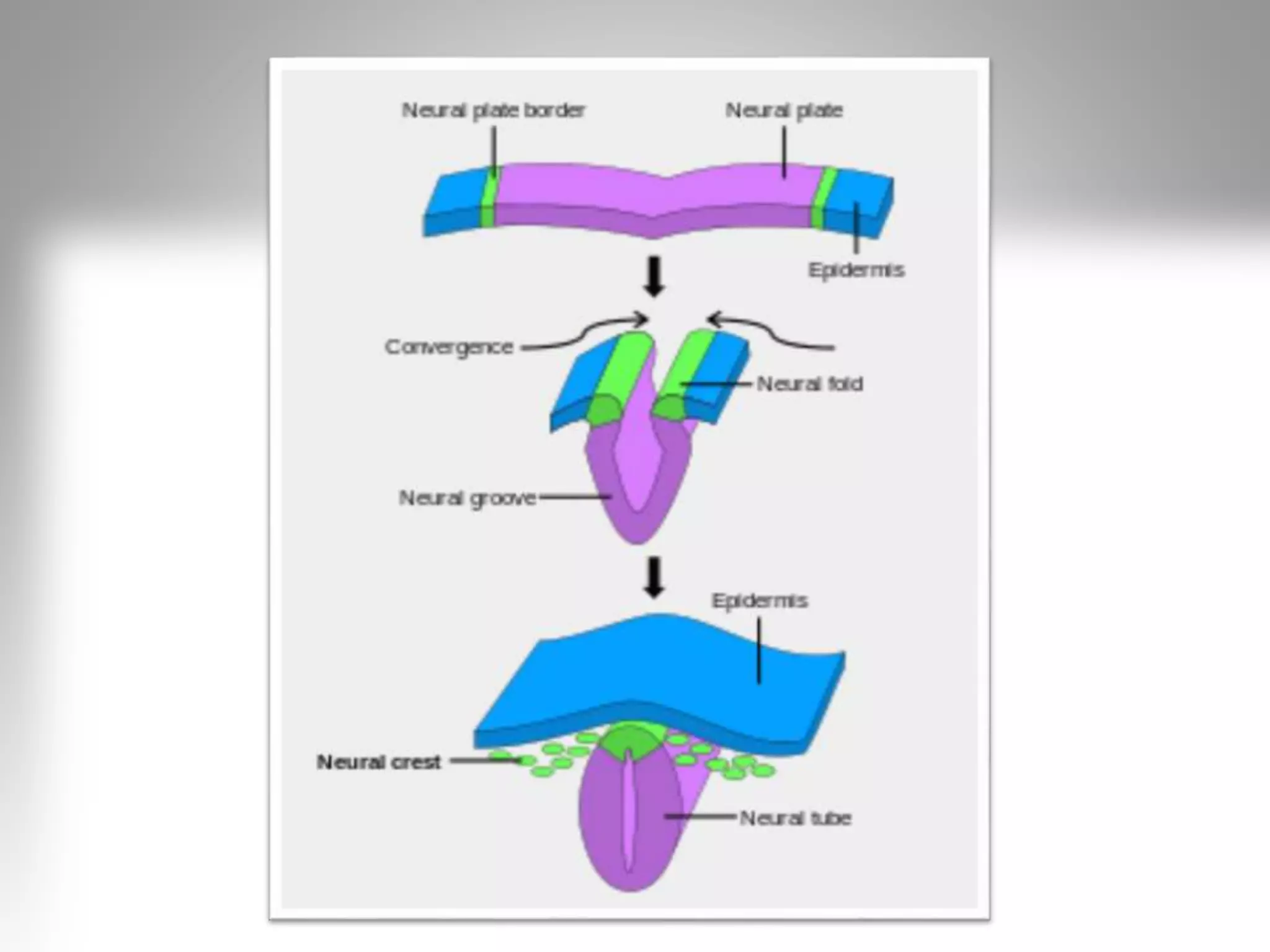
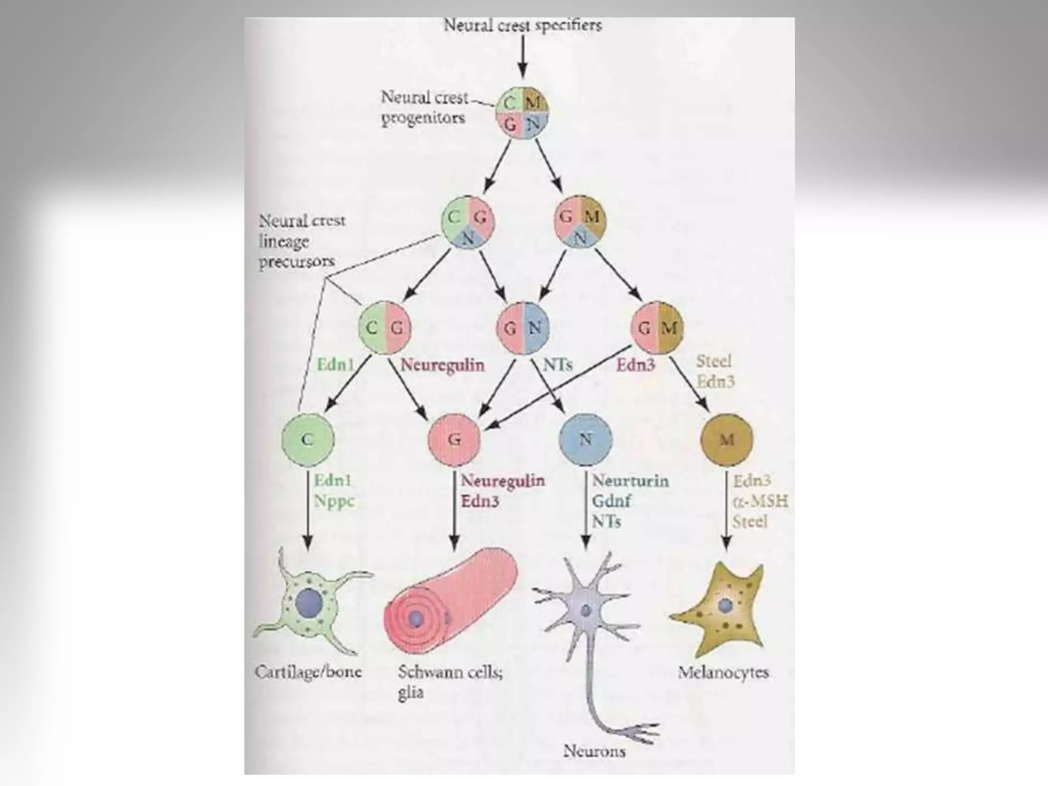

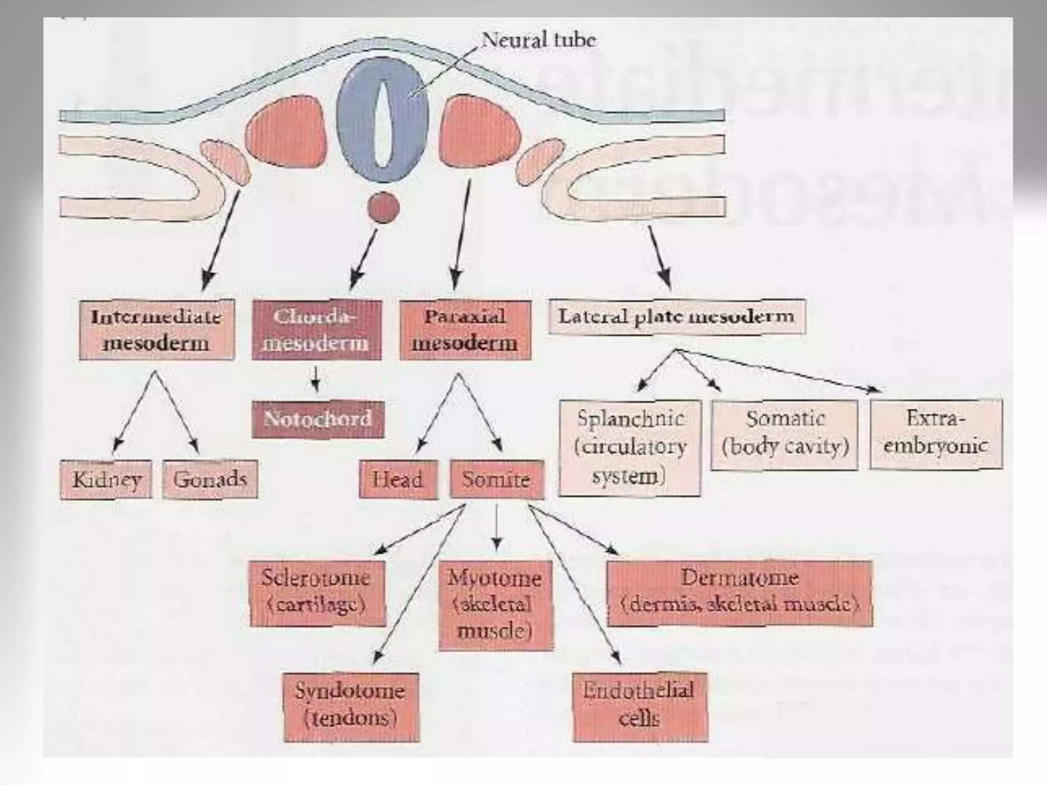


Neural crest cells arise from the ectoderm layer and give rise to diverse cell lineages including melanocytes, craniofacial cartilage and bone, smooth muscle, and neurons. They have multipotent capability, migrate throughout the embryo, and are precisely regulated. The neural crest can be divided into four main regions that develop into different structures and tissues. Somites form from paraxial mesoderm and later split into dermatomes, myotomes, syndetomes, and sclerotomes which give rise to skin, skeletal muscle, tendons and cartilage, and bone, respectively.
Introduction by M. Vharshini from Sri Ramachandra University.
Neural crest cells are temporary cells from the ectoderm, forming diverse lineages like melanocytes and neurons.
Neural crest cells have multipotent capability, migratory properties and regulatory abilities in embryo.
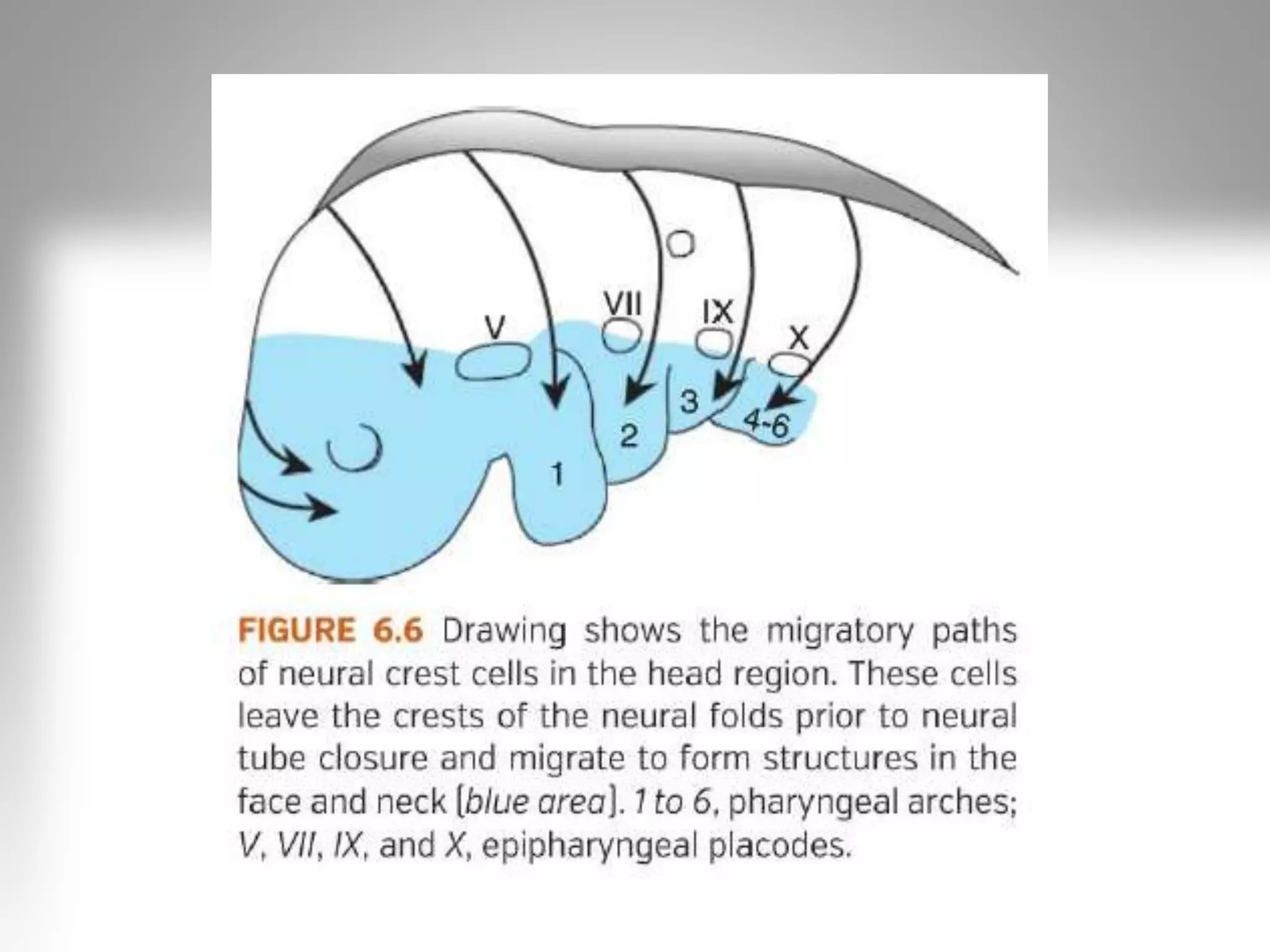
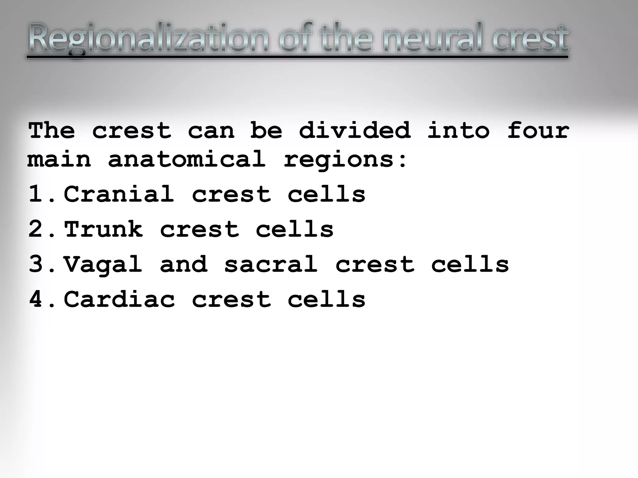
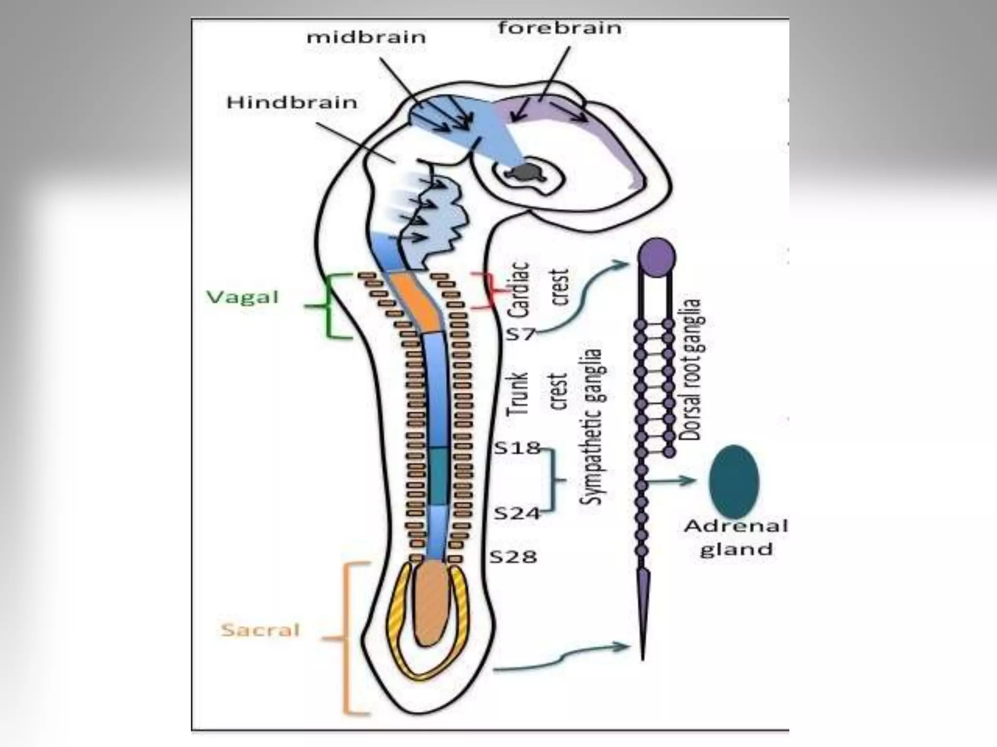
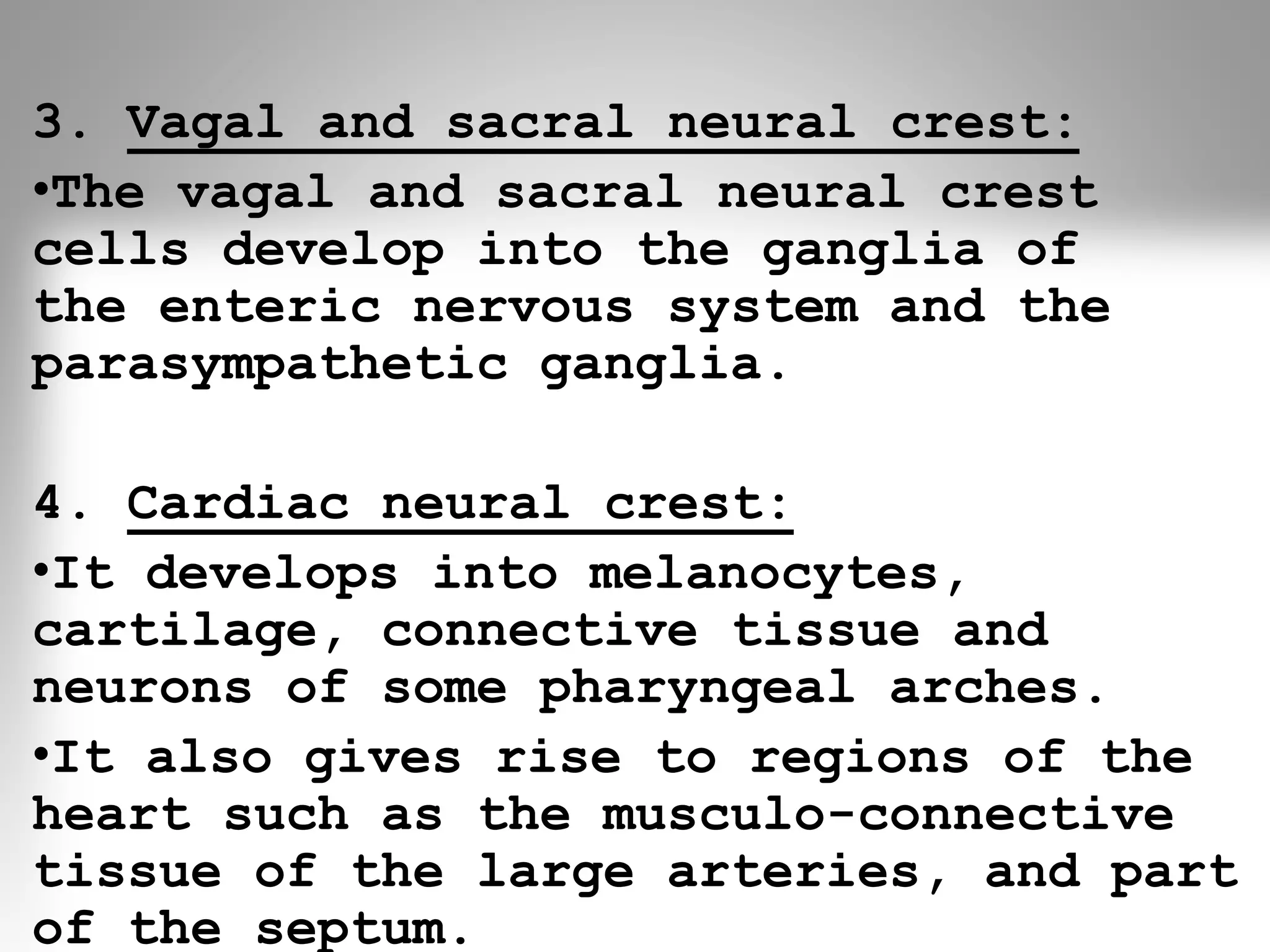
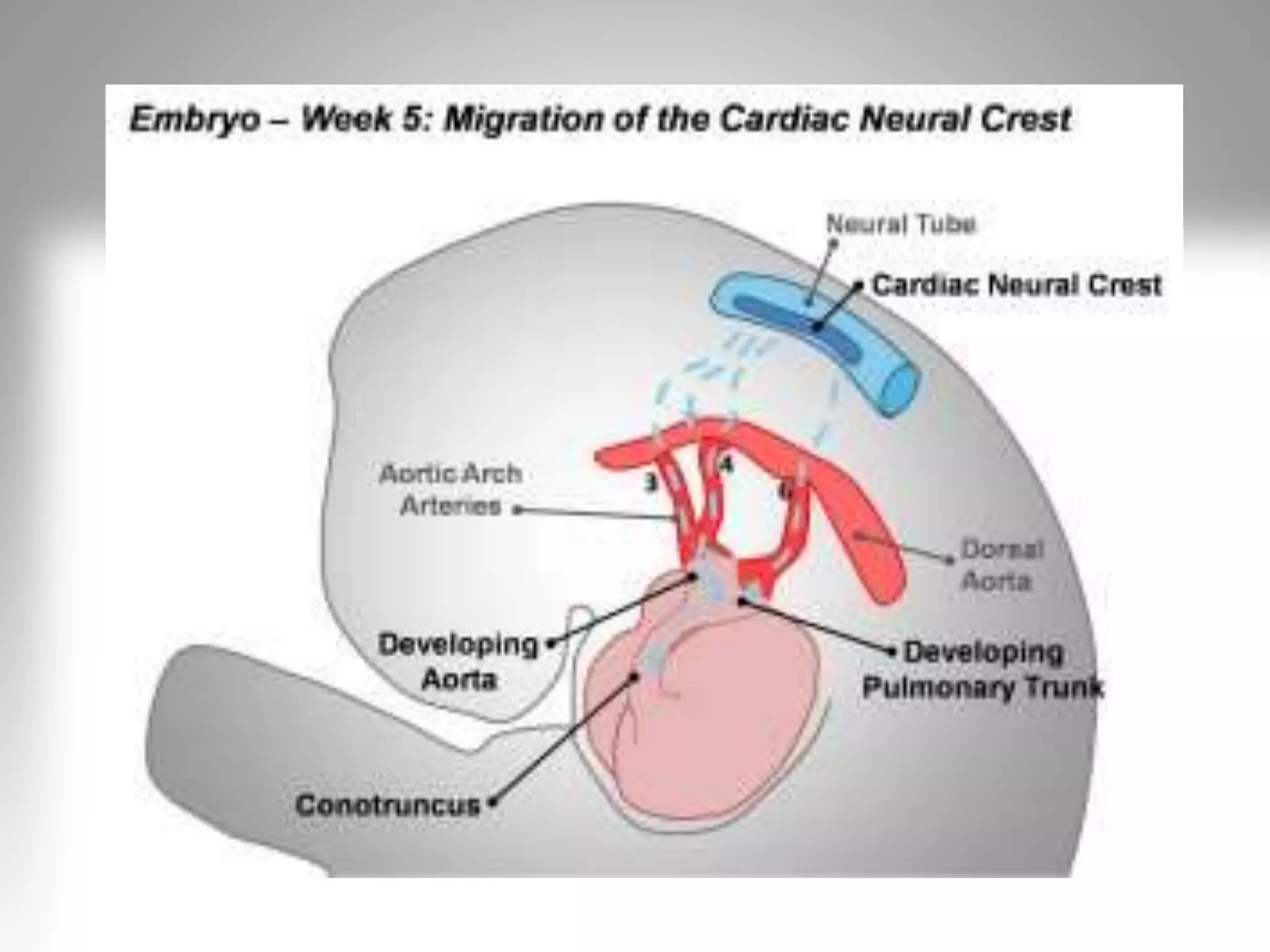
Neural crest divided into four anatomical regions: cranial, trunk, vagal & sacral, and cardiac.
Cranial crest cells form craniofacial structures including cartilage and contribute to the thymus.
Trunk neural crest forms melanocytes and sympathetic ganglia; crucial for various ectodermal structures.
Vagal/sacral crest cells develop enteric nervous system elements; cardiac crest forms tissues in pharyngeal arches.
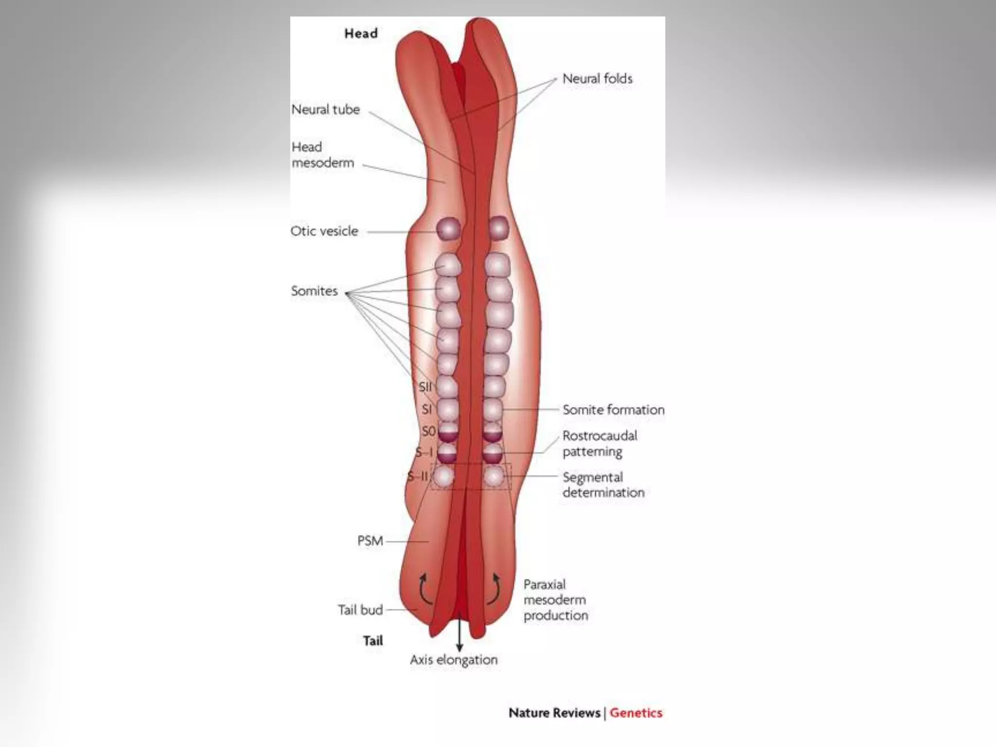
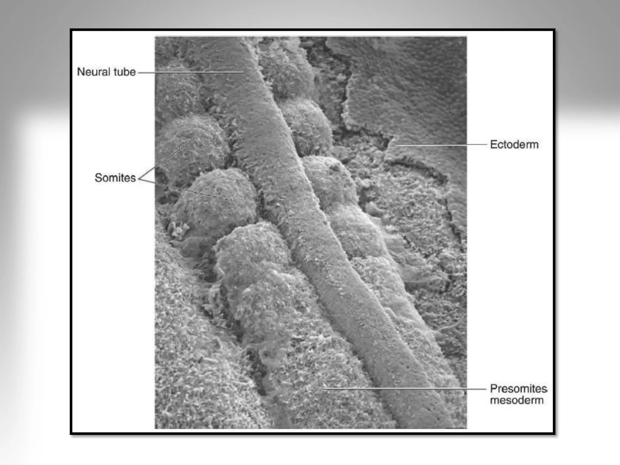
Mesoderm forms alongside ectoderm and endoderm; paraxial mesoderm separates to form somites.
Somites are mesodermal structures crucial for developing the vertebrate body plan, becoming dermis, muscles.
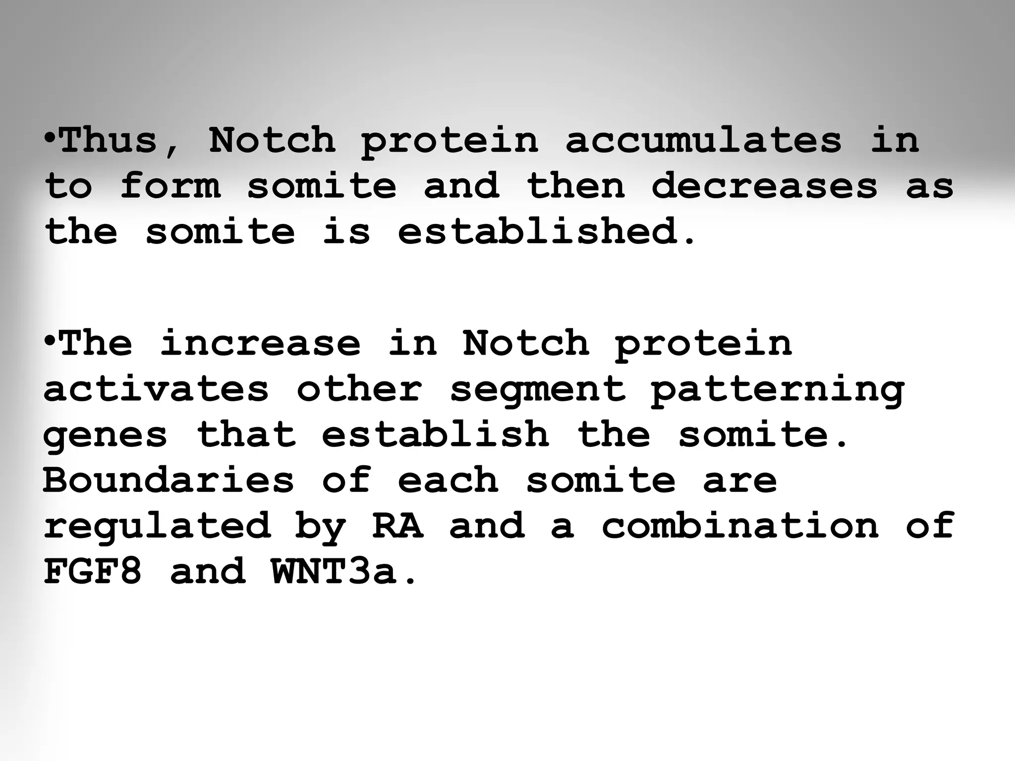
Somite segmentation relies on cyclic gene expression (NOTCH, WNT) regulating somite creation and patterning.
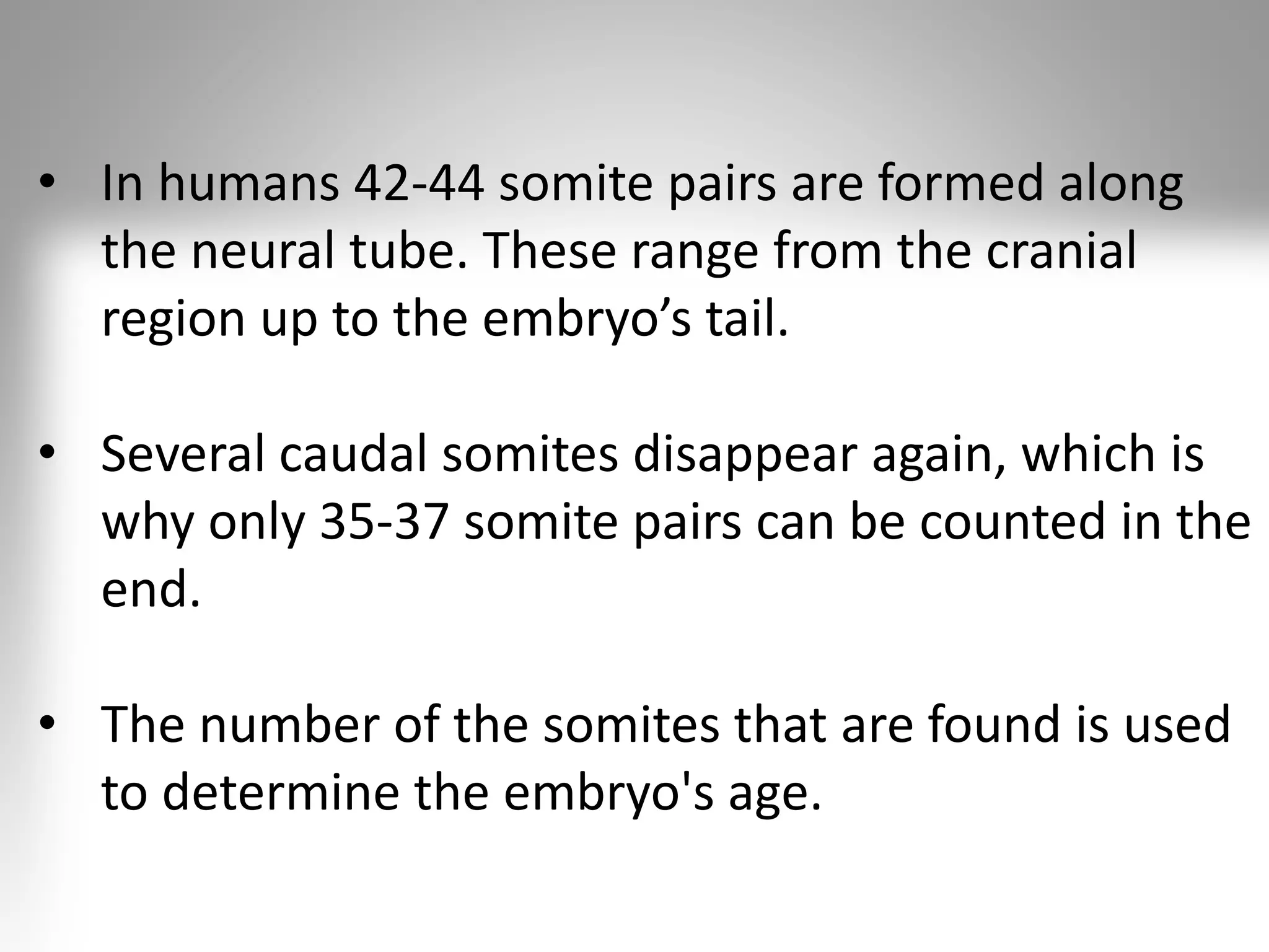
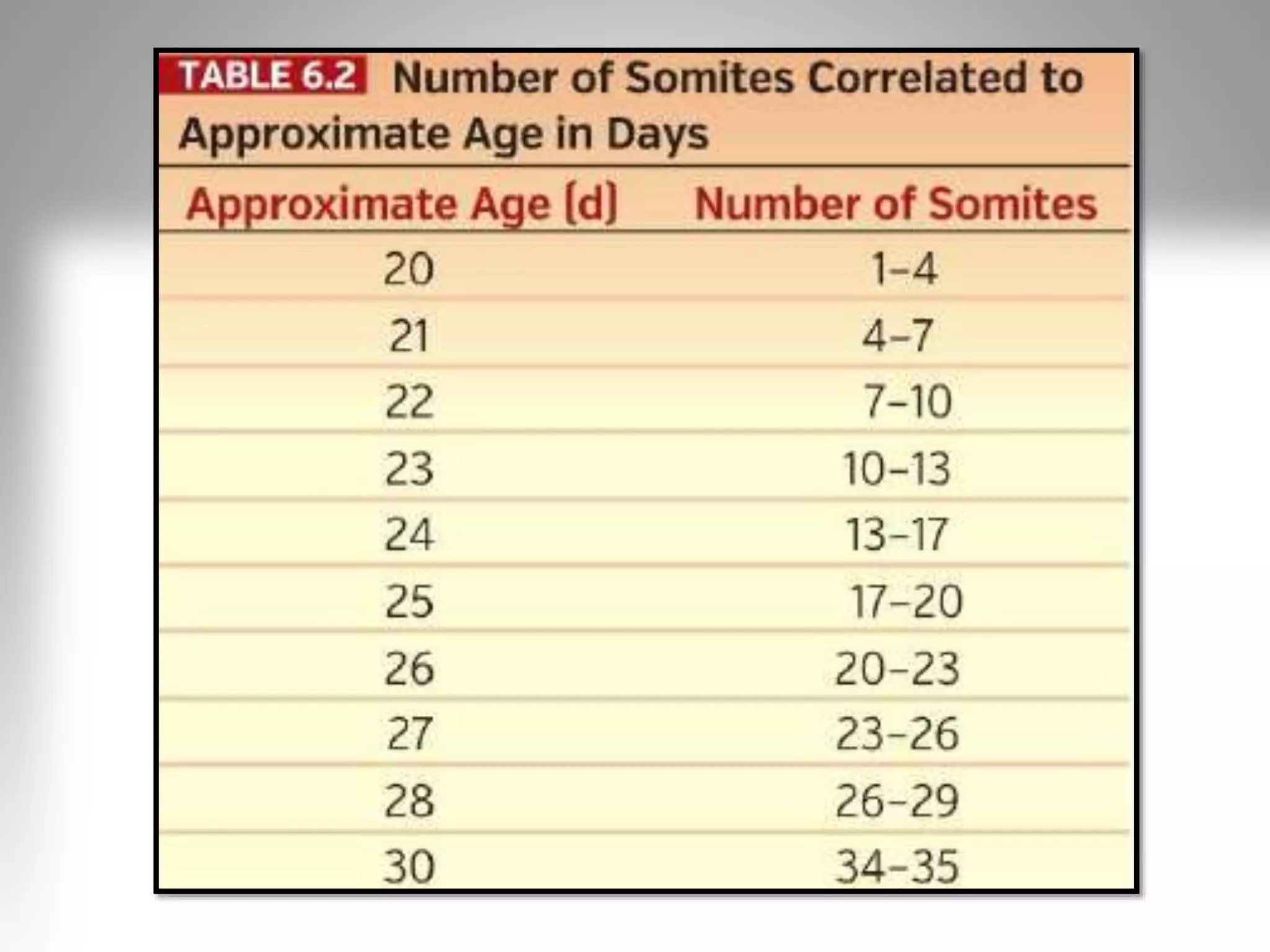
Humans have 42-44 somite pairs; 35-37 persist at maturity, used for determining embryo age.
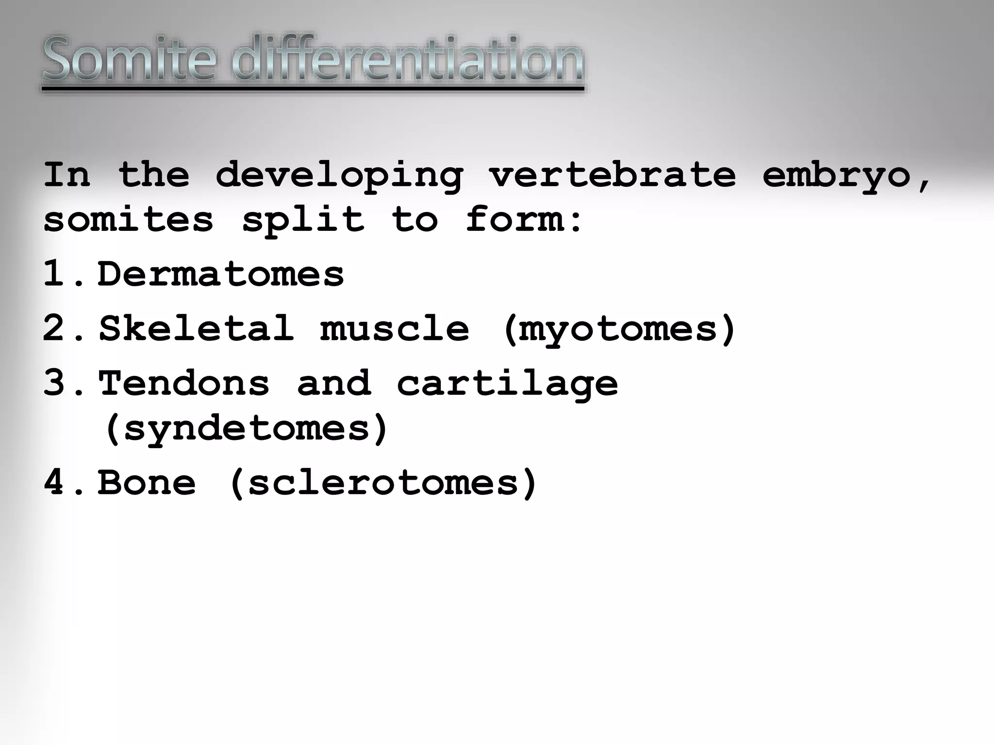
Somites develop into dermatomes, myotomes, syndetomes, and sclerotomes, each forming vital structures.
Dermatomes, from dorsal somites, arise early to form skin, fat, and connective tissues in the trunk and neck.
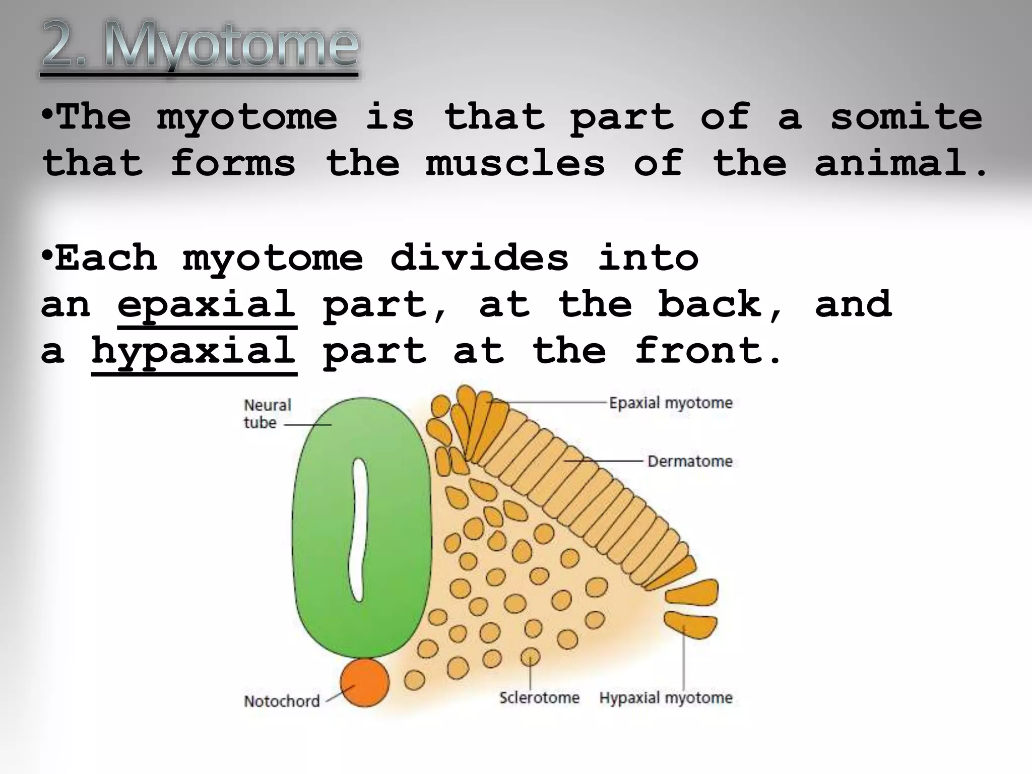
Myotomes form muscles; hypaxial and epaxial divisions contribute to thoracic and abdominal muscles.
Sclerotomes differentiate into vertebrae, rib cartilage, and part of the occipital bone.


