CELL DIVISION- Decoding Cell Division: The Dance of Life's Continuity

Decoding Cell Division: The Dance of Life's Continuity Step into the mesmerizing world of cell division with our illuminating SlideShare presentation. From the elegant choreography of mitosis to the intricacies of meiosis, witness the remarkable processes that underpin life's continuity and diversity. In this captivating presentation, we delve deep into the mechanisms of cell division, unraveling the stages and significance of mitosis and meiosis. Explore how cells meticulously replicate their DNA, segregate their chromosomes, and orchestrate their division to ensure the transmission of genetic information with precision and fidelity. Through vivid illustrations, clear explanations, and real-world examples, we illuminate the significance of cell division in growth, development, and reproduction. Gain a newfound understanding of how errors in cell division can lead to diseases like cancer and genetic disorders, and learn about the cutting-edge research driving advancements in this field. Whether you're a student, educator, or enthusiast of life sciences, our presentation offers valuable insights into one of the most fundamental processes of life. Join us as we unravel the mysteries of cell division and marvel at the beauty and complexity of nature's continuity. Don't miss this opportunity to deepen your knowledge and appreciation of cell biology. Embark on a journey into the heart of cell division and discover the dance of life's continuity unfolding before your eyes.

Recommended
More Related Content
Similar to CELL DIVISION- Decoding Cell Division: The Dance of Life's Continuity
Similar to CELL DIVISION- Decoding Cell Division: The Dance of Life's Continuity (20)
Recently uploaded
Recently uploaded (20)
CELL DIVISION- Decoding Cell Division: The Dance of Life's Continuity
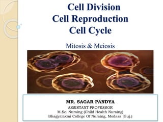
- 1. Cell Division Cell Reproduction Cell Cycle Mitosis & Meiosis MR. SAGAR PANDYA ASSISTANT PROFESSOR M.Sc. Nursing (Child Health Nursing) Bhagyalaxmi College Of Nursing, Modasa (Guj.)
- 2. THE CELL CYCLE /CELL DIVISION /CELL REPRODUCTION •The CELL CYCLE is an ordered sequence of events in which a body cell duplicates its contents and divides in two new cell (daughter cells). •The CELL CYCLE is the period between two cell division. •Also known as cell division or cell reproduction. •Three types of cell division, or cell reproduction Prokaryotes (bacteria) Binary fission divides forming two new identical cells Eukaryotes oMitosis -Cell or organism growth. -Replacement or repair of damaged cells. oMeiosis
- 3. Why do cells divide? 1: DNA Overload ◦ If cells grow without limit, an “information crisis” would develop ◦ DNA cannot serve the needs of the increasing size of cell 2: Exchange of materials ◦ Food and oxygen have to cross membrane very quickly ◦ Waste must get out ◦ If cell is too large, this occurs too slowly and cell will die. 3: For growth , repair & reproduction.
- 4. PROKARYOTIC CELL DIVISION Binary fission ◦ 3 main steps: 1: DNA Replication—DNA is copied, resulting in 2 identical chromosomes. 2: Chromosome Segregation—2 chromosomes separate, move towards ends (poles) of cell. 3: Cytokinesis—cytoplasm divides, forming 2 cells. ◦ Each new daughter cell is genetically identical to parent cell. •Prokaryotes have no nucleus. •They have a single circular chromosome. •Prokaryotes simply divide their cells in two by binary fission.
- 5. Eukaryotes Cell Division Eukaryotes must divide their nucleus (and other organelles such as mitochondria) in preparation for cell division (mitosis or meiosis) Before the nucleus divides the genetic material replicates (duplicates) Cell cycle has two parts: ◦ Growth and preparation (Interphase) ◦ Cell division (M Phase- Mitosis or Meiosis) Nuclear division (Mitosis/Karyokinesis) Cytokinesis (Cytoplasm division)
- 6. INTERPHASE INTERPHASE: Period of growth and DNA replication between cell divisions. Three phases: ◦ G1 Phase (First Gap Phase): Cell increases in size and volume. Cell metabolically active. Duplicates organelles and cytosolic components. Centrosome replication begins. ◦ S Phase (Synthesis of DNA): Replication of chromosomes (DNA replication / duplication). ◦ G2 Phase (Second Gap Phase): Organelles double. New cytoplasm forms. Cell growth continues (Enzymes and proteins are synthesized). Centrosome replication completed.
- 8. Terminology Chromatin Material- Thin threadlike fibrous form of DNA and proteins in nucleus (uncondensed chromosomes). Chromatid- Single condensed chromosome (condensed chromosome). Sister chromatids- Identical structure of homologus condensed chromosome. Centromere – Point where sister chromatid / two chromatids are joined together.
- 9. • Kinetochore- At the outside of each centromere is a protein complex (attachment site for mitotic spindle). • Diploid- Two sets of chromosomes (2n),in humans 23 pairs or 46 total. • Haploid- One set of chromosome (n),in humans 23 chromosomes (Gametes or sex chromosome). • Homologous pair- Each chromosome in pair are identical (Carry genes from same trait). only one pair is different- sex chromosome (X or Y)
- 10. • Tetrad- Four chromatids (2 homologous sister chromatid pairs) form a structure during meiosis. • Synapsis - Synapsis (also called syndesis) is the pairing of two homologous chromosomes that occurs during meiosis. • Crossing-over - The exchange of genetic material between homologous chromosomes (In prophase-I of meiosis). •Genetic recombination – It is result of Crossing-over - that is, the formation of new combinations of genes.
- 12. MITOSIS (SOMATIC CELL DIVISION) •The process of cell division which results in the production of two daughter cells from a single parent cell. •The daughter cells are identical to one another and to the original parent cell. •Process that divides cell nucleus to produce two new nuclei each with a complete set of chromosomes. •Mitosis is the basis of asexual reproduction.
- 13. Mitosis complete by two process- 1) NUCLEAR DIVISION (KARYOKINESIS) 4 sub-phases: 1st – Prophase 2nd – Metaphase 3rd – Anaphase 4th – Telophase followed by: 2) CYTOPLASMIC DIVISION (CYTOKINESIS) Interphase Cytokinesis Nuclear division
- 14. 1) NUCLEAR DIVISION (KARYOKINESIS) Nuclear division is most important part of eukaryotic cell division. In Mitosis ,as noted earlier, is the distribution of two sets of chromosomes into two separate nuclei equally. Biologists divide the process into four phases: Prophase, Metaphase, Anaphase, Telophase.
- 15. •Chromosome condense or visible (Chromatin materials/fibers to chromatids to sister chromatid pair). •Two homologous chromatids attached at a constricted region called a centromere (holds the chromatid pair together). •Centrioles migrate to the poles (only in animals). •Microtubules form (Mitotic spindle/Spindle fiber) from centrioles. Prophase
- 16. •At the outside of each centromere is a protein complex known as kinetochore. •Microtubules attach to the kinetochore for the pulling and pushing of chromosomes. •The nuclear envelope breaks down or starts disappear.
- 17. Metaphase Chromosomes line up at the center of the cell through the pulling pushing movement of mitotic spindle (Microtubules). The centomeres of the chromatids pairs at the exact center of the mitotic spindle. This midpoint makes a imaginary line called metaphase plate.
- 18. • Centromeres split out (Sister chromatids sreparate). • Spindle fibers pull one set of chromosomes (chromatids) to each pole. • Free spindle fibers lengthen and push poles of cell apart. Anaphase
- 19. Telophase • The final stage of mitosis. • Chromosomes uncoil (chromatids to chromatin material). • Spindle fibers disappears. • Nuclear envelopes form around both groups of chromosomes. • Cytokinesis occurs, enclosing each daughter nucleus into a separate cell. • Cleavage furrow forms on plasma membrane ( A constricted ring at the center of plasma membrane due to actin protein).
- 20. 2) CYTOPLASMIC DIVISION (CYTOKINESIS) Occurs at end of Mitosis. Cytokinesis through the formation of cleavage furrow (A ring of microtubules contract , pinching the cell in half). Division of the cytoplasm to form 2 new daughter cells. Organelles are divided. Daughter cells are genetically identical.
- 21. Cytokinesis Cleavage furrow Contracting ring of microfilaments Daughter cells Cleavage furrow Cells return to interphase
- 23. MeIOSIS (REPRODUCTIVE CELL DIVISION /GERMCELL DIVISION) •Meiosis is the type of cell division by which germ cells (eggs and sperm) are produced. •One parent cell produces four daughter cells. •Daughter cells have half the number of chromosomes found in the original parent cell. •Meiosis shuffles genes in new combinations. A division of the nucleus/cell that reduces chromosome number by half (forms four haploid cells from a diploid cell)
- 24. •Meiosis results in genetically different cells. •Meiosis and fertilization are the basis of sexual reproduction. •During meiosis, DNA replicates once, but the nucleus divides twice. •Completed in two stages- Meiosis-I or Meiosis-II. •Four sub-phases described for each stages.
- 25. Meiosis complete in two stages- 1) MEIOSIS-I A) NUCLEAR DIVISION (KARYOKINESIS) 4 sub-phases: Prophase-I Metaphase-I Anaphase-I Telophase-I followed by: B) CYTOPLASMIC DIVISION-I (CYTOKINESIS-I) Followed by: 2) MEIOSIS-II A) NUCLEAR DIVISION (KARYOKINESIS) 4 sub-phases: Prophase-II Metaphase-II Anaphase-II Telophase-II followed by: B) CYTOPLASMIC DIVISION-II (CYTOKINESIS-II)
- 27. Sex cells divide to produce GAMETES (sperm or egg). Gametes have HALF the numbers of chromosomes. Occurs only in GONADS (testes or ovaries). Male: SPERMATOGENESIS – Formation of sperm. Female: OOGENESIS – Formation of egg or ova/ovum Heredity : Way of transferring the genetic information to offspring. Chromosome theory of heredity: “Chromosomes carry genes”. Gene : Unit of heredity.
- 28. Spermatogenesis 2n=46 Human sex cell Diploid (2n) n=23 n=23 meiosis I n=23 n=23 n=23 n=23 sperm haploid (n) meiosis II 4 sperm cells are produced from each primary spermatocyte. Primary Spermatocyte Secondary Spermatocyte Secondary Spermatocyte
- 29. Oogenesis 2n=46 Human sex cell Diploid (2n) n=23 n=23 Meiosis I n=23 egg Haploid (n) Meiosis II 29 Polar Bodies (die) *** The polar bodies die… only one ovum (egg) is produced from each primary oocyte. Primary oocyte Secondary oocyte Ova ***One ova or three polar bodies produce from each primary oocyte
- 32. 1) MEIOSIS-I A) NUCLEAR DIVISION (KARYOKINESIS) Prophase-I Longest and most complex phase 90% of the meiotic process is spent in prophase-I. Chromosomes condense. Nuclear envelop & nucleolus disappears. Centromeres moves to opposite poles. Synapsis occurs: Homologous chromosomes come together to form a tetrad. Tetrad is two chromosomes or four chromatids (sister and nonsister chromatids). Crossing over (variation) may occur between non sister chromatids at the chiasmata. Crossing over: Segment of non sister chromatids break and reattach to the other chromatid. Chiasmata (Chiasma): The site of crossing over.
- 33. Prophase I - Synapsis Homologous chromosomes sister chromatids sister chromatids Tetrad
- 34. Homologous Chromosomes Pair of chromosomes (maternal and paternal) that are similar in shape and size. Homologous pairs (tetrads) carry genes controlling the same inherited traits. Each locus (position of a gene) is in the same position on homologues. Humans have 23 pairs of homologous chromosomes. a. 22 pairs of autosomes b. 01 pair of sex chromosomes
- 35. Crossing Over Creates variation (diversity) in the offspring’s traits. Nonsister chromatids Chiasmata: site of crossing over Variation (Genetic recombination) Tetrad
- 36. Prophase I
- 37. Metaphase-I Shortest phase Tetrads or homologous chromosomes move to center of cell. Tetrads align on the metaphase plate. Metaphase plate
- 39. Anaphase-I • Microtubules of spindle shorten. •Homologous chromosomes separate and move towards the poles. •Sister chromatids remain attached at their centromeres. Sister chromatids remain attached Homologous chromosomes separate
- 40. Telophase-I Each poles now has haploid (n) set of chromosome. Nuclear envelopes form around each set of chromosomes. Two daughter nuclei formed. Each new nucleus is now haploid. Sister chromatids are no longer identical because of crossing over. Sister chromatids uncoiled. Cytokinesis occurs and two haploid daughter cells
- 41. Telophase I & Cytokinesis I
- 42. B) CYTOPLASMIC DIVISION-I (CYTOKINESIS-I) Occurs at end of Meiosis-I. Cytokinesis through the formation of cleavage furrow (A ring of microtubules contract , pinching the cell in half). Constricted ring forms due to activation of actins protein present in plasma membrane. Division of the cytoplasm to form 2 new daughter cells. Organelles are divided. Daughter cells (haploid) are genetically different from parent cell. Undergo further division continue in meiosis-II.
- 43. Cytokinesis Cleavage furrow Contracting ring of microfilaments Daughter cells Cleavage furrow Undergo further division continue in meiosis-II.
- 45. 2) MEIOSIS-II Daughter cells undergo a second division of meiosis is meiosis-II. No interphase-II. Very short stage. No DNA replication. Meiosis-II is similar to mitosis. In meiosis-II 4 haploid cells forms from 2 haploid cells (daughter cells) formed in meiosis-I. Meiosis-II also complete in two processes: A)Nuclear division (Karyokinesis) B)Cytoplasmic division-II (Cytokinesis –II) Meiosis II Meiosis -I
- 46. Prophase-II A) NUCLEAR DIVISION (KARYOKINESIS) Same as Prophase in mitosis: *Nucleus & nucleolus disappear *Chromosomes condense *Chromatids makes sister chromatid (attached at centromere) *Centriole moves to opposite poles *Spindle forms *Spindle attached to sister chromatids (through kinatochore)
- 47. Prophase II
- 48. Metaphase-II Sister chromatids move to the center. Chromosomes (not homologous) line up at equator. Forms metaphase plate.
- 49. Anaphase-II Same as Anaphase in mitosis. Centromeres divide and sister chromatids migrate separately to each pole. Individual chromosomes are pulled to poles. Microtubules of spindle shorten.
- 50. Telophase-II Same as telophase in mitosis. The final stage of meiosis. Cell division is complete. Four haploid daughter cells are obtained. Chromosomes uncoil (chromatids to chromatin material). Spindle fibers disappears. Nuclear envelopes form around four groups of chromosomes. Nuclei and Nucleoli reforms. FOUR HAPLOID DAUGHTER cells are produced Called GAMETES (eggs and sperm) Cytokinesis occurs, enclosing each daughter nucleus into a separate cell. Cleavage furrow forms on plasma membrane ( A constricted ring at the center of plasma membrane due to actins protein).
- 51. Telophase II & Cytokinesis II
- 52. B) CYTOPLASMIC DIVISION-II (CYTOKINESIS-II) Occurs at end of Meiosis-II or meiosis. Cytokinesis through the formation of cleavage furrow (A ring of microtubules contract , pinching the cell in half). Constricted ring forms due to activation of actins protein present in plasma membrane. Division of the cytoplasm to forms 4 new daughter cells from both haploid cells. Organelles are divided. Daughter cells (haploid) are genetically different from parent cell. Meiosis division is completed.
- 53. Centrosomes (with centriole pairs) Centrioles Sites of crossing over Spindle Tetrad Nuclear envelope Chromatin Sister chromatids Fragments of the nuclear envelope Centromere (with a kinetochore) Spindle microtubules attached to a kinetochore Metaphase plate Homologous chromosomes separate Sister chromatids remain attached Chromosomes duplicate Prophase I Metaphase I Anaphase I INTERPHASE: MEIOSIS I: Homologous chromosomes separate
- 54. Prophase II Metaphase II Anaphase II MEIOSIS II: Sister chromatids separate Sister chromatids separate Haploid daughter cells forming Telophase II and Cytokinesis II