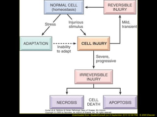
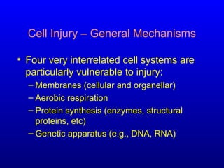
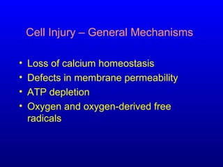
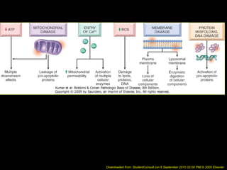
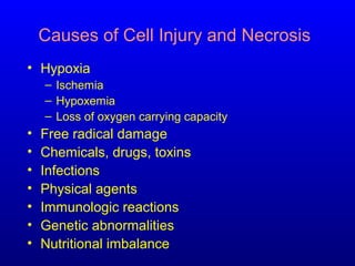
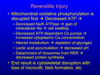
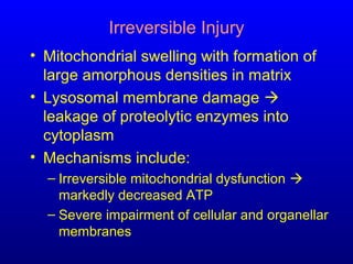
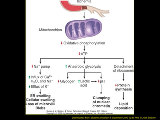
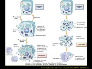
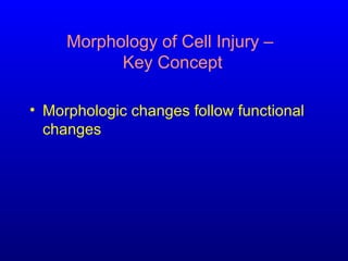
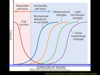
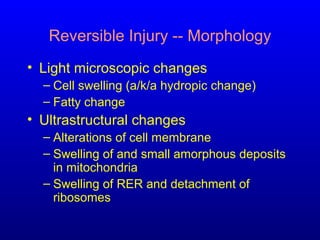
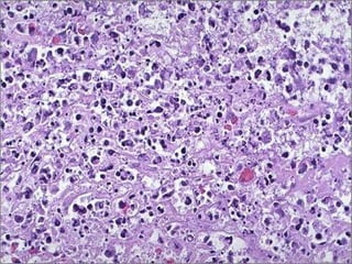
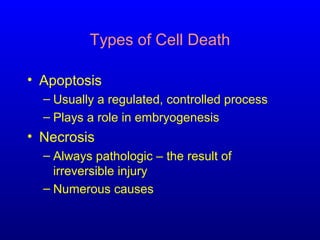
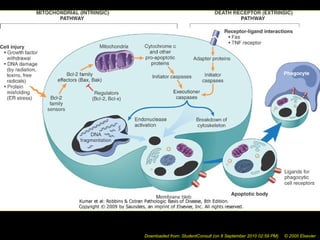
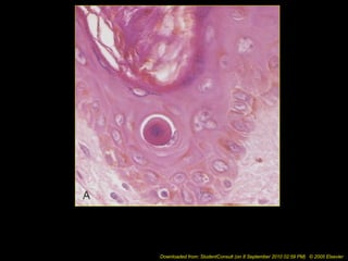
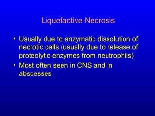
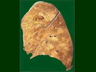
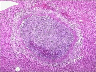
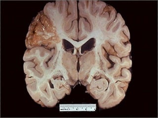
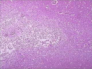
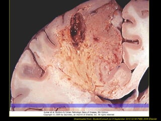
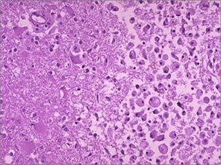
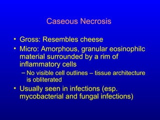
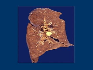
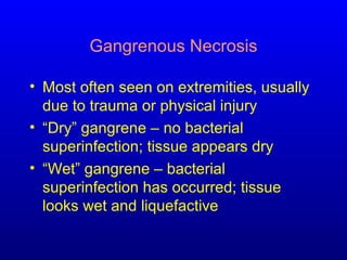
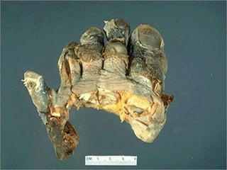
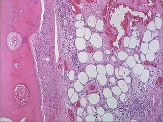
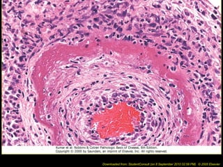
Cell Injury I – Cell Injury and
Cell Death
Dept. of Pathology
Key Concepts
• Normal cells have a fairly narrow range
of function or steady state:
Homeostasis
• Excess physiologic or pathologic stress
may force the cell to a new steady
state: Adaptation
• Too much stress exceeds the cell’s
adaptive capacity: Injury
Downloaded from: StudentConsult (on 8 September 2010 02:58 PM) © 2005 Elsevier
Key Concepts (cont’d)
• Cell injury can be reversible or
irreversible
• Reversibility depends on the type,
severity and duration of injury
• Cell death is the result of irreversible
injury
Cell Injury – General Mechanisms
• Four very interrelated cell systems are
particularly vulnerable to injury:
– Membranes (cellular and organellar)
– Aerobic respiration
– Protein synthesis (enzymes, structural
proteins, etc)
– Genetic apparatus (e.g., DNA, RNA)
Cell Injury – General Mechanisms
• Loss of calcium homeostasis
• Defects in membrane permeability
• ATP depletion
• Oxygen and oxygen-derived free
radicals
Downloaded from: StudentConsult (on 8 September 2010 02:58 PM) © 2005 Elsevier
Causes of Cell Injury and Necrosis
• Hypoxia
– Ischemia
– Hypoxemia
– Loss of oxygen carrying capacity
• Free radical damage
• Chemicals, drugs, toxins
• Infections
• Physical agents
• Immunologic reactions
• Genetic abnormalities
• Nutritional imbalance
Reversible Injury
• Mitochondrial oxidative phosphorylation is
disrupted first  Decreased ATP 
– Decreased Na/K ATPase  gain of
intracellular Na  cell swelling
– Decreased ATP-dependent Ca pumps 
increased cytoplasmic Ca concentration
– Altered metabolism  depletion of glycogen
– Lactic acid accumulation  decreased pH
– Detachment of ribosomes from RER 
decreased protein synthesis
• End result is cytoskeletal disruption with
loss of microvilli, bleb formation, etc
Irreversible Injury
• Mitochondrial swelling with formation of
large amorphous densities in matrix
• Lysosomal membrane damage 
leakage of proteolytic enzymes into
cytoplasm
• Mechanisms include:
– Irreversible mitochondrial dysfunction 
markedly decreased ATP
– Severe impairment of cellular and organellar
membranes
Downloaded from: StudentConsult (on 8 September 2010 02:58 PM) © 2005 Elsevier
Funky mitochondria
Cell Injury
• Membrane damage and loss of calcium
homeostasis are most crucial
• Some models of cell death suggest that
a massive influx of calcium “causes”
cell death
• Too much cytoplasmic calcium:
– Denatures proteins
– Poisons mitochondria
– Inhibits cellular enzymes
Downloaded from: StudentConsult (on 8 September 2010 02:58 PM) © 2005 Elsevier
Clinical Correlation
• Injured membranes are leaky
• Enzymes and other proteins that escape
through the leaky membranes make their
way to the bloodstream, where they can
be measured in the serum
Free Radicals
• Free radicals have an unpaired electron
in their outer orbit
• Free radicals cause chain reactions
• Generated by:
– Absorption of radiant energy
– Oxidation of endogenous constituents
– Oxidation of exogenous compounds
Examples of Free Radical Injury
• Chemical (e.g., CCl4, acetaminophen)
• Inflammation / Microbial killing
• Irradiation (e.g., UV rays  skin cancer)
• Oxygen (e.g., exposure to very high
oxygen tension on ventilator)
• Age-related changes
Mechanism of Free Radical Injury
• Lipid peroxidation  damage to cellular
and organellar membranes
• Protein cross-linking and fragmentation
due to oxidative modification of amino
acids and proteins
• DNA damage due to reactions of free
radicals with thymine
Downloaded from: StudentConsult (on 8 September 2010 02:58 PM) © 2005 Elsevier
Morphology of Cell Injury –
Key Concept
• Morphologic changes follow functional
changes
© 2005 Elsevier Downloaded from: StudentConsult (on 8 September 2010 02:58 PM)
Reversible Injury -- Morphology
• Light microscopic changes
– Cell swelling (a/k/a hydropic change)
– Fatty change
• Ultrastructural changes
– Alterations of cell membrane
– Swelling of and small amorphous deposits
in mitochondria
– Swelling of RER and detachment of
ribosomes
Irreversible Injury -- Morphology
• Light microscopic changes
– Increased cytoplasmic eosinophilia (loss of
RNA, which is more basophilic)
– Cytoplasmic vacuolization
– Nuclear chromatin clumping
• Ultrastructural changes
– Breaks in cellular and organellar membranes
– Larger amorphous densities in mitochondria
– Nuclear changes
Irreversible Injury – Nuclear Changes
• Pyknosis
– Nuclear shrinkage and increased basophilia
• Karyorrhexis
– Fragmentation of the pyknotic nucleus
• Karyolysis
– Fading of basophilia of chromatin
Karyolysis & karyorrhexis --
micro
Types of Cell Death
• Apoptosis
– Usually a regulated, controlled process
– Plays a role in embryogenesis
• Necrosis
– Always pathologic – the result of
irreversible injury
– Numerous causes
Apoptosis
• Involved in many processes, some
physiologic, some pathologic
– Programmed cell death during
embryogenesis
– Hormone-dependent involution of organs in
the adult (e.g., thymus)
– Cell deletion in proliferating cell populations
– Cell death in tumors
– Cell injury in some viral diseases (e.g.,
hepatitis)
Apoptosis – Morphologic Features
• Cell shrinkage with increased
cytoplasmic density
• Chromatin condensation
• Formation of cytoplasmic blebs and
apoptotic bodies
• Phagocytosis of apoptotic cells by
adjacent healthy cells
Downloaded from: StudentConsult (on 8 September 2010 02:59 PM) © 2005 Elsevier
Downloaded from: StudentConsult (on 8 September 2010 02:58 PM) © 2005 Elsevier
Downloaded from: StudentConsult (on 8 September 2010 02:59 PM) © 2005 Elsevier
Apoptosis – Micro
Types of Necrosis
• Coagulative (most common)
• Liquefactive
• Caseous
• Fat necrosis
• Gangrenous necrosis
Coagulative Necrosis
• Cell’s basic outline is preserved
• Homogeneous, glassy eosinophilic
appearance due to loss of cytoplasmic
RNA (basophilic) and glycogen (granular)
• Nucleus may show pyknosis, karyolysis
or karyorrhexis
Downloaded from: StudentConsult (on 8 September 2010 02:58 PM) © 2005 Elsevier
Splenic infarcts -- gross
Infarcted bowel -- gross
Myocardium photomic
Adrenal infarct -- Micro
3 stages of coagulative
necrosis (L to R) -- micro
Liquefactive Necrosis
• Usually due to enzymatic dissolution of
necrotic cells (usually due to release of
proteolytic enzymes from neutrophils)
• Most often seen in CNS and in
abscesses
Lung abscesses (liquefactive
necrosis) -- gross
Liver abscess -- micro
Liquefactive necrosis -- gross
Liquefactive necrosis of brain
-- micro
Downloaded from: StudentConsult (on 8 September 2010 02:58 PM)© 2005 Elsevier
Macrophages cleaning
liquefactive necrosis -- micro
Caseous Necrosis
• Gross: Resembles cheese
• Micro: Amorphous, granular eosinophilc
material surrounded by a rim of
inflammatory cells
– No visible cell outlines – tissue architecture
is obliterated
• Usually seen in infections (esp.
mycobacterial and fungal infections)
Caseous necrosis -- gross
Downloaded from: StudentConsult (on 8 September 2010 02:58 PM) © 2005 Elsevier
Extensive caseous necrosis
-- gross
Caseous necrosis -- micro
Enzymatic Fat Necrosis
• Results from hydrolytic action of lipases
on fat
• Most often seen in and around the
pancreas; can also be seen in other
fatty areas of the body, usually due to
trauma
• Fatty acids released via hydrolysis react
with calcium to form chalky white areas
 “saponification”
Enzymatic fat necrosis of
pancreas -- gross
Downloaded from: StudentConsult (on 8 September 2010 02:58 PM) © 2005 Elsevier
Fat necrosis -- micro
Gangrenous Necrosis
• Most often seen on extremities, usually
due to trauma or physical injury
• “Dry” gangrene – no bacterial
superinfection; tissue appears dry
• “Wet” gangrene – bacterial
superinfection has occurred; tissue
looks wet and liquefactive
Gangrene -- gross
Wet gangrene -- gross
Gangrenous necrosis -- micro
Fibrinoid Necrosis
• Usually seen in the walls of blood
vessels (e.g., in vasculitides)
• Glassy, eosinophilic fibrin-like material
is deposited within the vascular walls
Downloaded from: StudentConsult (on 8 September 2010 02:58 PM) © 2005 Elsevier

Pathology cellinjuryi

  • 1.
    Cell Injury I– Cell Injury and Cell Death Dept. of Pathology
  • 2.
    Key Concepts • Normalcells have a fairly narrow range of function or steady state: Homeostasis • Excess physiologic or pathologic stress may force the cell to a new steady state: Adaptation • Too much stress exceeds the cell’s adaptive capacity: Injury
  • 3.
    Downloaded from: StudentConsult(on 8 September 2010 02:58 PM) © 2005 Elsevier
  • 4.
    Key Concepts (cont’d) •Cell injury can be reversible or irreversible • Reversibility depends on the type, severity and duration of injury • Cell death is the result of irreversible injury
  • 5.
    Cell Injury –General Mechanisms • Four very interrelated cell systems are particularly vulnerable to injury: – Membranes (cellular and organellar) – Aerobic respiration – Protein synthesis (enzymes, structural proteins, etc) – Genetic apparatus (e.g., DNA, RNA)
  • 6.
    Cell Injury –General Mechanisms • Loss of calcium homeostasis • Defects in membrane permeability • ATP depletion • Oxygen and oxygen-derived free radicals
  • 7.
    Downloaded from: StudentConsult(on 8 September 2010 02:58 PM) © 2005 Elsevier
  • 8.
    Causes of CellInjury and Necrosis • Hypoxia – Ischemia – Hypoxemia – Loss of oxygen carrying capacity • Free radical damage • Chemicals, drugs, toxins • Infections • Physical agents • Immunologic reactions • Genetic abnormalities • Nutritional imbalance
  • 9.
    Reversible Injury • Mitochondrialoxidative phosphorylation is disrupted first  Decreased ATP  – Decreased Na/K ATPase  gain of intracellular Na  cell swelling – Decreased ATP-dependent Ca pumps  increased cytoplasmic Ca concentration – Altered metabolism  depletion of glycogen – Lactic acid accumulation  decreased pH – Detachment of ribosomes from RER  decreased protein synthesis • End result is cytoskeletal disruption with loss of microvilli, bleb formation, etc
  • 10.
    Irreversible Injury • Mitochondrialswelling with formation of large amorphous densities in matrix • Lysosomal membrane damage  leakage of proteolytic enzymes into cytoplasm • Mechanisms include: – Irreversible mitochondrial dysfunction  markedly decreased ATP – Severe impairment of cellular and organellar membranes
  • 11.
    Downloaded from: StudentConsult(on 8 September 2010 02:58 PM) © 2005 Elsevier
  • 12.
  • 13.
    Cell Injury • Membranedamage and loss of calcium homeostasis are most crucial • Some models of cell death suggest that a massive influx of calcium “causes” cell death • Too much cytoplasmic calcium: – Denatures proteins – Poisons mitochondria – Inhibits cellular enzymes
  • 14.
    Downloaded from: StudentConsult(on 8 September 2010 02:58 PM) © 2005 Elsevier
  • 15.
    Downloaded from: StudentConsult(on 8 September 2010 02:58 PM) © 2005 Elsevier
  • 16.
    Downloaded from: StudentConsult(on 8 September 2010 02:58 PM) © 2005 Elsevier
  • 17.
    Clinical Correlation • Injuredmembranes are leaky • Enzymes and other proteins that escape through the leaky membranes make their way to the bloodstream, where they can be measured in the serum
  • 18.
    Free Radicals • Freeradicals have an unpaired electron in their outer orbit • Free radicals cause chain reactions • Generated by: – Absorption of radiant energy – Oxidation of endogenous constituents – Oxidation of exogenous compounds
  • 19.
    Examples of FreeRadical Injury • Chemical (e.g., CCl4, acetaminophen) • Inflammation / Microbial killing • Irradiation (e.g., UV rays  skin cancer) • Oxygen (e.g., exposure to very high oxygen tension on ventilator) • Age-related changes
  • 20.
    Mechanism of FreeRadical Injury • Lipid peroxidation  damage to cellular and organellar membranes • Protein cross-linking and fragmentation due to oxidative modification of amino acids and proteins • DNA damage due to reactions of free radicals with thymine
  • 21.
    Downloaded from: StudentConsult(on 8 September 2010 02:58 PM) © 2005 Elsevier
  • 22.
    Morphology of CellInjury – Key Concept • Morphologic changes follow functional changes
  • 23.
    © 2005 ElsevierDownloaded from: StudentConsult (on 8 September 2010 02:58 PM)
  • 24.
    Reversible Injury --Morphology • Light microscopic changes – Cell swelling (a/k/a hydropic change) – Fatty change • Ultrastructural changes – Alterations of cell membrane – Swelling of and small amorphous deposits in mitochondria – Swelling of RER and detachment of ribosomes
  • 25.
    Irreversible Injury --Morphology • Light microscopic changes – Increased cytoplasmic eosinophilia (loss of RNA, which is more basophilic) – Cytoplasmic vacuolization – Nuclear chromatin clumping • Ultrastructural changes – Breaks in cellular and organellar membranes – Larger amorphous densities in mitochondria – Nuclear changes
  • 26.
    Irreversible Injury –Nuclear Changes • Pyknosis – Nuclear shrinkage and increased basophilia • Karyorrhexis – Fragmentation of the pyknotic nucleus • Karyolysis – Fading of basophilia of chromatin
  • 27.
  • 28.
    Types of CellDeath • Apoptosis – Usually a regulated, controlled process – Plays a role in embryogenesis • Necrosis – Always pathologic – the result of irreversible injury – Numerous causes
  • 29.
    Apoptosis • Involved inmany processes, some physiologic, some pathologic – Programmed cell death during embryogenesis – Hormone-dependent involution of organs in the adult (e.g., thymus) – Cell deletion in proliferating cell populations – Cell death in tumors – Cell injury in some viral diseases (e.g., hepatitis)
  • 30.
    Apoptosis – MorphologicFeatures • Cell shrinkage with increased cytoplasmic density • Chromatin condensation • Formation of cytoplasmic blebs and apoptotic bodies • Phagocytosis of apoptotic cells by adjacent healthy cells
  • 31.
    Downloaded from: StudentConsult(on 8 September 2010 02:59 PM) © 2005 Elsevier
  • 32.
    Downloaded from: StudentConsult(on 8 September 2010 02:58 PM) © 2005 Elsevier
  • 33.
    Downloaded from: StudentConsult(on 8 September 2010 02:59 PM) © 2005 Elsevier
  • 34.
  • 35.
    Types of Necrosis •Coagulative (most common) • Liquefactive • Caseous • Fat necrosis • Gangrenous necrosis
  • 36.
    Coagulative Necrosis • Cell’sbasic outline is preserved • Homogeneous, glassy eosinophilic appearance due to loss of cytoplasmic RNA (basophilic) and glycogen (granular) • Nucleus may show pyknosis, karyolysis or karyorrhexis
  • 37.
    Downloaded from: StudentConsult(on 8 September 2010 02:58 PM) © 2005 Elsevier
  • 38.
  • 39.
  • 40.
  • 41.
  • 42.
    3 stages ofcoagulative necrosis (L to R) -- micro
  • 43.
    Liquefactive Necrosis • Usuallydue to enzymatic dissolution of necrotic cells (usually due to release of proteolytic enzymes from neutrophils) • Most often seen in CNS and in abscesses
  • 44.
  • 45.
  • 46.
  • 47.
  • 48.
    Downloaded from: StudentConsult(on 8 September 2010 02:58 PM)© 2005 Elsevier
  • 49.
  • 50.
    Caseous Necrosis • Gross:Resembles cheese • Micro: Amorphous, granular eosinophilc material surrounded by a rim of inflammatory cells – No visible cell outlines – tissue architecture is obliterated • Usually seen in infections (esp. mycobacterial and fungal infections)
  • 51.
  • 52.
    Downloaded from: StudentConsult(on 8 September 2010 02:58 PM) © 2005 Elsevier
  • 53.
  • 54.
  • 55.
    Enzymatic Fat Necrosis •Results from hydrolytic action of lipases on fat • Most often seen in and around the pancreas; can also be seen in other fatty areas of the body, usually due to trauma • Fatty acids released via hydrolysis react with calcium to form chalky white areas  “saponification”
  • 56.
    Enzymatic fat necrosisof pancreas -- gross
  • 57.
    Downloaded from: StudentConsult(on 8 September 2010 02:58 PM) © 2005 Elsevier
  • 58.
  • 59.
    Gangrenous Necrosis • Mostoften seen on extremities, usually due to trauma or physical injury • “Dry” gangrene – no bacterial superinfection; tissue appears dry • “Wet” gangrene – bacterial superinfection has occurred; tissue looks wet and liquefactive
  • 60.
  • 61.
  • 62.
  • 63.
    Fibrinoid Necrosis • Usuallyseen in the walls of blood vessels (e.g., in vasculitides) • Glassy, eosinophilic fibrin-like material is deposited within the vascular walls
  • 64.
    Downloaded from: StudentConsult(on 8 September 2010 02:58 PM) © 2005 Elsevier