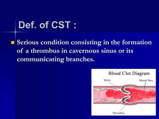
CAVERNOUS SINUS TROMBOSIS
hollow spaces located
under bottom of brain ,
behind each eye socket
◦ Medial : pituitary above, sphenoidal below
◦ Lateral : temporal lobe
◦ Anterior : SOF
◦ Posterior : petrous apex
◦ Superior : optic chiasm
Relations:
Contents
 oculomotor nerve (CN III)
 trochlear nerve (CN IV)
 ophthalmic nerve (CNV1)
 maxillary nerve (CNV2)
 abducens nerve (CN VI)
Connection :
Dangerous area of face
Def. of CST :
 Serious condition consisting in the formation
of a thrombus in cavernous sinus or its
communicating branches.
Etiology:
 Along lateral sinus and petrosal sinuses from
middle ear infections
 •Along venous plexus surrounding ICA from
middle ear
 A dental infection from osteomyelitis of
maxilla
Extension of bacteria
Clinical picture :
 male patient 60 years old, Diabetic 15 year
 presented to dentist with 1-week history
toothache and subsequent painful right
cheek
case study
Imaging Studies
Laboratory test
 CBC
 ESR
 blood cultures
 sinus culture
To identify an infectious primary source
Complication
 Vision problem
Lung : pulmonary embolism
 Brain : triggers a stroke
 Orbital cellulitis :
CST : Bil., papillodema, dilated pupil,
decreased periocular sensation
Preseptal cellulitis : no proptosis
 Orbital apex syndrome : more visual loss,
opthalmoplegia, less proptosis, periorbital
odema
MANAGEMENT
MANAGEMENT
Prognosis
 100% Mortality prior to effective antimicrobials.
death is due to sepsis or CNS infection.
 With aggressive management , the mortality
rate is now less than 30%
Questions?

Cavernous sinus thrombosis

Editor's Notes

  • #5 C ICA
  • #6 C ICA
  • #8 C ICA
  • #15 Mri confirm its presence and to differentiate it from alternatives such as orbital cellulitis.