1) Cavernous sinus thrombosis is a blood clot that forms within the cavernous sinus, which is a venous channel within the skull.
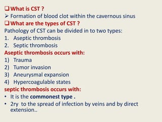
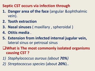
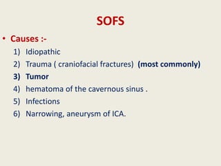
2) It can be aseptic (caused by trauma, tumors, or hypercoagulable states) or septic (caused by bacterial infection spreading from nearby areas like the face, sinuses, or ears).
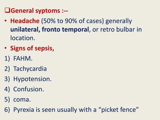
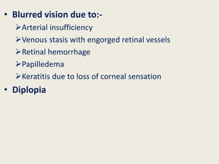
3) The most common symptoms are headaches, signs of sepsis, and ocular issues like proptosis, ophthalmoplegia, ptosis, and blurred vision. Complications can include orbital abscesses, meningitis, or extensions of the clot or infection.
2. What is cavernous sinus ?
intracranial Venous channels
It is formed by the splitting of the Dura matter.
• The 2 cavernous sinus are interconnected by small
anterior and posterior inter cavernous channels
• The lateral wall of cavernous sinus is composed
of 2 layers
1. Superficial layer of dura – dura matter on the medial
side of middle cranial fossa
2. Deep layer of dura – formed by sheath of :
Oculomotor 3
Trochlea 4
Trigeminal(Ophthalmic and Maxillary). 5
3.
4. Cavernous sinus receives blood from the following:
• Face, eyes and maxilla
superior and inferior ophthalmic veins
Pterigoid plexus via emissary veins
• Anteriorly
Sphenoparietal vein
Superficial middle cerebral vein
• Posteriorly
Superior petrosal sinus
Inferior petrosal sinus • It drains into
1. Transverse sinuses
2. Internal jugular vein
5.
6.
7. What is CST ?
Formation of blood clot within the cavernous sinus
What are the types of CST ?
Pathology of CST can be divided in to two types:
1. Aseptic thrombosis
2. Septic thrombosis
Aseptic thrombosis occurs with:
1) Trauma
2) Tumor invasion
3) Aneurysmal expansion
4) Hypercoagulable states
septic thrombosis occurs with:
• It is the commonest type .
• 2ry to the spread of infection by veins and by direct
extension..
8. Septic CST occurs via infection through
1. Danger area of the face (angular &ophthalmic
vein).
2. Tooth extraction
3. Nasal sinuses ( maxillary , spheroidal )
4. Otitis media .
5. Extension from infected internal jugular vein,
lateral sinus or petrosal sinus
What is The most commonly isolated organisms
causing CST ?
1) Staphylococcus aurous (about 70%)
2) Streptococcus species (about 20%)..
9.
10.
11. General syptoms :--
• Headache (50% to 90% of cases) generally
unilateral, fronto temporal, or retro bulbar in
location.
• Signs of sepsis,
1) FAHM.
2) Tachycardia
3) Hypotension.
4) Confusion.
5) coma.
6) Pyrexia is seen usually with a “picket fence”
13. • Blurred vision due to:-
Arterial insufficiency
Venous stasis with engorged retinal vessels
Retinal hemorrhage
Papilledema
Keratitis due to loss of corneal sensation
• Diplopia
14.
15. What are the complications of CST ??
• Orbital abscess .
• Septicemia
• Intracranial extension of infection may result in:-
Meningitis,
Encephalitis,
Brain abscess
• Extension of the thrombus to other sinuses
• Cortical vein thrombosis can result in:-
Hemorrhagic infarction
hemiplegia
16. Differential Diagnosis
1) THS ( painful Ophthalmoplegia ) , no affect on ON.
2) OAS (cranial nerve palsy +optic nerve dysfunction).
3) Myositis .
4) Orbital cellulitis
5) Orbital tumor
6) Orbital pseudo tumor .
21. Medical Management
ANTIBIOTICS
• Broad spectrum iv antibiotics – 3 to 4 weeks but if
evidence of intracranial suppuration then 6 to 8 weeks.
i.e.
1. Ceftriaxone IV
2. Metronidazole IV
ANTICOAGULANTS .(ASAP )
• The proposed benefit is to dissolve the clot E.g. heparin .
STEROID THERAPY
• Helps reduce level of inflammation
22. Surgical Management
SURGICAL MANAGEMENT
• Surgical intervention should be directed at the
primary source of the infection and the
surrounding areas of involvement.
• Incision and drainage should be done ASAP.
26. SUPERIOR ORBITAL FISSURE
• 22 mm long
• It communicate ( ) the orbit & the middle cranial
fossa
• Lateral superior part of the fissure is narrower
than the medial inferior part.
27. SOFS
• Causes :-
1) Idiopathic
2) Trauma ( craniofacial fractures) (most commonly)
3) Tumor
4) hematoma of the cavernous sinus .
5) Infections
6) Narrowing, aneurysm of ICA.
28. Clinical picture :- DOPPE ( diplopia , Ophthalmoplegia , ptosis , Proptosis ).
It occurs as a result of inflammation & Compression of
adjacent nervous tissue
• Diplopia
• Orbital pain .
• Lid ptosis
– Sympathetic fibers in CS – Mullers muscle
– Efferent fibers Superior Oculomotor Nerve – Levetor
palpebrae superioris muscle
• Ophthalmoplegia
– Impairment of cranial nerves III, IV, and VI.
• Anesthesia of the forehead and upper eyelid.
• Proptosis
– loss of tone of EOM muscles ..
29.
30.
31. Investigations
1) FBP & ESR
2) Blood culture and sensitivity
3) CT/MRI Brain and orbit
4) Angiogram
35. Treatment
1. Exploration .
2. Treatment of the cause
3. Steroids
4. Antibiotics
5. surgical intervention .
a) Orbitotomy.
b) Orbital decompression .