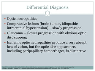
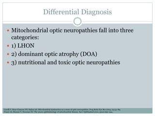
A D E W I J A Y A , M D – J U L Y 2 0 2 1
Leber’s Hereditary
Optic Neuropathy (LHON)
Introduction
 Maternally inherited disease characterized by severe
visual loss, which usually does not manifest until
young adulthood
 First formally defined by Theodor Leber in 1871
 Mitochondrial DNA (mtDNA) mutation
Carelli V, Ross-Cisneros FN, Sadun AA: Mitochondrial dysfunction as a cause of optic neuropathies. Prog Retin Eye Res 2004, 23:53–89.
Fraser JA, Biousse V, Newman NJ: The neuro-ophthalmology of mitochondrial disease. Surv Ophthalmol 2010, 55(4):299–334.
Epidemiology
 Prevalence  1 : 30.000 – 50.000
 Male (80-90%) >> Female
 Second or third decade of life
 Environmental factors may trigger the visual loss but
do not fully explain why only certain individuals
within a family become symptomatic.
Fraser JA, Biousse V, Newman NJ: The neuro-ophthalmology of mitochondrial disease. Surv Ophthalmol 2010, 55(4):299–334.
Pathophysiology
 mtDNA mutation, which is a necessary determinant
but not sufficient to lead to visual loss
 Bioenergetic defect & Chronic oxidative stress lead to
changes in mitochondrial membrane potential,
lowering the threshold for the mitochondrial
permeability transition pore (MPTP) opening and
initiating mitochondrially driven apoptosis
 Histopathology: a dramatic loss of retinal ganglion
cells and their axons
Carelli V, Ross-Cisneros FN, Sadun AA: Mitochondrial dysfunction as a cause of optic neuropathies. Prog Retin Eye Res 2004, 23:53–89.
Fraser JA, Biousse V, Newman NJ: The neuro-ophthalmology of mitochondrial disease. Surv Ophthalmol 2010, 55(4):299–334.
Sadun AA, Win PH, Ross-Cisneros FN, et al.: Leber’s hereditary optic neuropathy differentially affects smaller axons in the optic nerve. Trans Am Ophthalmol Soc 2000, 98:223–
232. discussion 232–235.
Clinical Presentation
 The patient classically presents with painless,
subacute loss of vision in one eye  progress to
bilateral in weeks or months later (usually less than a
year)
 The visual acuity is usually worse than 20/400. loss
of color vision
 Optic nerve dysfunctions: large and dense central or
cecocentral scotomas on visual fields
Barboni P, Carbonelli M, Savini G, et al.: Natural history of Leber’s hereditary optic neuropathy: longitudinal analysis of the retinal nerve fiber layer by optical coherence
tomography. Ophthalmology 2010, 117(3):623–627.
Sadun AA, Salomao SR, Berezovsky A, et al.: Subclinical carriers and conversions in Leber hereditary optic neuropathy: a prospective psychophysical study. Trans Am
Ophthalmol Soc 2006, 104:51–61.
Barboni P, Savini G, Valentino ML, et al.: Retinal nerve fiber layer evaluation by optical coherence tomography in Leber’s hereditary optic neuropathy. Ophthalmology
2005, 112:120–126.
Clinical Presentation
 Fundus examination in LHON may show
telangiectatic capillaries and pseudoedema of the
optic disc with surrounding swelling of the retinal
nerve fibre layers  progressing to optic atrophy
 LHON is usually permanent, although a subgroup of
patients may spontaneously recover some visual
acuity. This recovery is particularly frequent with the
14484/ND6 mutation
Sadun AA, La Morgia C, Carelli V. Leber’s hereditary optic neuropathy. Current treatment options in neurology. 2011 Feb;13(1):109-17.
Diagnosis
 Clinical diagnosis
 Confirmation can be made by blood testing of the mtDNA to
reveal one of the three common mutations (3460/ND1 ;
11778/ND4 ; 14484/ND6-milder)
 Complete mtDNA sequence analysis may be recommended if
the clinical diagnosis of LHON remains as a strong indication
despite negative most common mutations
 DNA testing of primary LHON mutations is especially useful
in atypical presentations or in the absence of a clear family
history of LHON or optic atrophy of unknown etiology limited
to the maternal side of the pedigree
 Ophthalmologic and psychophysical tests
 MRI
Sadun AA, La Morgia C, Carelli V. Leber’s hereditary optic neuropathy. Current treatment options in neurology. 2011 Feb;13(1):109-17.
T2 hyperintensity in both the optic nerves
(left) and the optic chiasm (right)
Differential Diagnosis
 Optic neuropathies
 Compressive lesions (brain tumor, idiopathic
intracranial hypertension) – slowly progression
 Glaucoma – slower progression with obvious optic
disc cupping
 Ischemic optic neuropathies produce a very abrupt
loss of vision, but the optic disc appearance,
including peripapillary hemorrhages, is distinctive
Carelli V, Ross-Cisneros FN, Sadun AA: Mitochondrial dysfunction as a cause of optic neuropathies. Prog Retin Eye Res 2004, 23:53–89.
Fraser JA, Biousse V, Newman NJ: The neuro-ophthalmology of mitochondrial disease. Surv Ophthalmol 2010, 55(4):299–334.
Differential Diagnosis
 Mitochondrial optic neuropathies fall into three
categories:
 1) LHON
 2) dominant optic atrophy (DOA)
 3) nutritional and toxic optic neuropathies
Carelli V, Ross-Cisneros FN, Sadun AA: Mitochondrial dysfunction as a cause of optic neuropathies. Prog Retin Eye Res 2004, 23:53–89.
Fraser JA, Biousse V, Newman NJ: The neuro-ophthalmology of mitochondrial disease. Surv Ophthalmol 2010, 55(4):299–334.
Dominant Optic Atrophy (DOA)
 The disease segregation in DOA will involve paternal
as well as maternal transmission
 Furthermore, the visual loss occurs at a younger age
(usually before age 10)
 Progresses slowly over many years, often leveling off
at 20/100 or 20/ 200
Carelli V, Ross-Cisneros FN, Sadun AA: Mitochondrial dysfunction as a cause of optic neuropathies. Prog Retin Eye Res 2004, 23:53–89.
Fraser JA, Biousse V, Newman NJ: The neuro-ophthalmology of mitochondrial disease. Surv Ophthalmol 2010, 55(4):299–334.
Nutritional and Toxic Optic Neuropathies
 Folate and vitamin B deficiencies are usually
associated with a very poor diet over a long course
 There may also be an associated anemia
 Toxic agents that can produce a mitochondrial optic
neuropathy include several antibiotics
Sadun AA, Carelli V, Salomao SR, et al.: A very large Brazilian pedigree with 11778 Leber’s hereditary optic neuropathy. Trans Am Ophthalmol Soc 2002, 100:169–178. discussion 178–179.
Sadun AA, Carelli V, Salomao SR, et al.: Extensive investigation of a large Brazilian pedigree of 11778/haplogroup J Leber hereditary optic neuropathy. Am J Ophthalmol 2003, 136:231–238.
Kirkman MA, Yu-Wai-Man P, Korsten A, et al.: Geneenvironment interactions in Leber hereditary optic neuropathy. Brain 2009, 132:2317–2326.
Differential Diagnosis
Carelli V, Carbonelli M, de Coo I, et al. International consensus statement on the clinical and therapeutic management of Leber hereditary optic neuropathy. J Neuro-
Ophthalmol. 2017;37:371-381.
Bennett JL. Optic neuritis. Continuum. 2019;25:1236-1264.
Antibiotics and drugs Toxins
 Ethambutol
 Aminoglycosides
Chloramphenicol
 Linezolid
 Zidovudine (AZT) and
other antiretroviral
drugs
 Smoke (including
tobacco)
 Ethanol
 Pesticides
 Cyanide
 Methanol
Risk Factors
Sadun AA, La Morgia C, Carelli V. Leber’s hereditary optic neuropathy. Current treatment options in neurology. 2011 Feb;13(1):109-17.
Treatment
 There are currently no proven treatments to prevent
or reverse the optic neuropathy in LHON
 Experimental therapeutic strategies
 Education
Sadun AA, La Morgia C, Carelli V. Leber’s hereditary optic neuropathy. Current treatment options in neurology. 2011 Feb;13(1):109-17.
Treatment
 Idebenone is a short-chain benzoquinone that
improves the shuttling of electrons from complexes I
and II directly to complex III in the mitochondrial
electron transport chain.
 It is approved in Europe to treat visual impairment
in adults and adolescents with LHON
 The EMA-approved dose for idebenone is 900
mg/day, divided into three 300-mg doses daily for at
least 18-24 months. The most common adverse
events are gastrointestinal, and the majority of them
are mild
Gaier ED, Boudreault K, Nakata I, et al. Diagnostic genetic testing for patients with bilateral optic neuropathy and comparison of clinical features according
to OPA1 mutation status. Mol Vision. 2017;23:548560.
Raxone® (idebenone) Product Information. European Medicines Agency (EMA). Published September 8, 2015. Updated August 8, 2020. Accessed March 31, 2021.
https://www.ema.europa.eu/en/documents/product-information/raxone-epar-product-information_en.pdf
Recent Treatment Development
Zuccarelli M, Vella-Szijj J, Serracino-Inglott A, Borg JJ. Treatment of Leber’s hereditary optic neuropathy: An overview of recent developments. European Journal of
Ophthalmology. 2020 Nov;30(6):1220-7.
THANK YOU

Leber’s Hereditary Optic Neuropathy

  • 1.
    A D EW I J A Y A , M D – J U L Y 2 0 2 1 Leber’s Hereditary Optic Neuropathy (LHON)
  • 2.
    Introduction  Maternally inheriteddisease characterized by severe visual loss, which usually does not manifest until young adulthood  First formally defined by Theodor Leber in 1871  Mitochondrial DNA (mtDNA) mutation Carelli V, Ross-Cisneros FN, Sadun AA: Mitochondrial dysfunction as a cause of optic neuropathies. Prog Retin Eye Res 2004, 23:53–89. Fraser JA, Biousse V, Newman NJ: The neuro-ophthalmology of mitochondrial disease. Surv Ophthalmol 2010, 55(4):299–334.
  • 3.
    Epidemiology  Prevalence 1 : 30.000 – 50.000  Male (80-90%) >> Female  Second or third decade of life  Environmental factors may trigger the visual loss but do not fully explain why only certain individuals within a family become symptomatic. Fraser JA, Biousse V, Newman NJ: The neuro-ophthalmology of mitochondrial disease. Surv Ophthalmol 2010, 55(4):299–334.
  • 4.
    Pathophysiology  mtDNA mutation,which is a necessary determinant but not sufficient to lead to visual loss  Bioenergetic defect & Chronic oxidative stress lead to changes in mitochondrial membrane potential, lowering the threshold for the mitochondrial permeability transition pore (MPTP) opening and initiating mitochondrially driven apoptosis  Histopathology: a dramatic loss of retinal ganglion cells and their axons Carelli V, Ross-Cisneros FN, Sadun AA: Mitochondrial dysfunction as a cause of optic neuropathies. Prog Retin Eye Res 2004, 23:53–89. Fraser JA, Biousse V, Newman NJ: The neuro-ophthalmology of mitochondrial disease. Surv Ophthalmol 2010, 55(4):299–334. Sadun AA, Win PH, Ross-Cisneros FN, et al.: Leber’s hereditary optic neuropathy differentially affects smaller axons in the optic nerve. Trans Am Ophthalmol Soc 2000, 98:223– 232. discussion 232–235.
  • 5.
    Clinical Presentation  Thepatient classically presents with painless, subacute loss of vision in one eye  progress to bilateral in weeks or months later (usually less than a year)  The visual acuity is usually worse than 20/400. loss of color vision  Optic nerve dysfunctions: large and dense central or cecocentral scotomas on visual fields Barboni P, Carbonelli M, Savini G, et al.: Natural history of Leber’s hereditary optic neuropathy: longitudinal analysis of the retinal nerve fiber layer by optical coherence tomography. Ophthalmology 2010, 117(3):623–627. Sadun AA, Salomao SR, Berezovsky A, et al.: Subclinical carriers and conversions in Leber hereditary optic neuropathy: a prospective psychophysical study. Trans Am Ophthalmol Soc 2006, 104:51–61. Barboni P, Savini G, Valentino ML, et al.: Retinal nerve fiber layer evaluation by optical coherence tomography in Leber’s hereditary optic neuropathy. Ophthalmology 2005, 112:120–126.
  • 6.
    Clinical Presentation  Fundusexamination in LHON may show telangiectatic capillaries and pseudoedema of the optic disc with surrounding swelling of the retinal nerve fibre layers  progressing to optic atrophy  LHON is usually permanent, although a subgroup of patients may spontaneously recover some visual acuity. This recovery is particularly frequent with the 14484/ND6 mutation Sadun AA, La Morgia C, Carelli V. Leber’s hereditary optic neuropathy. Current treatment options in neurology. 2011 Feb;13(1):109-17.
  • 7.
    Diagnosis  Clinical diagnosis Confirmation can be made by blood testing of the mtDNA to reveal one of the three common mutations (3460/ND1 ; 11778/ND4 ; 14484/ND6-milder)  Complete mtDNA sequence analysis may be recommended if the clinical diagnosis of LHON remains as a strong indication despite negative most common mutations  DNA testing of primary LHON mutations is especially useful in atypical presentations or in the absence of a clear family history of LHON or optic atrophy of unknown etiology limited to the maternal side of the pedigree  Ophthalmologic and psychophysical tests  MRI Sadun AA, La Morgia C, Carelli V. Leber’s hereditary optic neuropathy. Current treatment options in neurology. 2011 Feb;13(1):109-17.
  • 8.
    T2 hyperintensity inboth the optic nerves (left) and the optic chiasm (right)
  • 9.
    Differential Diagnosis  Opticneuropathies  Compressive lesions (brain tumor, idiopathic intracranial hypertension) – slowly progression  Glaucoma – slower progression with obvious optic disc cupping  Ischemic optic neuropathies produce a very abrupt loss of vision, but the optic disc appearance, including peripapillary hemorrhages, is distinctive Carelli V, Ross-Cisneros FN, Sadun AA: Mitochondrial dysfunction as a cause of optic neuropathies. Prog Retin Eye Res 2004, 23:53–89. Fraser JA, Biousse V, Newman NJ: The neuro-ophthalmology of mitochondrial disease. Surv Ophthalmol 2010, 55(4):299–334.
  • 10.
    Differential Diagnosis  Mitochondrialoptic neuropathies fall into three categories:  1) LHON  2) dominant optic atrophy (DOA)  3) nutritional and toxic optic neuropathies Carelli V, Ross-Cisneros FN, Sadun AA: Mitochondrial dysfunction as a cause of optic neuropathies. Prog Retin Eye Res 2004, 23:53–89. Fraser JA, Biousse V, Newman NJ: The neuro-ophthalmology of mitochondrial disease. Surv Ophthalmol 2010, 55(4):299–334.
  • 11.
    Dominant Optic Atrophy(DOA)  The disease segregation in DOA will involve paternal as well as maternal transmission  Furthermore, the visual loss occurs at a younger age (usually before age 10)  Progresses slowly over many years, often leveling off at 20/100 or 20/ 200 Carelli V, Ross-Cisneros FN, Sadun AA: Mitochondrial dysfunction as a cause of optic neuropathies. Prog Retin Eye Res 2004, 23:53–89. Fraser JA, Biousse V, Newman NJ: The neuro-ophthalmology of mitochondrial disease. Surv Ophthalmol 2010, 55(4):299–334.
  • 12.
    Nutritional and ToxicOptic Neuropathies  Folate and vitamin B deficiencies are usually associated with a very poor diet over a long course  There may also be an associated anemia  Toxic agents that can produce a mitochondrial optic neuropathy include several antibiotics Sadun AA, Carelli V, Salomao SR, et al.: A very large Brazilian pedigree with 11778 Leber’s hereditary optic neuropathy. Trans Am Ophthalmol Soc 2002, 100:169–178. discussion 178–179. Sadun AA, Carelli V, Salomao SR, et al.: Extensive investigation of a large Brazilian pedigree of 11778/haplogroup J Leber hereditary optic neuropathy. Am J Ophthalmol 2003, 136:231–238. Kirkman MA, Yu-Wai-Man P, Korsten A, et al.: Geneenvironment interactions in Leber hereditary optic neuropathy. Brain 2009, 132:2317–2326.
  • 13.
    Differential Diagnosis Carelli V,Carbonelli M, de Coo I, et al. International consensus statement on the clinical and therapeutic management of Leber hereditary optic neuropathy. J Neuro- Ophthalmol. 2017;37:371-381. Bennett JL. Optic neuritis. Continuum. 2019;25:1236-1264.
  • 14.
    Antibiotics and drugsToxins  Ethambutol  Aminoglycosides Chloramphenicol  Linezolid  Zidovudine (AZT) and other antiretroviral drugs  Smoke (including tobacco)  Ethanol  Pesticides  Cyanide  Methanol Risk Factors Sadun AA, La Morgia C, Carelli V. Leber’s hereditary optic neuropathy. Current treatment options in neurology. 2011 Feb;13(1):109-17.
  • 15.
    Treatment  There arecurrently no proven treatments to prevent or reverse the optic neuropathy in LHON  Experimental therapeutic strategies  Education Sadun AA, La Morgia C, Carelli V. Leber’s hereditary optic neuropathy. Current treatment options in neurology. 2011 Feb;13(1):109-17.
  • 16.
    Treatment  Idebenone isa short-chain benzoquinone that improves the shuttling of electrons from complexes I and II directly to complex III in the mitochondrial electron transport chain.  It is approved in Europe to treat visual impairment in adults and adolescents with LHON  The EMA-approved dose for idebenone is 900 mg/day, divided into three 300-mg doses daily for at least 18-24 months. The most common adverse events are gastrointestinal, and the majority of them are mild Gaier ED, Boudreault K, Nakata I, et al. Diagnostic genetic testing for patients with bilateral optic neuropathy and comparison of clinical features according to OPA1 mutation status. Mol Vision. 2017;23:548560. Raxone® (idebenone) Product Information. European Medicines Agency (EMA). Published September 8, 2015. Updated August 8, 2020. Accessed March 31, 2021. https://www.ema.europa.eu/en/documents/product-information/raxone-epar-product-information_en.pdf
  • 17.
    Recent Treatment Development ZuccarelliM, Vella-Szijj J, Serracino-Inglott A, Borg JJ. Treatment of Leber’s hereditary optic neuropathy: An overview of recent developments. European Journal of Ophthalmology. 2020 Nov;30(6):1220-7.
  • 18.