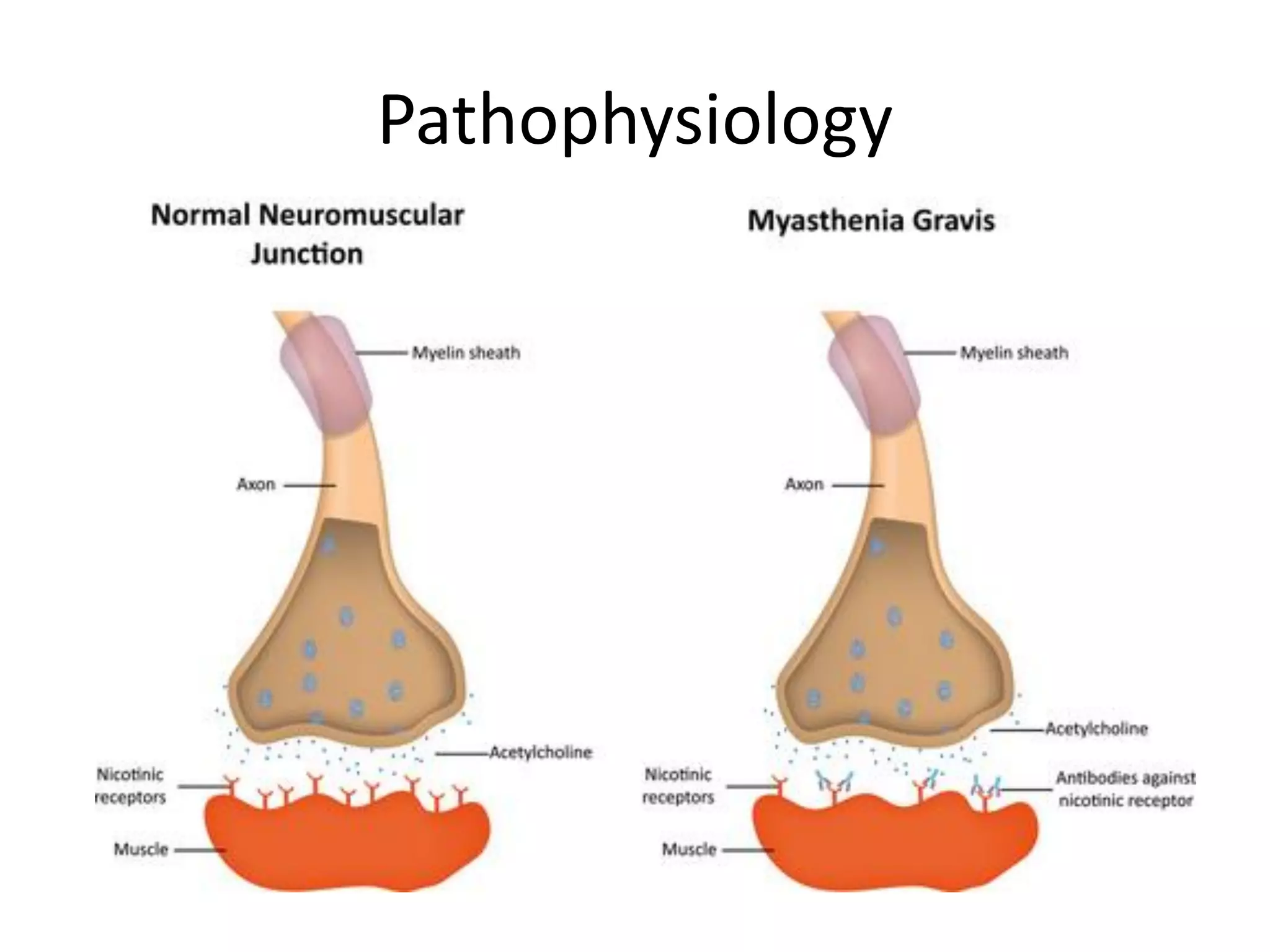
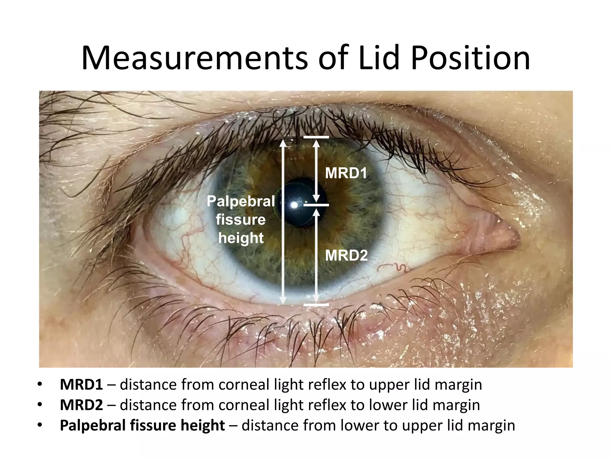
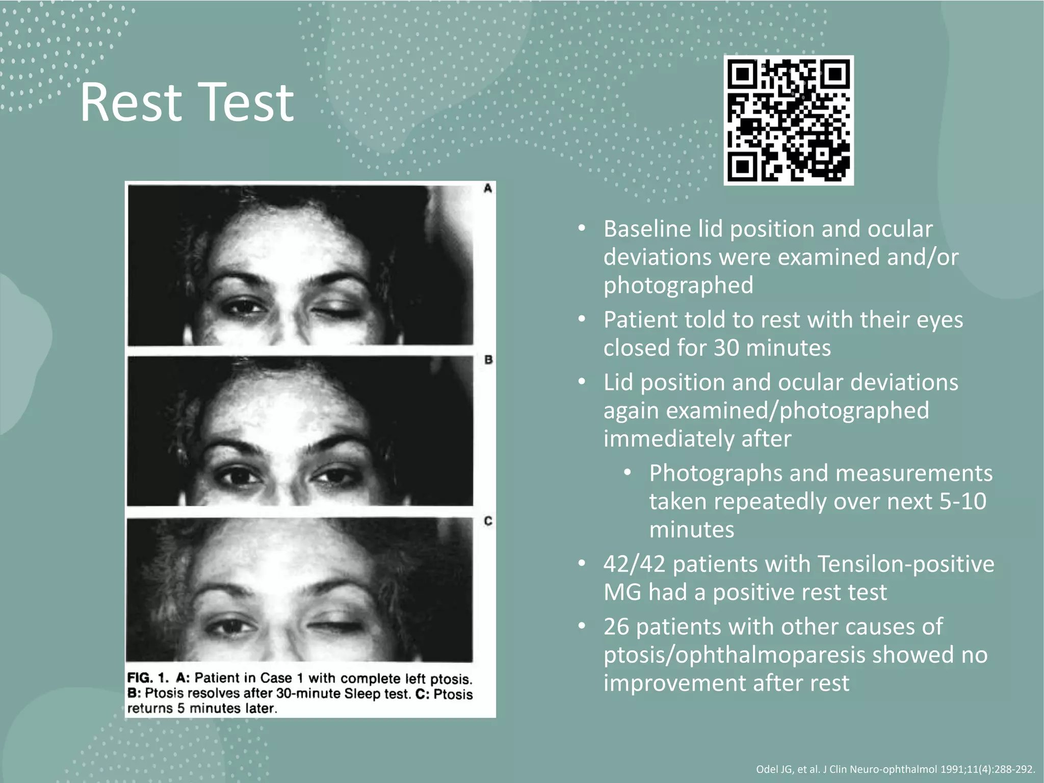
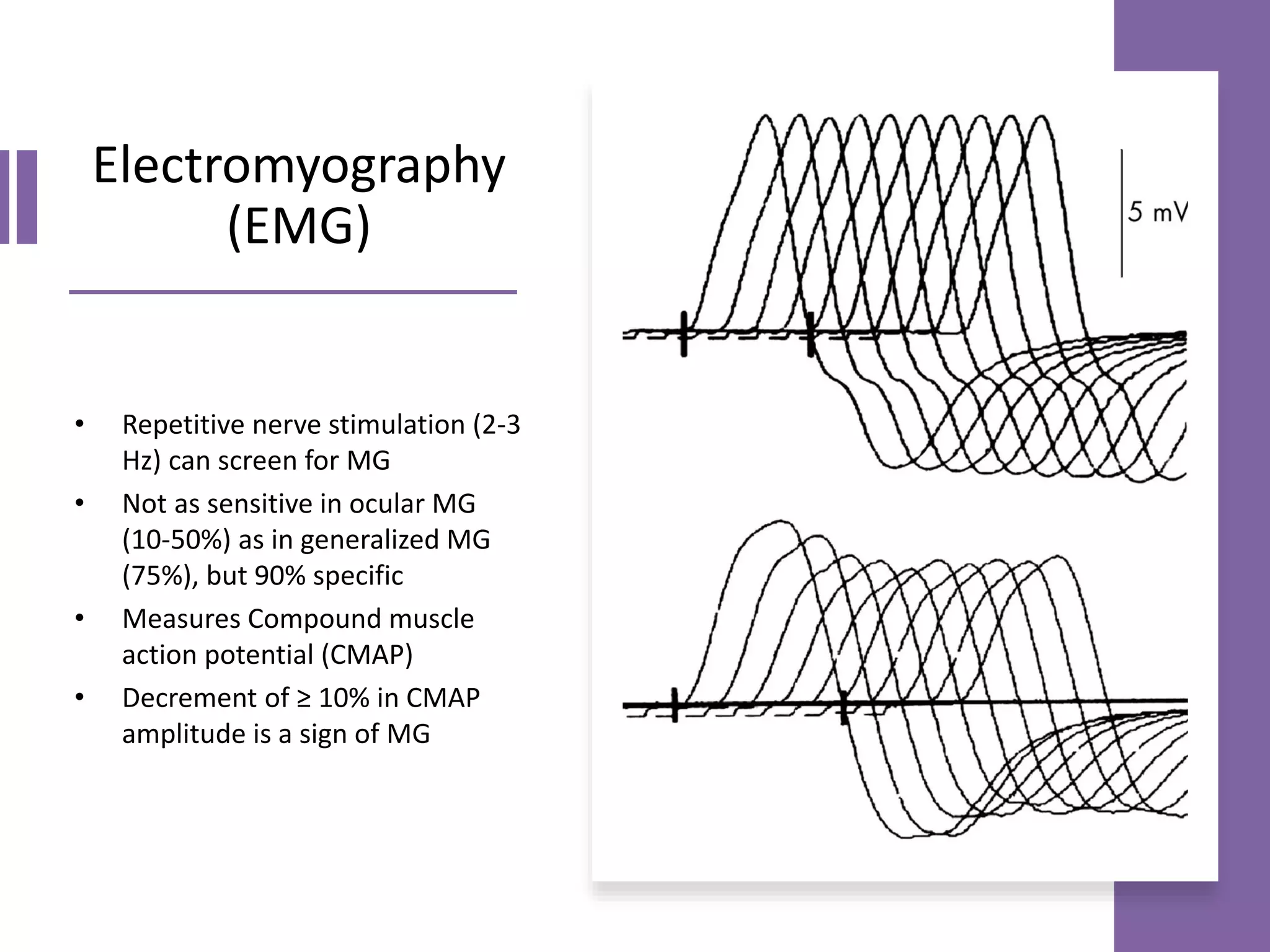
This document provides an overview of ocular myasthenia gravis (OMG), including pathophysiology, clinical presentation, diagnostic testing, and management. Key points include: OMG is caused by antibodies impairing acetylcholine receptors, causing muscle weakness that worsens with use. Common symptoms are fluctuating ptosis and diplopia. Diagnosis involves history, exam findings like fatigable ptosis, and may include tests like ice pack, Tensilon/neostigmine, EMG. Treatment focuses on pyridostigmine and immunosuppression with prednisone and azathioprine. Thymectomy may be considered for generalized cases under age 50.
This document provides information about ocular myasthenia gravis (OMG). It begins by defining myasthenia gravis and OMG, which is when symptoms are isolated to eye muscles. It then discusses the pathogenesis, including that OMG is often associated with acetylcholine receptor antibodies. Clinical features of OMG include ptosis, diplopia, and fatigable eye muscle weakness. The document contrasts OMG with generalized myasthenia gravis and reviews tests to diagnose OMG, including ice pack, Tensilon, antibody, and electrophysiological testing. It concludes with discussing prognosis, treatment considerations like immunotherapy and thymectomy, and symptomatic treatments for ptosis and diplopia.
SIXTH CRANIAL NERVE PALSY- Diagnosis and managementDrArvindMorya
Cover the fixing eye
Examiner: Note movement of uncovered eye
- Esotropia in primary position
- Exotropia on looking towards affected side
- No movement on looking towards normal side
- Quantifies the deviation
- Helps in diagnosis and management
Past pointing test
- Patient asked to point with finger to target
- Deviation of finger from target indicates diplopia
- Helps in localizing the diplopia
Diplopia charting
- Patient asked to draw position of second image
- Helps in localizing and quantifying diplopia
Worth 4 dot test
- Assesses binocular single vision
- Loss of fusion indicates diplop
This document discusses the anatomy and function of the oculomotor nerve (cranial nerve 3). It describes the nucleus and course of the nerve, causes of lesions, clinical features of total oculomotor nerve palsy, treatment options which may include surgery or monitoring, and differential diagnoses. History, examination, and investigations are outlined to evaluate patients presenting with oculomotor nerve palsies.
Optic neuritis is inflammation of the optic nerve that commonly presents as sudden, unilateral vision loss. It affects young females predominantly and is often the initial presentation of multiple sclerosis. Treatment with intravenous steroids followed by oral steroids accelerates visual recovery but provides no long-term benefit over oral steroids alone. Early treatment of multiple sclerosis after an initial demyelinating event such as optic neuritis can reduce future relapse rates and disability progression.
This document provides an overview of nystagmus, including its definition, terminology, types, causes, and characteristics. Nystagmus is defined as a repetitive, to-and-fro movement of the eyes. It is classified as congenital or acquired, and includes types such as infantile nystagmus, spasmus nutans, end point nystagmus, vestibular nystagmus, optokinetic nystagmus, downbeat nystagmus, and nystagmus associated with strabismus. The document discusses the mechanisms, features, and treatments of different forms of nystagmus.
Chronic progressive external ophthalmoplegiaPS Deb
Chronic progressive external ophthalmoplegia (CPEO) is a descriptive term for a heterogeneous group of disorders characterized by chronic, progressive, bilateral, and usually symmetric ocular motility deficit and ptosis, without pain, proptosis and pupil involvement. Commonly a syndrome of Mitochondrial Cytopathy.
This document provides an overview of sixth nerve palsy, including:
- The sixth cranial nerve innervates the lateral rectus muscle to enable eye abduction. Sixth nerve palsy results in limited ability to turn the eye outward.
- Causes of sixth nerve palsy include idiopathic, vascular issues like hypertension and diabetes, trauma, and tumors. Symptoms include esotropia and diplopia.
- Diagnosis involves assessing eye movement limitations and diplopia. Treatment options include occlusion to control diplopia, botulinum toxin injection, or strabismus surgery if no spontaneous recovery occurs. Prognosis is generally good, with many cases recovering spontaneously in
The document describes the anatomy and causes of various cranial nerve palsies. It discusses the third, fourth, sixth, and trochlear nerves. For each nerve, it outlines the nuclear location, anatomical course, common causes of palsy for adults and children, associated signs and symptoms, and important diagnostic considerations. Evaluation may include medical history, examination of eye movements and pupil function, and neuroimaging in certain cases to identify potentially compressive lesions.
This document summarizes various types of gaze palsies, including their causes and clinical presentations. Supranuclear gaze palsies can result from lesions in areas that control eye movements, like the brainstem or cerebral cortex. Clinical exams help localize lesions and differentiate organic from functional disorders. Specific syndromes discussed include Parinaud's syndrome, progressive supranuclear palsy, internuclear ophthalmoplegia, and one-and-a-half syndrome. The document provides details on symptoms, locations of lesions, and distinguishing features of different supranuclear gaze palsy conditions.
This document provides information on key anatomical structures of the fundus including:
1) It describes several structures that can be seen on examination of the fundus including the optic disc, macula, retinal blood vessels, and peripheral retina.
2) It explains features of the optic disc including that it is insensitive, round or oval in shape, and contains the lamina cribrosa through which ganglion cell axons exit the eye.
3) The macula is described as being located temporally from the optic disc, containing the highest concentration of cones, and responsible for photopic vision and color vision.
This document provides an overview of optic atrophy, including:
1. It defines optic atrophy as degeneration of the optic nerve due to damage to the visual pathways from the retina to the lateral geniculate body.
2. It classifies optic atrophy based on whether damage originates in the retina or more centrally, and by cause. Primary optic atrophy occurs without swelling, while secondary involves prior swelling.
3. Causes of primary optic atrophy include optic neuritis, compression, hereditary conditions, toxins, trauma, and multiple sclerosis. Secondary optic atrophy follows conditions like papilledema.
4. Treatment focuses on the underlying cause, with vitamins sometimes used
This document discusses approaches to vision loss. It defines various patterns of vision loss including transient monocular, persistent monocular, and binocular vision loss. It describes causes of transient monocular vision loss such as amaurosis fugax which can be due to circulatory, ocular, or neurologic factors. Key factors in evaluating vision loss include whether it is monocular or binocular, the pattern and degree of loss, tempo of onset, and associated symptoms. Common causes of sudden monocular vision loss discussed include central retinal artery occlusion and optic neuritis.
Thyroid eye disease (TED), also known as Graves' ophthalmopathy, is an autoimmune disorder affecting the eyes that is commonly associated with Graves' disease and hyperthyroidism. It causes inflammation and swelling of the muscles and fatty tissues behind the eyes. The document discusses the epidemiology, risk factors, pathogenesis, clinical features including proptosis, lid retraction, optic neuropathy, and restrictive myopathy, as well as treatments such as steroids, radiation, orbital decompression surgery, and eyelid surgery. Management involves treatment of both the eye symptoms and any underlying thyroid abnormalities.
Primary optic atrophy occurs due to direct damage to the optic nerve and results in chalky white disc color with well defined margins and normal cupping and vessels. Secondary optic atrophy follows conditions like papilledema that cause swelling first, resulting in a filled cup and dirty white color. Consecutive optic atrophy occurs after other retinal conditions and shows waxy pallor, normal cup and grossly thinned vessels.
This document provides an overview of blepharoptosis (ptosis) including its classification, evaluation, and case study. It begins with an introduction to ptosis and eyelid anatomy. It then covers the types of ptosis such as congenital, acquired, and pseudo ptosis. The evaluation section details the history taking, inspection, measurements including levator function, margin reflex distance, and margin crease distance. It also discusses ancillary tests and differential diagnosis. Overall, the document serves as a guide for clinicians to classify and evaluate different types of ptosis.
This document provides information about idiopathic intracranial hypertension (IIH), including its symptoms, pathophysiology, risk factors, diagnosis, and treatment. IIH is characterized by increased intracranial pressure without a tumor or other identifiable cause. Common symptoms include headache, nausea, visual disturbances, and papilledema. The main risk factors are obesity, particularly in women of childbearing age. The cause is unknown but may involve increased cerebrospinal fluid production or impaired cerebral venous drainage. Diagnosis involves evaluating signs and symptoms of increased intracranial pressure and ruling out other potential causes through imaging and lumbar puncture.
This document discusses various systemic diseases that can manifest ocularly and be detected on eye examination. It covers 10 categories of disease including congenital, traumatic, vascular, neoplastic, autoimmune, idiopathic, infectious, metabolic/endocrine, and drugs/toxins. For each category, specific diseases are described along with their characteristic ocular signs and symptoms. The importance of comprehensive eye exams for evaluating systemic health is emphasized.
This document provides information on how to evaluate blepharoptosis (drooping of the upper eyelid) clinically. It discusses classifications of ptosis as congenital or acquired. Evaluation involves measuring eyelid positions, assessing levator function and other muscle movements. Tests like phenylephrine, ice and edrophonium help determine the cause. Thorough history, examination and documentation including photographs are needed before considering surgical management of ptosis.
This document provides information about nystagmus, including its definition, mechanisms, causes, types, and clinical features. Some key points:
- Nystagmus is a periodic rhythmic oscillation of the eyes, characterized by a slow drift in one direction followed by a fast corrective movement. It can be caused by disturbances of vision, eye movements, or the vestibulo-ocular reflex.
- Types of nystagmus include jerk nystagmus, pendular nystagmus, see-saw nystagmus, convergence-retraction nystagmus, and various forms of congenital or acquired nystagmus.
- Causes can include lesions of the
This document discusses papilledema, which is swelling of the optic disc due to increased intracranial pressure. It defines papilledema and outlines its causes, signs, symptoms, grading, histopathology, investigations, differential diagnosis, and treatment. The main points are that papilledema is caused by increased intracranial pressure, it can be graded on a scale from 0-5 based on severity, and treatment involves addressing the underlying cause of pressure increase as well as surgical decompression in severe cases to prevent vision loss.
This document discusses supranuclear pathways and lesions that can affect eye movements. It begins with the fundamentals of extraocular movements and anatomy of cortical and brainstem centers that control eye movements. It then covers the basic types of eye movements like saccades, smooth pursuit, vestibular-ocular reflex, and vergence movements. It provides a step-wise approach to evaluating eye movement disorders and localizing lesions based on the type of eye movement affected. Supranuclear lesions can cause bilateral eye movement involvement, while specific brainstem lesions impact horizontal or vertical eye movements or specific eye movement types like saccades or vestibular-ocular reflex.
Optic atrophy refers to degeneration of the optic nerve resulting in vision loss and changes in the appearance of the optic disc. It can be primary, secondary, consecutive, or glaucomatous. Primary optic atrophy is caused by lesions along the visual pathway and results in a chalky white disc. Secondary optic atrophy follows optic nerve swelling and has a dirty grey-white appearance. Consecutive optic atrophy is caused by retinal or choroidal disease and leaves a waxy pallor. Glaucomatous optic atrophy is characterized by deep cupping of the disc. Diagnosis involves imaging and visual field testing while treatment focuses on the underlying cause.
Diplopia, or double vision, can be caused by ocular misalignment or optical abnormalities. The document discusses various types of diplopia including monocular and binocular diplopia. It describes how to evaluate a patient with diplopia through history, physical exam, and tests to determine the underlying cause which may be supranuclear, nuclear, internuclear, infranuclear, restrictive or orbital issues. Key examination findings that help localize the source of diplopia are discussed.
This document discusses papilloedema, which refers to optic disc swelling caused by increased intracranial pressure. It defines various types of optic disc swelling including unilateral, bilateral, and papilloedema. It also discusses pseudo-papilloedema. The causes, signs, symptoms, diagnostic workup, and grading scale of papilloedema are explained in detail. Treatment options for increased intracranial pressure including idiopathic intracranial hypertension involve medical management with carbonic anhydrase inhibitors and weight loss as well as potential surgical interventions.
This document provides information on anterior ischemic optic neuropathy (AION), which is the most common cause of acute optic neuropathy in older age groups. It can be divided into two types: arteritic AION, which is due to giant cell arteritis; and non-arteritic AION, which makes up most cases. Both types present with sudden painless vision loss and optic disc swelling. Arteritic AION carries a worse prognosis and requires high-dose steroid treatment to prevent loss of vision in the fellow eye. Non-arteritic AION has a variable course but generally a poor rate of recovery without any proven effective treatments.
The visual pathway begins in the retina and passes through the optic nerves, optic chiasm, optic tracts, lateral geniculate nucleus, optic radiations, and terminates in the occipital lobe. Lesions along this pathway can cause various visual field defects including hemianopias and quadrantanopias depending on the location of the lesion. Lesions in the optic nerve cause blindness on the affected side while lesions in the optic chiasm or tracts cause incongruous homonymous hemianopias. Lesions of the optic radiations or visual cortex cause congruous homonymous defects.
This document discusses various types of ischemic optic neuropathy (ION). It begins by introducing ION as a major cause of vision loss and outlines its classification into anterior and posterior forms. It then details the anatomy and vascular supply of the optic nerve, risk factors for ION such as nocturnal blood pressure changes, and the pathogenesis involving hypoperfusion and axonal swelling. Non-arteritic anterior ION is described as the most common type, while posterior ION and arteritic forms are less prevalent but can involve vascular inflammation. The document outlines signs, investigations, management approaches including steroids, and variable prognoses for the different ION types.
Horner's syndrome and Internuclear ophthalmoplegiaAnkit Raiyani
1. The document describes a case of a 26-year-old male presenting with progressive weakness in his right upper limb over 5 years and difficulty walking due to tightness in his right lower limb for 3 months. On examination, he showed signs of Horner's syndrome and LMN weakness in his right upper limb with UMN involvement in his right lower limb.
2. Imaging revealed basilar invagination and syringohydromyelia extending from C2-D9. He was diagnosed with a CV junction anomaly (basilar invagination) with dorsal syringohydromyelia.
3. The document then discusses Horner's syndrome, its localization, and
This document provides an overview of neuromuscular junction disorders with a focus on Myasthenia Gravis. It discusses the anatomy and physiology of the neuromuscular junction. It then outlines the approach to evaluating and diagnosing neuromuscular junction disorders including medical history, examination findings, and diagnostic testing. Specific details are provided on the immunopathology, subtypes, clinical presentation, physical findings, and treatment of Myasthenia Gravis. Treatment options discussed include cholinesterase inhibitors, immunotherapy, steroids, immunosuppressants, thymectomy, and crisis management. Considerations for pregnancy are also summarized.
Myasthenia gravis for students part two Pratap Tiwari
This document provides an overview of myasthenia gravis (MG), including its pathophysiology, classification, diagnostic approach, management, and prognosis. MG is an autoimmune disorder causing muscle weakness due to antibodies interfering with signal transmission at the neuromuscular junction. It is classified based on symptoms affecting ocular, bulbar, limb or respiratory muscles. Diagnosis involves testing for serum antibodies, electrophysiology, and response to medication. Management includes anticholinesterase medications, immunotherapies like steroids, plasmapheresis for crisis, and sometimes thymectomy. With advances in care, prognosis has improved greatly though crisis still requires intensive monitoring and support.
This document provides information about ocular myasthenia gravis (OMG). It begins by defining myasthenia gravis and OMG, which is when symptoms are isolated to eye muscles. It then discusses the pathogenesis, including that OMG is often associated with negative acetylcholine receptor antibody tests. Clinical features like ptosis, diplopia and fatigable weakness are outlined. Differential diagnoses and diagnostic tests are explained, including ice pack testing, Tensilon testing, and electrophysiological studies. The prognosis, risk of developing generalized myasthenia gravis, and treatment approaches are also summarized.
This document provides information about ocular myasthenia (OMG). It discusses how OMG is characterized by isolated weakness of the levator palpebrae and extraocular muscles, causing ptosis and diplopia. The document outlines the pathogenesis of OMG, including potential reasons why symptoms may remain localized to the eye muscles in some patients. It also describes the clinical features and examination findings of OMG, emphasizing signs of fatigable weakness like eyelid drooping with prolonged upgaze. Common conditions in the differential diagnosis of OMG are mentioned, such as thyroid eye disease and mitochondrial disorders.
Myasthenia gravis is an autoimmune disorder where antibodies target acetylcholine receptors at the neuromuscular junction, impairing muscle contraction. Symptoms include weakness of eye muscles and facial muscles that worsens with activity. Diagnosis involves tests like edrophonium testing showing improved strength or electrodiagnostic testing showing impaired neuromuscular transmission. Treatment focuses on symptomatic relief with anticholinesterases and immunosuppression with steroids, IVIG, or thymectomy. Lambert-Eaton syndrome is a related disorder where antibodies target calcium channels, impairing acetylcholine release.
This document describes a case of congenital myasthenia gravis in a 16-year-old boy who presented with difficulty walking and drooping eyelids since age 1. Investigations revealed response to pyridostigmine, indicating a postsynaptic defect. Congenital myasthenia is a rare inherited disorder of neuromuscular transmission causing weakness and fatigability. It is classified based on the location of the primary defect within the neuromuscular junction as presynaptic, synaptic, or postsynaptic. Diagnosis relies on clinical features like early onset, family history, electrodiagnostic findings, and response to treatment.
Optic Neuritis and OCT in Multiple Sclerosis neurophq8
This talk was given in the MS preceptorship day in Dasman Institute . It discusses the advances in the diagnosis of optic neuritis and value of optical coherence tomography in MS patients.
This document provides an overview of myasthenia gravis (MG), including its pathophysiology, clinical presentation, diagnosis, and treatment. MG is an autoimmune disorder caused by antibodies against acetylcholine receptors at the neuromuscular junction. This results in impaired signal transduction and muscle weakness/fatigability. Diagnosis involves testing for specific antibodies and demonstrating improvement of weakness with medication challenges like edrophonium (Tensilon test). Treatment includes acetylcholinesterase inhibitors, immunosuppression, plasmapheresis, IVIG, and sometimes thymectomy.
Optic neuritis is an inflammatory condition of the optic nerve that commonly affects people ages 20-50. It is more common in females. The document defines optic neuritis and discusses its various classifications, clinical features, diagnosis, relationship to multiple sclerosis, treatment, and prognosis. Treatment typically involves intravenous steroids like methylprednisolone to hasten recovery of vision, though long-term outcomes are generally good with many patients regaining near-normal vision. The presence of lesions on MRI increases the risk of developing multiple sclerosis within 10 years.
Optic Neuritis and OCT in Multiple Sclerosis neurophq8
An overview of the update in optic neuritis and the utility of OCT in multiple sclerosis presented at the MS perceptorship in Dasman Institute in April 13 , 2017
This document provides information about Myasthenia Gravis from Harvard Medical School and Massachusetts General Hospital. It details the history, clinical classification, presentation, differential diagnosis, treatment, and pathophysiology of MG. Key points include that MG is caused by loss of acetylcholine receptors at the neuromuscular junction preventing signal transmission, most patients initially present with ocular or bulbar symptoms, treatment involves cholinesterase inhibitors and corticosteroids like prednisone, and acetylcholine receptor antibodies are present in many cases but their level does not correlate with severity.
This document provides information on glaucoma medications. It discusses how most glaucoma medications are administered topically but can still be systemically absorbed. It recommends measures to minimize absorption like closing the eyes after instillation. It also categorizes different classes of glaucoma medications and discusses their common side effects. Prostaglandin agonists are highlighted as first-line treatment options but warnings are provided about potential side effects like pigmentation changes. The document aims to guide practitioners on appropriate treatment and management of glaucoma patients.
This document discusses disorders of the neuromuscular junction, focusing on Myasthenia Gravis and Lambert-Eaton Myasthenic Syndrome. It covers the pathophysiology, clinical features, diagnostic tests, and treatment for each condition. Myasthenia Gravis is an autoimmune disorder causing weakness through antibodies against acetylcholine receptors. It is treated with anticholinesterases, immunosuppressants, thymectomy, plasmapheresis or IVIg. Lambert-Eaton Myasthenic Syndrome features impaired acetylcholine release associated with cancer; it is treated by addressing any tumors plus immunosuppression or 3,4-diaminopyridine. Both can potentially cause respiratory failure requiring
Myasthenia Gravis is a neuromuscular disorder characterized by fluctuating weakness that worsens with activity and improves with rest. It results from antibodies blocking or lessening the effects of acetylcholine at the neuromuscular junction. Symptoms often begin with weakness of the eye muscles or face. While treatments can help control symptoms, there is currently no cure. Management involves anticholinesterase medications, immunosuppressants, plasmapheresis, thymectomy, and ventilatory support during myasthenic crises.
- Studies show that lowering IOP reduces risk of glaucoma progression and maintaining a steady IOP level over time is important. IOP fluctuation is associated with progressive visual field loss and is a stronger predictor of progression than mean IOP. Higher IOP fluctuation during office hours and over 24 hours is seen in glaucoma patients compared to normal subjects.
- Educating patients about their condition and treatment through tools like perimetry, OCT/GDx imaging can help improve compliance which is important for minimizing IOP fluctuations and risk of further optic nerve damage. Compliance is better with once or twice daily dosing compared to multiple medications or more frequent dosing.
Myasthenia Gravis is a chronic autoimmune disease that causes muscle weakness by interfering with signal transmission between nerves and muscles. It is characterized by drooping eyelids, double vision, and fatigue that worsens with sustained physical activity. The underlying cause is antibodies produced against acetylcholine receptors at the neuromuscular junction that inhibit muscle contraction. While there is no cure, symptoms can be managed through acetylcholine esterase inhibitors and surgery to remove the thymus gland if tumors are present.
This document summarizes a case report of a 22-year-old male patient diagnosed with Neuromyelitis Optica Spectrum Disorder (NMOSD). The patient presented with weakness in all four limbs, impaired vision, urinary incontinence, and dyspnea. Initial treatments including azathioprine and corticosteroids provided no improvement. The patient was then treated with a combination of dexamethasone, seven cycles of therapeutic plasma exchange, and physiotherapy, which significantly improved his symptoms and reduced his disability score. NMOSD is an autoimmune disorder targeting the optic nerve and spinal cord associated with antibodies to aquaporin-4. Diagnosis requires characteristic clinical and radiological features along with exclusion of alternative
This document provides an overview of the diagnostic workup and treatment for myasthenia gravis. Key points include:
- Common symptoms include fluctuating weakness that worsens with exertion and improves with rest. Physical exam looks for muscle weakness and fatigability.
- Diagnostic tests include acetylcholine receptor antibodies, repetitive nerve stimulation, and single fiber electromyography to confirm diagnosis.
- Treatment involves anticholinesterase medications to increase acetylcholine levels, immunosuppressants like prednisone and azathioprine to reduce antibody production, and sometimes thymectomy to remove the thymus gland.
This study compared the clinical manifestations of 71 patients with ocular myasthenia gravis (MG) to those with generalized MG. Patients with generalized MG had a higher rate of other autoimmune diseases and required long-term steroid treatment more often than those with ocular MG alone. Both groups experienced similar ophthalmic symptoms. The study recommends regular eye exams for all MG patients due to risks from autoimmune diseases and long-term steroid use.
This document discusses neuromuscular junction disorders, focusing on Myasthenia Gravis. It describes the pathogenesis and clinical presentation of MG, including that it causes fluctuating weakness in voluntary muscles due to antibodies against nicotinic acetylcholine receptors. It covers diagnostic tests for MG including response to cholinesterase inhibitors, repetitive nerve stimulation, and antibody testing. Treatments discussed include symptomatic therapies like anticholinesterase drugs as well as treatments that alter the disease course such as thymectomy and immunosuppressants.
Depositions and Degenerations of Conjuctiva and Cornea.docxIddi Ndyabawe
This document provides an overview of degenerative changes that can occur in the conjunctiva, cornea, and sclera. It describes age-related changes such as thinning and loss of transparency in the conjunctiva. Specific conditions discussed include pinguecula, pterygium, conjunctival concretions, and conjunctivochalasis. Corneal degenerations covered include Coats white ring, spheroidal degeneration, iron deposition, and calcific band keratopathy. The document also briefly outlines degenerations of the sclera and endothelium and their causes, signs, and treatments.
Central corneal ulcers are local epithelial defects with underlying corneal tissue degradation or inflammation. Common causes include bacterial and fungal infections related to contact lens use or exposure. Diagnosis involves history, exam noting epithelial loss and infiltrate size/density, and corneal cultures. Treatment begins with frequent topical fluoroquinolone or fortified antibiotics based on culture results. Referral is needed if unable to culture, no response to treatment, progressive lesion, or atypical infiltrate. Daily follow up is required until the lesion stabilizes, indicated by epithelial healing and resolution of symptoms and signs of infection.
1. Acute viral conjunctivitis is caused primarily by adenovirus and presents as a bilateral red eye with watery discharge. It affects both children and adults and has no genetic predisposition.
2. Diagnosis is based on symptoms and physical exam findings like injection and follicular reaction. Point of care immunoassays can rapidly detect adenovirus but culture and PCR are also options.
3. Treatment focuses on supportive care with artificial tears since antiviral medications are generally not effective. Most cases resolve spontaneously in 10 days but some may develop subepithelial infiltrates requiring follow up.
The science of refractive surgery by Dr. Iddi.pptxIddi Ndyabawe
This document provides an overview of refractive surgery and corneal optics. It discusses topics such as corneal biomechanics, imaging, and the effects of different keratorefractive surgical procedures. Laser refractive techniques like LASIK, PRK, and conductive keratoplasty are outlined. The importance of preoperative imaging and wavefront analysis is emphasized to detect contraindications like keratoconus and optimize outcomes. Key principles of excimer laser photoablation and factors influencing postoperative visual quality are also summarized.
Surgeries for glaucoma An Overview by Dr. Iddi.pptxIddi Ndyabawe
This document provides an overview of surgeries for glaucoma, including their evolution and mechanisms. It discusses non-filtering surgeries like iridotomy and trabeculotomy, as well as filtering surgeries like trabeculectomy. The document also covers combined cataract extraction and trabeculectomy, cyclo-destructive procedures, and the use of surgery in specific glaucoma subtypes. It aims to educate physicians on the various surgical options available to treat glaucoma.
This document summarizes a presentation on orbital surgery. It discusses various surgical approaches to the orbit including lid crease incisions, lateral orbitotomies, and endoscopic decompression. It also covers orbital decompression techniques like superior, medial, inferior and lateral decompression to treat conditions like Graves' orbitopathy. Potential complications of orbital surgery are discussed such as diplopia, optic neuropathy, and hypoesthesia, as well as techniques to avoid complications by careful patient evaluation, approach selection, exposure and hemostasis.
Lasers are commonly used in the treatment of glaucoma. ND:YAG lasers are well suited for procedures like peripheral iridotomy due to their wavelength of 1064nm which is absorbed by pigment in the iris but transmits through aqueous and lens. Laser trabeculoplasty procedures like ALT and more selectively SLT are used to lower intraocular pressure by modifying outflow pathways in the trabecular meshwork. Other laser applications include iridoplasty/gonioplasty to surgically treat angle closure glaucoma and malignant glaucoma, as well as revision of failed glaucoma surgeries through techniques like suturolysis. While highly effective, lasers require precision to avoid
Introduction to ocular trauma Dr. Iddi Slides.pptxIddi Ndyabawe
This document provides an introduction to ocular trauma, including common causes, populations affected, assessment of injuries, and management principles. It discusses various types of injuries like mechanical, chemical, thermal, radiation and injuries from organic and non-organic materials. Modes of injury include tools/machinery, assaults, sports activities and war injuries. Assessment involves history, examination, and investigations like x-ray and CT. Injuries can range from mild to severe, with moderate and severe cases threatening vision.
This document summarizes corneal collagen shrinkage and collagen crosslinking techniques. It discusses how collagen shrinkage was initially used to treat keratoconus through heating methods but had limitations due to necrosis. Collagen crosslinking was developed to strengthen corneal collagen through riboflavin and UV light exposure based on the Dresden Protocol. Variations including accelerated and customized protocols aim to treat thinner corneas and focal disease. While generally safe and effective for keratoconus, complications can include haze, infection, and continued progression requiring proper technique. New applications investigate refractive corrections and other corneal conditions.
Approach to a patient with ectropion, entropion, symblepharon.pptxIddi Ndyabawe
This document discusses approaches to treating ectropion, entropion, and symblepharon. It describes the anatomy and causes of ectropion and entropion, including involutional, cicatricial, paralytis, and congenital types. Clinical features, grading scales, and surgical management techniques are outlined for different types and severities of ectropion and entropion. Symblepharon is defined as an adhesion between conjunctival surfaces, with treatment involving conjunctival grafts or flaps.
This document outlines the procedure for enucleation to treat retinoblastoma. There are three main goals: save the child's life, keep the eye, and preserve vision. Enucleation is indicated for advanced intraocular retinoblastoma, when saving the globe has failed, or for a phthisical eye after chemotherapy. The procedure involves 30 steps, including confirming the correct eye, administering anesthesia, cutting the extraocular muscles, removing the globe, and closing the conjunctiva. Post-operative care includes antibiotics, steroids, checking for high-risk histopathology findings, and fitting an ocular prosthesis.
This document provides a 10 step standard operating procedure for cleansing and caring for ophthalmic surgical instruments:
1) Wipe off blood and debris using gauze and saline.
2) Soak instruments in jik water for 5 minutes to disinfect.
3) Clean instruments using soapy water and a soft toothbrush.
4) Rinse instruments with distilled water to remove soap and dry thoroughly before storage.
5) Lubricate hinged instruments to prevent corrosion.
Conjunctival lymphoma is a type of extranodal lymphoma that originates in the conjunctiva without involving lymph nodes. It represents about 2% of extranodal lymphomas and 8% of all conjunctival tumors. Risk factors include immunosuppression and chronic infections. Symptoms include a painless pink conjunctival mass. Diagnosis involves biopsy and imaging to identify the lymphoma and rule out systemic involvement. Treatment options include surgical excision when possible followed by chemotherapy, radiation, or monoclonal antibodies. Patients require lifelong follow-up to monitor for recurrence or progression to systemic lymphoma, which occurs in up to 28% of cases over 10 years.
1. Herpes simplex virus (HSV) and varicella zoster virus (VZV) can cause ocular infections and disease. HSV commonly causes blepharoconjunctivitis but can also lead to recurrent epithelial, stromal, or endothelial keratitis. VZV reactivation causes herpes zoster ophthalmicus (HZO), presenting with rash in the V1 dermatome and potentially causing keratitis.
2. HSV keratitis presentations include punctate epithelial erosions, dendritic ulcers, or stromal infiltrates and opacities. Treatment involves topical antivirals like acyclovir and topical steroids. Recurrent disease may
Episcleritis is a benign inflammation of the outer layer of the eyeball (episclera) that causes redness and mild discomfort. It typically affects young adults and has no known cause, though it can be associated with conditions like gout. Scleritis is a more serious inflammation of the white part of the eyeball (sclera) that can cause vision loss if untreated. It usually occurs in older patients and has been linked to autoimmune disorders and infections. Scleritis is classified based on location and severity, with necrotizing forms requiring strong steroids or immunosuppressants to prevent complications like scleral thinning. Blue sclera is an asymptomatic condition where the sclera
1. Blepharitis is an inflammation of the eyelid margins that can be seborrheic, staphylococcal, mixed, or parasitic in nature. Seborrheic blepharitis involves scales on the lid margins and lashes falling out easily. Staphylococcal blepharitis features ulcers and crusts on the lid margins that can lead to complications like conjunctivitis if not treated promptly with antibiotic ointment and eyedrops. Posterior blepharitis or meibomitis presents with foam-like secretions and clogged glands, treated by expressing the glands and using antibiotics. Parasitic blepharitis involves nits
Ophthalmia neonatorum is inflammation of the conjunctiva in infants less than 30 days old, usually caused by infections transmitted during or after birth. Common causes include gonorrhea, chlamydia, staph, and strep bacteria. Left untreated, gonorrheal ophthalmia can lead to corneal ulceration and blindness. Prophylactic treatment with antibiotics like tetracycline or erythromycin ointment immediately after birth can prevent most cases. For diagnosed infections, treatment involves topical antibiotics, antivirals, or antiseptics as well as systemic antibiotics depending on the causative agent. Prompt and complete treatment is important to resolve symptoms and prevent complications.
1. Xerophthalmia refers to all ocular manifestations of vitamin A deficiency, including structural changes to the conjunctiva, cornea, and retina as well as disorders of retinal rod and cone function.
2. It is caused by dietary vitamin A deficiency or impaired absorption and often accompanies protein-energy malnutrition and infections.
3. The WHO classification includes stages from night blindness to corneal scarring. Treatment involves local eye care, high-dose vitamin A supplementation, and treating underlying illnesses.
This document discusses ocular surface squamous neoplasia (OSSN), a spectrum of conjunctival and corneal epithelial dysplasia ranging from intraepithelial dysplasia to invasive squamous cell carcinoma. It affects mostly older males with a history of sun exposure or smoking. Diagnosis involves examining the eyes for white or gray lesions and confirming with biopsy. Treatment is complete surgical excision to prevent recurrence, with referral to an ocular oncology service for invasive or difficult to remove cases.
Basavarajeeyam is a Sreshta Sangraha grantha (Compiled book ), written by Neelkanta kotturu Basavaraja Virachita. It contains 25 Prakaranas, First 24 Chapters related to Rogas& 25th to Rasadravyas.
TEST BANK For Community Health Nursing A Canadian Perspective, 5th Edition by...Donc Test
TEST BANK For Community Health Nursing A Canadian Perspective, 5th Edition by Stamler, Verified Chapters 1 - 33, Complete Newest Version Community Health Nursing A Canadian Perspective, 5th Edition by Stamler, Verified Chapters 1 - 33, Complete Newest Version Community Health Nursing A Canadian Perspective, 5th Edition by Stamler Community Health Nursing A Canadian Perspective, 5th Edition TEST BANK by Stamler Test Bank For Community Health Nursing A Canadian Perspective, 5th Edition Pdf Chapters Download Test Bank For Community Health Nursing A Canadian Perspective, 5th Edition Pdf Download Stuvia Test Bank For Community Health Nursing A Canadian Perspective, 5th Edition Study Guide Test Bank For Community Health Nursing A Canadian Perspective, 5th Edition Ebook Download Stuvia Test Bank For Community Health Nursing A Canadian Perspective, 5th Edition Questions and Answers Quizlet Test Bank For Community Health Nursing A Canadian Perspective, 5th Edition Studocu Test Bank For Community Health Nursing A Canadian Perspective, 5th Edition Quizlet Test Bank For Community Health Nursing A Canadian Perspective, 5th Edition Stuvia Community Health Nursing A Canadian Perspective, 5th Edition Pdf Chapters Download Community Health Nursing A Canadian Perspective, 5th Edition Pdf Download Course Hero Community Health Nursing A Canadian Perspective, 5th Edition Answers Quizlet Community Health Nursing A Canadian Perspective, 5th Edition Ebook Download Course hero Community Health Nursing A Canadian Perspective, 5th Edition Questions and Answers Community Health Nursing A Canadian Perspective, 5th Edition Studocu Community Health Nursing A Canadian Perspective, 5th Edition Quizlet Community Health Nursing A Canadian Perspective, 5th Edition Stuvia Community Health Nursing A Canadian Perspective, 5th Edition Test Bank Pdf Chapters Download Community Health Nursing A Canadian Perspective, 5th Edition Test Bank Pdf Download Stuvia Community Health Nursing A Canadian Perspective, 5th Edition Test Bank Study Guide Questions and Answers Community Health Nursing A Canadian Perspective, 5th Edition Test Bank Ebook Download Stuvia Community Health Nursing A Canadian Perspective, 5th Edition Test Bank Questions Quizlet Community Health Nursing A Canadian Perspective, 5th Edition Test Bank Studocu Community Health Nursing A Canadian Perspective, 5th Edition Test Bank Quizlet Community Health Nursing A Canadian Perspective, 5th Edition Test Bank Stuvia
Muktapishti is a traditional Ayurvedic preparation made from Shoditha Mukta (Purified Pearl), is believed to help regulate thyroid function and reduce symptoms of hyperthyroidism due to its cooling and balancing properties. Clinical evidence on its efficacy remains limited, necessitating further research to validate its therapeutic benefits.
Osteoporosis - Definition , Evaluation and Management .pdfJim Jacob Roy
Osteoporosis is an increasing cause of morbidity among the elderly.
In this document , a brief outline of osteoporosis is given , including the risk factors of osteoporosis fractures , the indications for testing bone mineral density and the management of osteoporosis
8 Surprising Reasons To Meditate 40 Minutes A Day That Can Change Your Life.pptxHolistified Wellness
We’re talking about Vedic Meditation, a form of meditation that has been around for at least 5,000 years. Back then, the people who lived in the Indus Valley, now known as India and Pakistan, practised meditation as a fundamental part of daily life. This knowledge that has given us yoga and Ayurveda, was known as Veda, hence the name Vedic. And though there are some written records, the practice has been passed down verbally from generation to generation.
Histololgy of Female Reproductive System.pptxAyeshaZaid1
Dive into an in-depth exploration of the histological structure of female reproductive system with this comprehensive lecture. Presented by Dr. Ayesha Irfan, Assistant Professor of Anatomy, this presentation covers the Gross anatomy and functional histology of the female reproductive organs. Ideal for students, educators, and anyone interested in medical science, this lecture provides clear explanations, detailed diagrams, and valuable insights into female reproductive system. Enhance your knowledge and understanding of this essential aspect of human biology.
Does Over-Masturbation Contribute to Chronic Prostatitis.pptxwalterHu5
In some case, your chronic prostatitis may be related to over-masturbation. Generally, natural medicine Diuretic and Anti-inflammatory Pill can help mee get a cure.
ABDOMINAL TRAUMA in pediatrics part one.drhasanrajab
Abdominal trauma in pediatrics refers to injuries or damage to the abdominal organs in children. It can occur due to various causes such as falls, motor vehicle accidents, sports-related injuries, and physical abuse. Children are more vulnerable to abdominal trauma due to their unique anatomical and physiological characteristics. Signs and symptoms include abdominal pain, tenderness, distension, vomiting, and signs of shock. Diagnosis involves physical examination, imaging studies, and laboratory tests. Management depends on the severity and may involve conservative treatment or surgical intervention. Prevention is crucial in reducing the incidence of abdominal trauma in children.
1. Ocular Myasthenia Gravis
Devin D. Mackay, M.D.
Director of Neuro-Ophthalmology
Associate Professor of Neurology, Ophthalmology, and Neurological Surgery
Indiana University
2. Objectives
• Become familiar with typical history,
examination, and testing findings
associated with ocular myasthenia gravis
(OMG)
• Describe antibodies associated with
OMG
• Be familiar with special examination
techniques to elicit evidence of OMG
• Apply basic principles to the
management of patients with OMG
3. Introduction
• Myasthenia gravis is an autoimmune disorder
caused by antibody-mediated blockade of
neuromuscular transmission that causes skeletal
muscle fatigability and weakness
• The autoimmune attack impairs the function of
nicotinic post-synaptic acetylcholine receptors on
the muscle cell (motor end plate)
Image courtesy of Wikimedia Commons
6. Antibody Types
• Acetylcholine receptor antibodies
– Binding (the most sensitive, very
rare false +)
– Blocking
– Modulating
• Anti-LRP4 (1-3% of MG)
– Tend to have only mild to moderate
symptoms, younger
• Anti-MuSK (~ 8 % of MG, not isolated
ocular MG)
– Marked facial and bulbar
weakness, limb and ocular
weakness less common
• Seronegative
– About 6% of generalized MG
patients, up to 50% of OMG
– Serologic testing should be
repeated after 6-12 months
7. Question
• Which of the following muscles or muscle
groups is most likely to show weakness related
to myasthenia gravis?
a) Urinary bladder
b) Frontalis
c) Iris
d) Superior tarsal muscle
8. Epidemiology
– Higher prevalence in
women ages 20-40
– Higher prevalence in
men over age 60
Vincent A, et al. Myasthenia Gravis. Lancet 2001;357(9274):2122-2128.
• There is a bimodal age distribution in MG
patients with acetylcholine receptor
antibodies
9. History
• Fluctuating ptosis and diplopia are the cardinal
symptoms of ocular MG
– More than 50% of all MG patients first present with
ptosis and/or diplopia
• Other common symptoms include
– Weakness of bulbar muscles
• Fatigable chewing, dysarthria, dysphagia
• Speech may sound nasal with palatal muscle
weakness or hypophonic
– Facial muscle weakness
• Difficulty smiling, “myasthenic sneer” where
middle of lip rises but not corners of mouth,
orbicularis weakness
– Neck muscles
• Difficulty holding head up
10. Statistics
• In 15 – 25% of patients with MG, symptoms are
confined to the extraocular muscles
• 90% of patients with purely ocular MG 2 years after
symptom onset will never generalize
• 50-60% of patients with initially isolated ocular
involvement go on to generalized weakness, typically
within 3 years
• The majority of patients have ocular symptoms at
onset (even if they also have generalized weakness)
11. Measurements of Lid Position
• MRD1 – distance from corneal light reflex to upper lid margin
• MRD2 – distance from corneal light reflex to lower lid margin
• Palpebral fissure height – distance from lower to upper lid margin
MRD1
MRD2
Palpebral
fissure
height
12. Measuring Levator Function
• Useful as measure of levator muscle strength
• May help distinguish ptosis related to muscle weakness from
mechanical ptosis
Place ruler at upper lid margin
with patient looking down
Have patient look up and
measure the distance the upper
lid travelled between downgaze
and upgaze (16 mm in this case)
13. Fatigable Ptosis
with Sustained
Upgaze
• Have patient look up for 2 minutes
• Compare MRD1 and/or palpebral fissure height before and
after
• Does not exclude nonmyasthenic causes of ptosis
Figure from Walsh and Hoyt’s Clinical Neuro-Ophthalmology, The Essentials, 3rd Edition, p. 374
14. Enhancement
of Ptosis
• Manually elevating one eyelid causes the contralateral eyelid to
become more ptotic
• Explained by Hering law of equal innervation
• Manual eyelid elevation decreases effort needed to keep eyelid open
ipsilaterally, results in relaxation of contralateral levator
• Can also be see in congenital ptosis and other non-MG causes of ptosis
15. Cogan’s Lid
Twitch
• Elicit by having the patient look down for about 10-15 seconds,
then a vertical saccade to primary position
• The ptotic lid “overshoots”, followed by settling in the ptotic
position
• Caused by easy fatiguability and rapid recovery of myasthenic
muscle
16. Pseudo INO
• Common pattern of eye movement abnormality in MG
• Can be distinguished from INO by convergence testing
– Adduction improves with convergence in true INO
– Adduction does NOT improve with convergence in pseudo INO from MG
• Note slowing of the end of the adducting saccade in left eye,
characteristic of MG
17. “Peek” sign
• Orbicularis weakness in MG causes
eyelid closure weakness
• On gentle voluntary eyelid closure,
the lids initially appose without sclera
visible (B)
• Within two seconds, the palpebral
fissure widens, exposing sclera (C)
• Seen in 3/25 myasthenia patients and
only 1/50 patients with other
neuromuscular disorders with facial
weakness
• Can be the only sign of MG in some
patients
Osher RH, Griggs RC. Arch Ophthalmol 1979;97:677-679.
A
B
C
18. Ice Pack Test
• Reduced muscle temperature is thought to inhibit acetylcholinesterase activity
• Place ice pack on patient’s eyelids for two minutes
– Significant improvement of ptosis is suggestive of MG
– Ocular misalignment usually does not change, but may with longer ice duration
– May be uncomfortable for the patient
• For MG-related ptosis, sensitivity 92% and specificity 79%
• High negative predictive value of 94% makes ice pack test useful in helping rule out
MG-related ptosis
Liu and Chen. N Engl J Med 2016;375:e39.
19. Question
• Do you have access to edrophonium (Tensilon)
or neostigmine (Prostigmin) in your practice
location?
a) I have access to both
b) I have access to edrophonium (Tensilon), but not
neostigmine (Prostigmin)
c) I have access to neostigmine (Prostigmin), but
not edrophonium (Tensilon)
d) I do not have access to either
20. Edrophonium (Tensilon) Test
No longer available in the United States
•Rapid onset (< 30 seconds)
•Short duration (about 5 minutes)
Reversible acetylcholinesterase inhibitor injected intravenously
•Verify lid position and ocular alignment
•10 mg (1 cc) of edrophonium in syringe
•Test dose of 2 mg given IV
•Look for improvement in ptosis, ocular misalignment, and/or range of eye movements
•If improvement, test is positive
•If no improvement after 30 sec, administer 2 mg every 60 seconds until response of 10 mg total administered
Test procedure
•Have atropine available at the time of the test
Major side effects in 0.1% include bradycardia, asystole, syncope, seizures
•False positive tests also occur (botulism, ALS, Guillain-Barre syndrome, and others)
Sensitivity is 60-95% in ocular MG, so negative test does not definitively rule out MG
21. Neostigmine (Prostigmin) Test
• Longer duration of action than
edrophonium
– Especially helpful in patients
with diplopia without ptosis,
and children
– Allows time to measure
ocular alignment changes
• Procedure for administration
– Mix 0.6 mg atropine sulfate
and 1.5 mg neostigmine
(0.04 mg/kg for children, up
to 1.5 mg max) in a 3-cc
syringe and inject into
deltoid or gluteus muscle
– Change in lid position and/or
ocular alignment noted after
30-45 minutes
– Monitor with pulse oximetry
and cardiac monitoring for
two hours afterwards
• Sensitivity of 70-94%
Figure from Walsh and Hoyt’s Clinical Neuro-Ophthalmology, The Essentials, 3rd Edition, p. 380
22. Rest Test
• Baseline lid position and ocular
deviations were examined and/or
photographed
• Patient told to rest with their eyes
closed for 30 minutes
• Lid position and ocular deviations
again examined/photographed
immediately after
• Photographs and measurements
taken repeatedly over next 5-10
minutes
• 42/42 patients with Tensilon-positive
MG had a positive rest test
• 26 patients with other causes of
ptosis/ophthalmoparesis showed no
improvement after rest
Odel JG, et al. J Clin Neuro-ophthalmol 1991;11(4):288-292.
23. Diplopia
• First verify the diplopia is
binocular
• Useful to measure the
misalignment to document
variability
• Any pattern of misalignment is
possible
– Pseudo-INO is a common
pattern
• MG-related diplopia is typically
not well-treated with prisms or
strabismus surgery due to
variability
24. Diagnosis
• Made clinically with history
and exam
• Positive antibody testing
confirms the diagnosis, but
many cases are seronegative
– Only about 50% of patients with
OMG have detectable antibodies
• Pharmacologic testing no
longer used as commonly as
in past
• Confirmatory testing with
electrophysiology is helpful in
seronegative cases
25. What about
seronegative cases?
• Diagnosis typically secured
with characteristic clinical
findings, EMG, and expected
response to therapy
• If possible, advisable to seek
EMG confirmation prior to
using immunosuppressants
• Consider repeating antibody
testing in 6-12 months (about
15% later have positive
antibodies)
26. Electromyography
(EMG)
• Repetitive nerve stimulation (2-3
Hz) can screen for MG
• Not as sensitive in ocular MG
(10-50%) as in generalized MG
(75%), but 90% specific
• Measures Compound muscle
action potential (CMAP)
• Decrement of ≥ 10% in CMAP
amplitude is a sign of MG
27. Question
• Are you able to refer a patient for single fiber
electromyography (SFEMG) in your practice
location?
a) Yes
b) No
28. Single Fiber
Electromyography (EMG)
• More technically difficult than standard EMG
– Performed at specialized centers
• Often performed in frontalis muscle for
patients with possible ocular MG
• Electrode is positioned to record action
potentials (APs) from two muscle fibers
innervated by the same motor neuron
• The difference in latency between stimulus
and muscle APs in two muscle fibers is the
“jitter”
• Jitter increases with defects in transmission
at the neuromuscular junction
Figures from https://emedicine.medscape.com/article/1832855-overview#a1
Motor unit = muscle fibers innervated by one motor neuron
29. Mediastinal Imaging
• 10% of patients with
MG have a thymoma
– Prevalence
increases with
advancing age
• CT chest or similar
imaging should be
performed in all
patients with MG
Image courtesy of Wikimedia Commons
30. Thymectomy
• Thymus plays key role in inducing
AChR antibody production in MG
• All patients with MG and thymoma
should undergo thymectomy
• Early thymectomy recommended
for patients with early onset
generalized MG (age < 50)
– MG at age < 50 usually
associated with thymic
hyperplasia
• If no thymoma, thymectomy not
recommended in anti-MuSK and
anti-LRP4 MG cases, or purely
ocular cases
Image courtesy of Medscape.com
31. Question
• In which of the following patients with
myasthenia gravis would thymectomy be most
strongly recommended?
a) 60 year-old man with purely ocular myasthenia
gravis
b) 45 year-old woman with generalized myasthenia
gravis and + anti-MuSK antibodies
c) 30 year-old woman with generalized myasthenia
gravis
d) 70 year-old man with generalized myasthenia
and no thymoma
32. Symptomatic
Therapy
• Pyridostigmine is the preferred
drug for treatment of symptoms
– Dose is decided based on
effects on symptoms and
dose-dependent side effects
– Side effects include
diarrhea, abdominal
cramps, increased flatus,
nausea, urinary urgency,
increased sweating, and
increased salivation
– If symptoms are mild and in
near-remission, no other
drug therapy is necessary
33. Medications to Avoid in MG
Medications that may unmask of worsen MG
Anesthetic agents
Neuromuscular blocking agents
Aminoglycosides (e.g., gentamicin, neomycin,
tobramycin)
Fluoroquinolones (e.g., cipro, levofloxacin)
Ketolides (e.g., telithromycin)
Macrolides (e.g., azithromycin, erythromycin)
Beta blockers (e.g., atenolol, labetolol, metoprolol)
Procainamide
Quinidine
Botulinum toxin
Chloroquine
Hydroxychloroquine
Magnesium
Pencillamine
Quinine
Ophthalmic drugs
Betazolol
Echothiophate
Proparacaine
Timolol
Tropicamide
• Drugs that interfere with
neuromuscular
transmission can
exacerbate MG
• Even ophthalmic drops
have some systemic
absorption and may
exacerbate MG
• The effects are not enough
to cause symptoms in
most normal patients, but
affect patients with MG
34. Immunosuppressive
Therapy
• Most patients need immunosuppressive
therapy
• First line treatment includes a combination
of prednisone/prednisolone and
azathioprine (2-3 mg/kg)
– There is weak evidence that alternate
day dosing can reduce side effects, and
usually does not cause disease
fluctuation
– Prednisone usually increased up to 60 to
80 mg on alternate days
– After stable control of symptoms,
prednisone can be slowly tapered to a
baseline of usually 10 – 40 mg on
alternate days
– Prednisolone monotherapy appears to
reduce the risk of generalization of
ocular MG
Image courtesy of healthline.com
35. Immunosuppressive
Therapy
• Mycophenolate mofetil can also be
considered
• Alternatives include methotrexate,
cyclosporine, and tacrolimus
• Rituximab may be recommended for severe
symptoms
Image courtesy of healthline.com
36. Treatment of Severe
Exacerbations
• Need to be hospitalized
• May require intensive care
• IVIG or plasma exchange
• Supportive care with treatment
of precipitating event (e.g.,
infection, etc.)
• Long-term immunosuppression
should be intensified after
patient improves
37. References
• Gilhus, NE. Myasthenia Gravis. N Engl J Med 2016;375:2570-2581.
• Osher RH, Griggs RC. Orbicularis Fatigue. The ‘Peek’ Sign of Myasthenia
Gravis. Arch Ophthalmol 1979;97:677-679.
• Odel JG, Winterkorn JMS, Behrens MM. The Sleep Test for Myasthenia
Gravis. A Safe Alternative to Tensilon. J Clin Neuro-ophthalmol
1991;11(4):288-292.
• Walsh & Hoyt’s Clinical Neuro-Ophthalmology: The Essentials. Miller NR,
Subramanian PS, Patel VR, editors, 3rd edition. Published by Lippincott
Williams & Wilkins, 2015.