Amblyopia
Paediatric Eye Examination
How do I make a diagnosis of Amblyopia
Causes
• Strabismic: misaligned eyes
• Refractive:
• By anisometropia
• Difference of a certain degree of nearsightedness, farsightedness, or astigmatis
• By significant amount of equal refractive error in both eyes
• Deprivational:
• By deprivation of vision early in life by vision-obstructing disorders
• Congenital cataract
Pathophysiology
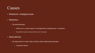
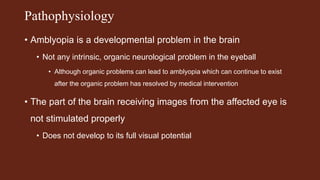
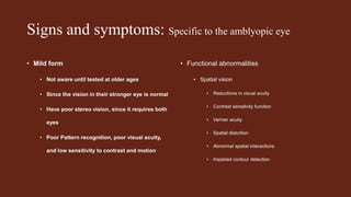
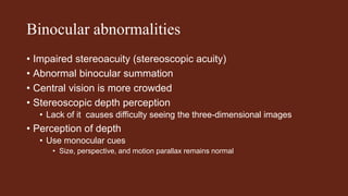
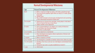
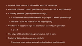
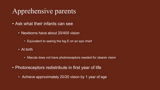
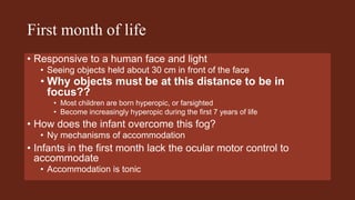
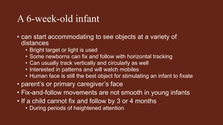
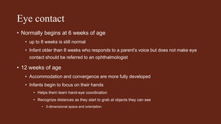
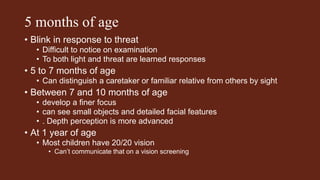
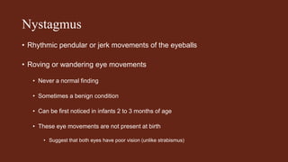
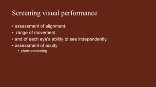
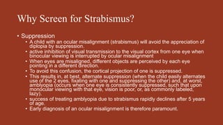
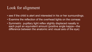
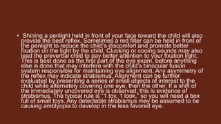
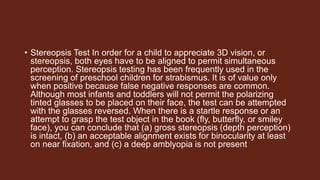
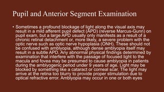
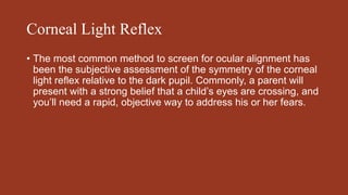
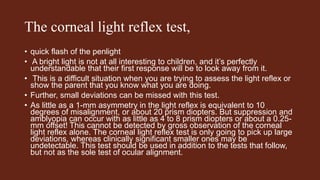
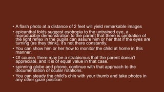
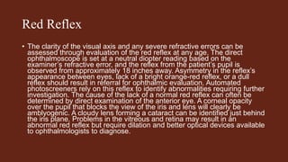
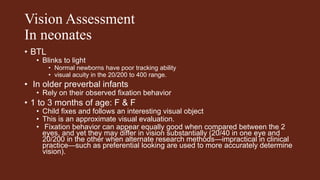
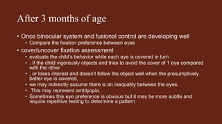
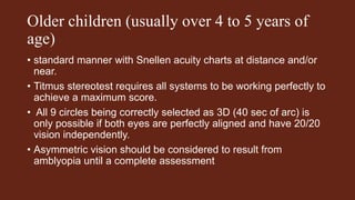
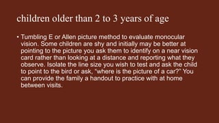
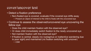
• Amblyopia is a developmental problem in the brain
• Not any intrinsic, organic neurological problem in the eyeball
• Although organic problems can lead to amblyopia which can continue to exist
after the organic problem has resolved by medical intervention
• The part of the brain receiving images from the affected eye is
not stimulated properly
• Does not develop to its full visual potential
Signs and symptoms: Specific to the amblyopic eye
• Mild form
• Not aware until tested at older ages
• Since the vision in their stronger eye is normal
• Have poor stereo vision, since it requires both
eyes
• Poor Pattern recognition, poor visual acuity,
and low sensitivity to contrast and motion
• Functional abnormalities
• Spatial vision
• Reductions in visual acuity
• Contrast sensitivity function
• Vernier acuity
• Spatial distortion
• Abnormal spatial interactions
• Impaired contour detection
Binocular abnormalities
• Impaired stereoacuity (stereoscopic acuity)
• Abnormal binocular summation
• Central vision is more crowded
• Stereoscopic depth perception
• Lack of it causes difficulty seeing the three-dimensional images
• Perception of depth
• Use monocular cues
• Size, perspective, and motion parallax remains normal
• Likely to be reached later in children who were born prematurely
• Premature infants of 30 weeks, gestational age at birth will blink in response to light
• A pupillary light reflex (pupillary constriction to light)
• Can be noted even in premature babies as young as 31 weeks, gestational age
• Newborn’s pupils will be small and will respond poorly
• Constriction in response to light can be difficult to see without magnification
• It is brief
• Use bright light to elicit this reflex, preferably in a dimly lit room
• Pupils that dilate rather than constrict with light
• Paradoxical response that requires investigation by an ophthalmologist
Apprehensive parents
• Ask what their infants can see
• Newborns have about 20/400 vision
• Equivalent to seeing the big E on an eye chart
• At birth
• Macula does not have photoreceptors needed for clearer vision
• Photoreceptors redistribute in first year of life
• Achieve approximately 20/20 vision by 1 year of age
First month of life
• Responsive to a human face and light
• Seeing objects held about 30 cm in front of the face
• Why objects must be at this distance to be in
focus??
• Most children are born hyperopic, or farsighted
• Become increasingly hyperopic during the first 7 years of life
• How does the infant overcome this fog?
• Ny mechanisms of accommodation
• Infants in the first month lack the ocular motor control to
accommodate
• Accommodation is tonic
A 6-week-old infant
• can start accommodating to see objects at a variety of
distances
• Bright target or light is used
• Some newborns can fix and follow with horizontal tracking
• Can usually track vertically and circularly as well
• Interested in patterns and will watch mobiles
• Human face is still the best object for stimulating an infant to fixate
• parent’s or primary caregiver’s face
• Fix-and-follow movements are not smooth in young infants
• If a child cannot fix and follow by 3 or 4 months
• During periods of heightened attention
Eye contact
• Normally begins at 6 weeks of age
• up to 8 weeks is still normal
• Infant older than 8 weeks who responds to a parent’s voice but does not make eye
contact should be referred to an ophthalmologist
• 12 weeks of age
• Accommodation and convergence are more fully developed
• Infants begin to focus on their hands
• Helps them learn hand-eye coordination
• Recognize distances as they start to grab at objects they can see
• 3-dimensional space and orientation
5 months of age
• Blink in response to threat
• Difficult to notice on examination
• To both light and threat are learned responses
• 5 to 7 months of age
• Can distinguish a caretaker or familiar relative from others by sight
• Between 7 and 10 months of age
• develop a finer focus
• can see small objects and detailed facial features
• . Depth perception is more advanced
• At 1 year of age
• Most children have 20/20 vision
• Can’t communicate that on a vision screening
Amblyopia:
Poor vision? Inadequate stimulation??
Lack of appropriate synapses and cells in visual cortex
• Nobel Prize in 1981
• Identified the relationship between optimal visual stimulation
and cortical neuronal cell and network development
• Earlier in childhood the visual development is handicapped
• More profound the effect on development in the visual cortex
Stimulation of the central vision &
Amblyopia
• Any condition that interferes (served by the macula and fovea)
• Peripheral vision, is not susceptible to amblyopia
• Prior to 3 months of age, the binocular fusion system in the midbrain has
not developed sufficiently to hold the 2 eyes in parallel alignment
• variety of strabismus conditions may be observed,
• all of which disappear after 3 months of age
• unless there is a more serious underlying condition
Oculodigital reflex
• Sign of poor visual development
• Forceful or constant eye rubbing,
• ? An attempt by the infant to stimulate the nerves in the back of the eye
• To fire manually because they are not responding to light.
• In addition to intrinsic problems with the eye or brain,
• Deprivation of visual stimuli can lead to poor visual development
• Cataract or a ptotic lid that prevents light from getting to the
Opsoclonus
• Nystagmus can be confused with opsoclonus,
• Flutterlike, random eye movement that may be normal in neonates
• Rarely, opsoclonus can be a sign of neuroblastoma
• Rapid and unpredictable eye movements noted in opsoclonus
• ?? caused by a paraneoplastic process in neuroblastoma,
• Can also cause myoclonus and ataxia
Nystagmus
• Dissociated nystagmus
• One eye has a greater amplitude or
frequency—may be a sign of a chiasmal
glioma or of a condition termed spasmus
nutans
. Spasmus nutans is a benign condition
• Nystagmus
• Head nodding
• Torticollis
• The nystagmus in spasmus nutans
• Shimmering (small amplitude and high
frequency)
• Asymmetrical and variable depending on
head position
• Onset for spasmus nutans
• is usually within the first 2 years of life
• Usually spontaneously resolves by age 3 or 4
years
Nystagmus
• Rhythmic pendular or jerk movements of the eyeballs
• Roving or wandering eye movements
• Never a normal finding
• Sometimes a benign condition
• Can be first noticed in infants 2 to 3 months of age
• These eye movements are not present at birth
• Suggest that both eyes have poor vision (unlike strabismus)
Setting-sun sign
• Sunsetting, a tonic downward movement of both eyes
• Healthy newborns exhibit transient vertical ocular misalignment
• Not be confused with the true setting-sun sign of hydrocephalus
• Both ocular globes are deviated downward
• Upper lids are retracted
• White sclera may be visible above the iris
Children under 9 years
• When presented with the diplopia resulting from misaligned eye
• suppress the image in the deviated eye, and that eye becomes
amblyopic
common problems that lead to the
development of amblyopia
Screening visual performance
• assessment of alignment,
• range of movement,
• and of each eye’s ability to see independently.
• assessment of acuity
• photoscreening
• The suggested order of examination will progress from the least
threatening, remote assessment, to a closer interaction. A
thorough screening will require only 1 to 2 minutes. The 5 key
maneuvers
• Corneal light reflex 2. Ocular rotations 3. Bruckner test 4.
Cover test 5. Stereopsis test
Why Screen for Strabismus?
• Suppression
• A child with an ocular misalignment (strabismus) will avoid the appreciation of
diplopia by suppression.
• active inhibition of visual transmission to the visual cortex from one eye when
binocular viewing is interrupted by ocular misalignment
• When eyes are misaligned, different objects are perceived by each eye
pointing in a different direction.
• To avoid this confusion, the cortical projection of one is suppressed.
• This results in, at best, alternate suppression (when the child easily alternates
use of the 2 eyes, fixating with one and suppressing the other) and, at worst,
amblyopia (occurs when one eye is consistently suppressed, such that upon
monocular viewing with that eye, vision is poor, or, as commonly labeled,
lazy).
• success of treating amblyopia due to strabismus rapidly declines after 5 years
of age.
• Early diagnosis of an ocular misalignment is therefore paramount.
Look for alignment
• see if the child is alert and interested in his or her surroundings.
• Examine the reflection of the overhead lights on the corneas
• Symmetric pupillary light reflex slightly displaced nasally in
each pupil an equivalent amount (positive angle kappa—the
difference between the anatomic and visual axis of the eye)
• Shining a penlight held in front of your face toward the child will also
provide the best reflex. Sometimes a red filter can be held in front of
the penlight to reduce the child’s discomfort and promote better
fixation on the light by the child. Clucking or cooing sounds may also
lead the preverbal child to pay better attention to your fixation light.
This is best done as the first part of the eye exam, before anything
else is done that may interfere with the child’s binocular fusion
system responsible for maintaining eye alignment. Any asymmetry of
the reflex may indicate strabismus. Alignment can be further
evaluated by presenting a series of small objects of interest to the
child while alternately covering one eye, then the other. If a shift of
the immediately uncovered eye is observed, this is evidence of
strabismus. The typical rule is “1 toy, 1 look,” so you will need a box
full of small toys. Any detectable strabismus may be assumed to be
causing amblyopia to develop in the less favored eye.
Ocular Rotations
• It is equally important to capture a photo in right and left gaze,
using the spinning light globe to secure the child’s attention.
Forms of strabismus exist where the eyes are straight in
primary position but deviated in other positions of gaze (eg,
Duane’s syndrome and fourth cranial nerve palsy) (Figure 3-2).
Therefore, don’t be too reassuring until you get a photo of right
and left gaze. Take care to present only one stimulus at a time.
Voicing a noisy stimulus in one direction and then holding a
fixation target in another quickly confuses the child. If the child
is under 1 year of age, the status of ocular rotations can be
quickly assessed by generating rotations or an optokinetic
nystagmoid movement with either of the following 2 methods.
Doll’s Eyes
• While holding the child with a hand under each armpit and each
thumb steadying the chin, the child is turned right, left, up, and
down while the examiner uses his or her face and sounds to
attract his or her attention. Observe the symmetry of ocular
rotation into these 4 fields of gaze created as the child
maintains fixation on your face. Noisy targets (you) are helpful
when the goal is to stimulate ocular rotations, but not to assess
acuity
Room-based Optokinetic Nystagnus
• To elicit an optokinetic nystagmus (OKN), support the child under the
arms as described previously, steady the chin with your thumbs, and
rotate yourself along with the child. Observe the child’s eye
movement as he or she views the fixed objects in the room over your
shoulder (Figure 3-3). Observe the ocular rotation as the child fixes
on an object and follows it (slow pursuit movement), then refixates
(fast saccadic movement) on the next target. This maneuver is
valuable in the 4- to 6-month-old with poor fixation reflexes and
delayed visual maturation (DVM), as opposed to poor acuity. A child
with true vision loss will not be able to demonstrate this reflex, and a
child who appears to have vision loss as deduced by visual
inattention can be proven to be sighted if he or she can demonstrate
this room-based OKN
Bruckner Test
• Best performed after dilation but adequately done in a dimly lit room,
this test offers an assessment of the relative intensity of the red light
reflex, reflecting off the fundus and through the pupil, when viewed
through the direct ophthalmoscope. Look at the pupillary red reflex
from both eyes simultaneously from a distance of approximately 3
feet through your direct ophthalmoscope set to 0. Holding the
spinning globe near his or her face will keep attention during your
observation. Assess the relative size, color, and brightness of the 2
reflexes. Any significant asymmetry warrants referral. The American
Academy of Pediatrics publishes this testing method as part of a
“See Red” campaign and the site can be accessed at
http://aappolicy.aappublications.org/cgi/content/full/
pediatrics;122/6/1401. Another consumer web site is
www.knowtheglow.org, which encourages parents to examine the
red reflex seen in most family photos.
Cover Test
• The gold standard for the diagnosis of a constant ocular misalignment is the cover test. Again, the
key to success is the use of an interesting target. An eye is covered and any shift in ocular position
by the uncovered eye to find the target you present is a positive test. Get the interest of the child,
then cover the right while observing the left, and then switch hands and cover the left while you
observe the right. You may want to use different targets for each eye due to the child’s attention
span. Stickers of cartoon characters on a pencil are inexpensive and can be readily interchanged.
The success of the test is not only dependent on the ability of the target to maintain attention during
the test, but also on the quality of the cover used. Typically, a child will not tolerate a black paddle-
type eye occluder, and using your thumb as the cover while the hand steadies the forehead may be
more successful (Figure 3-4). The cover can’t be so interesting as to distract attention from the
target. This test screens for alignment at near fixation, the most important position for visual
development. Because strabismus may exist only on distance fixation (eg, intermittent exotropia),
diagnosis of these conditions may have to await their ability to cooperate with fixation on distant
projected targets while the cover test is performed. The cover test simultaneously screens for
possible amblyopia, suspected when the child consistently objects to covering one eye and
demonstrates a strong preference for viewing with only one eye on multiple trials. Equal objection to
cover is not diagnostic of amblyopia, but rather of a symmetric preference for seeing with either eye
in the absence of amblyopia.
• Stereopsis Test In order for a child to appreciate 3D vision, or
stereopsis, both eyes have to be aligned to permit simultaneous
perception. Stereopsis testing has been frequently used in the
screening of preschool children for strabismus. It is of value only
when positive because false negative responses are common.
Although most infants and toddlers will not permit the polarizing
tinted glasses to be placed on their face, the test can be attempted
with the glasses reversed. When there is a startle response or an
attempt to grasp the test object in the book (fly, butterfly, or smiley
face), you can conclude that (a) gross stereopsis (depth perception)
is intact, (b) an acceptable alignment exists for binocularity at least
on near fixation, and (c) a deep amblyopia is not present
Pupil and Anterior Segment Examination
• Sometimes a profound blockage of light along the visual axis may
result in a mild afferent pupil defect (APD) (reverse Marcus-Gunn) on
pupil exam, but a large APD usually only manifests as a result of a
chronic retinal detachment or, more likely, a severe problem with the
optic nerve such as optic nerve hypoplasia (ONH). These should not
be confused with amblyopia, although dense amblyopia itself may
result in a subtle APD. Any abnormal physical findings determined by
examination that interfere with the passage of focused light to the
macula and fovea may be presumed to cause amblyopia in patients
during the amblyogenic period under 9 years of age. Light may be
blocked by something like a cataract or corneal scar. The light may
arrive at the retina too blurry to provide proper stimulation due to
optical refractive error. Amblyopia may occur in one or both eyes
Corneal Light Reflex
• The most common method to screen for ocular alignment has
been the subjective assessment of the symmetry of the corneal
light reflex relative to the dark pupil. Commonly, a parent will
present with a strong belief that a child’s eyes are crossing, and
you’ll need a rapid, objective way to address his or her fears.
The corneal light reflex test,
• quick flash of the penlight
• A bright light is not at all interesting to children, and it’s perfectly
understandable that their first response will be to look away from it.
• This is a difficult situation when you are trying to assess the light reflex or
show the parent that you know what you are doing.
• Further, small deviations can be missed with this test.
• As little as a 1-mm asymmetry in the light reflex is equivalent to 10
degrees of misalignment, or about 20 prism diopters. But suppression and
amblyopia can occur with as little as 4 to 8 prism diopters or about a 0.25-
mm offset! This cannot be detected by gross observation of the corneal
light reflex alone. The corneal light reflex test is only going to pick up large
deviations, whereas clinically significant smaller ones may be
undetectable. This test should be used in addition to the tests that follow,
but not as the sole test of ocular alignment.
• A flash photo at a distance of 2 feet will yield remarkable images
• epicanthal folds suggest esotropia to the untrained eye, a
reproducible demonstration to the parent that there is centration of
the light reflex in the pupils can assure him or her that if the eyes are
turning (as they think), it’s not there constantly.
• You can show him or her how to monitor the child at home in this
manner.
• Of course, there may be a strabismus that the parent doesn’t
appreciate, and it is of equal value in that case.
• spinning globe and phone, continue with that approach to the
documentation of ocular rotations.
• You can steady the child’s chin with your thumb and take photos in
any other gaze position
Red Reflex
• The clarity of the visual axis and any severe refractive errors can be
assessed through evaluation of the red reflex at any age. The direct
ophthalmoscope is set at a neutral diopter reading based on the
examiner’s refractive error, and the reflex from the patient’s pupil is
observed from approximately 18 inches away. Asymmetry in the reflex’s
appearance between eyes, lack of a bright orange-red reflex, or a dull
reflex should result in referral for ophthalmic evaluation. Automated
photoscreeners rely on this reflex to identify abnormalities requiring further
investigation. The cause of the lack of a normal red reflex can often be
determined by direct examination of the anterior eye. A corneal opacity
over the pupil that blocks the view of the iris and lens will clearly be
amblyogenic. A cloudy lens forming a cataract can be identified just behind
the iris plane. Problems in the vitreous and retina may result in an
abnormal red reflex but require dilation and better optical devices available
to ophthalmologists to diagnose.
Vision Assessment
In neonates
• BTL
• Blinks to light
• Normal newborns have poor tracking ability
• visual acuity in the 20/200 to 400 range.
• In older preverbal infants
• Rely on their observed fixation behavior
• 1 to 3 months of age: F & F
• Child fixes and follows an interesting visual object
• This is an approximate visual evaluation.
• Fixation behavior can appear equally good when compared between the 2
eyes, and yet they may differ in vision substantially (20/40 in one eye and
20/200 in the other when alternate research methods—impractical in clinical
practice—such as preferential looking are used to more accurately determine
vision).
After 3 months of age
• Once binocular system and fusional control are developing well
• Compare the fixation preference between eyes
• cover/uncover fixation assessment
• evaluate the child’s behavior while each eye is covered in turn
• . If the child vigorously objects and tries to avoid the cover of 1 eye compared
with the other
• , or loses interest and doesn’t follow the object well when the presumptively
better eye is covered,
• we may indirectly assume there is an inequality between the eyes.
• This may represent amblyopia.
• Sometimes this eye preference is obvious but it may be more subtle and
require repetitive testing to determine a pattern
Older children (usually over 4 to 5 years of
age)
• standard manner with Snellen acuity charts at distance and/or
near.
• Titmus stereotest requires all systems to be working perfectly to
achieve a maximum score.
• All 9 circles being correctly selected as 3D (40 sec of arc) is
only possible if both eyes are perfectly aligned and have 20/20
vision independently.
• Asymmetric vision should be considered to result from
amblyopia until a complete assessment
children older than 2 to 3 years of age
• Tumbling E or Allen picture method to evaluate monocular
vision. Some children are shy and initially may be better at
pointing to the picture you ask them to identify on a near vision
card rather than looking at a distance and reporting what they
observe. Isolate the line size you wish to test and ask the child
to point to the bird or ask, “where is the picture of a car?” You
can provide the family a handout to practice with at home
between visits.
cover/uncover test
• Detect a fixation preference
• Non-fixated eye is covered, evaluate if the covered eye drifts away
• Present an object of interest to the child to fixate with the uncovered eye
• Continue to assess the observed/uncovered eye uncovering the
fellow eye
• Does the child maintain fixation with the observed eye?
• Or does child immediately switch fixation to the newly uncovered eye
• Not maintain fixation with the observed eye?
• If fixation is central, steady (no nystagmus or inattentive wandering due
to poor sight) and maintained (no fixation switching with uncover)
• CSM

amblyopia.pptx

  • 1.
    Amblyopia Paediatric Eye Examination Howdo I make a diagnosis of Amblyopia
  • 2.
    Causes • Strabismic: misalignedeyes • Refractive: • By anisometropia • Difference of a certain degree of nearsightedness, farsightedness, or astigmatis • By significant amount of equal refractive error in both eyes • Deprivational: • By deprivation of vision early in life by vision-obstructing disorders • Congenital cataract
  • 3.
    Pathophysiology • Amblyopia isa developmental problem in the brain • Not any intrinsic, organic neurological problem in the eyeball • Although organic problems can lead to amblyopia which can continue to exist after the organic problem has resolved by medical intervention • The part of the brain receiving images from the affected eye is not stimulated properly • Does not develop to its full visual potential
  • 4.
    Signs and symptoms:Specific to the amblyopic eye • Mild form • Not aware until tested at older ages • Since the vision in their stronger eye is normal • Have poor stereo vision, since it requires both eyes • Poor Pattern recognition, poor visual acuity, and low sensitivity to contrast and motion • Functional abnormalities • Spatial vision • Reductions in visual acuity • Contrast sensitivity function • Vernier acuity • Spatial distortion • Abnormal spatial interactions • Impaired contour detection
  • 5.
    Binocular abnormalities • Impairedstereoacuity (stereoscopic acuity) • Abnormal binocular summation • Central vision is more crowded • Stereoscopic depth perception • Lack of it causes difficulty seeing the three-dimensional images • Perception of depth • Use monocular cues • Size, perspective, and motion parallax remains normal
  • 7.
    • Likely tobe reached later in children who were born prematurely • Premature infants of 30 weeks, gestational age at birth will blink in response to light • A pupillary light reflex (pupillary constriction to light) • Can be noted even in premature babies as young as 31 weeks, gestational age • Newborn’s pupils will be small and will respond poorly • Constriction in response to light can be difficult to see without magnification • It is brief • Use bright light to elicit this reflex, preferably in a dimly lit room • Pupils that dilate rather than constrict with light • Paradoxical response that requires investigation by an ophthalmologist
  • 8.
    Apprehensive parents • Askwhat their infants can see • Newborns have about 20/400 vision • Equivalent to seeing the big E on an eye chart • At birth • Macula does not have photoreceptors needed for clearer vision • Photoreceptors redistribute in first year of life • Achieve approximately 20/20 vision by 1 year of age
  • 9.
    First month oflife • Responsive to a human face and light • Seeing objects held about 30 cm in front of the face • Why objects must be at this distance to be in focus?? • Most children are born hyperopic, or farsighted • Become increasingly hyperopic during the first 7 years of life • How does the infant overcome this fog? • Ny mechanisms of accommodation • Infants in the first month lack the ocular motor control to accommodate • Accommodation is tonic
  • 10.
    A 6-week-old infant •can start accommodating to see objects at a variety of distances • Bright target or light is used • Some newborns can fix and follow with horizontal tracking • Can usually track vertically and circularly as well • Interested in patterns and will watch mobiles • Human face is still the best object for stimulating an infant to fixate • parent’s or primary caregiver’s face • Fix-and-follow movements are not smooth in young infants • If a child cannot fix and follow by 3 or 4 months • During periods of heightened attention
  • 11.
    Eye contact • Normallybegins at 6 weeks of age • up to 8 weeks is still normal • Infant older than 8 weeks who responds to a parent’s voice but does not make eye contact should be referred to an ophthalmologist • 12 weeks of age • Accommodation and convergence are more fully developed • Infants begin to focus on their hands • Helps them learn hand-eye coordination • Recognize distances as they start to grab at objects they can see • 3-dimensional space and orientation
  • 12.
    5 months ofage • Blink in response to threat • Difficult to notice on examination • To both light and threat are learned responses • 5 to 7 months of age • Can distinguish a caretaker or familiar relative from others by sight • Between 7 and 10 months of age • develop a finer focus • can see small objects and detailed facial features • . Depth perception is more advanced • At 1 year of age • Most children have 20/20 vision • Can’t communicate that on a vision screening
  • 13.
    Amblyopia: Poor vision? Inadequatestimulation?? Lack of appropriate synapses and cells in visual cortex • Nobel Prize in 1981 • Identified the relationship between optimal visual stimulation and cortical neuronal cell and network development • Earlier in childhood the visual development is handicapped • More profound the effect on development in the visual cortex
  • 14.
    Stimulation of thecentral vision & Amblyopia • Any condition that interferes (served by the macula and fovea) • Peripheral vision, is not susceptible to amblyopia • Prior to 3 months of age, the binocular fusion system in the midbrain has not developed sufficiently to hold the 2 eyes in parallel alignment • variety of strabismus conditions may be observed, • all of which disappear after 3 months of age • unless there is a more serious underlying condition
  • 15.
    Oculodigital reflex • Signof poor visual development • Forceful or constant eye rubbing, • ? An attempt by the infant to stimulate the nerves in the back of the eye • To fire manually because they are not responding to light. • In addition to intrinsic problems with the eye or brain, • Deprivation of visual stimuli can lead to poor visual development • Cataract or a ptotic lid that prevents light from getting to the
  • 16.
    Opsoclonus • Nystagmus canbe confused with opsoclonus, • Flutterlike, random eye movement that may be normal in neonates • Rarely, opsoclonus can be a sign of neuroblastoma • Rapid and unpredictable eye movements noted in opsoclonus • ?? caused by a paraneoplastic process in neuroblastoma, • Can also cause myoclonus and ataxia
  • 17.
    Nystagmus • Dissociated nystagmus •One eye has a greater amplitude or frequency—may be a sign of a chiasmal glioma or of a condition termed spasmus nutans . Spasmus nutans is a benign condition • Nystagmus • Head nodding • Torticollis • The nystagmus in spasmus nutans • Shimmering (small amplitude and high frequency) • Asymmetrical and variable depending on head position • Onset for spasmus nutans • is usually within the first 2 years of life • Usually spontaneously resolves by age 3 or 4 years
  • 18.
    Nystagmus • Rhythmic pendularor jerk movements of the eyeballs • Roving or wandering eye movements • Never a normal finding • Sometimes a benign condition • Can be first noticed in infants 2 to 3 months of age • These eye movements are not present at birth • Suggest that both eyes have poor vision (unlike strabismus)
  • 19.
    Setting-sun sign • Sunsetting,a tonic downward movement of both eyes • Healthy newborns exhibit transient vertical ocular misalignment • Not be confused with the true setting-sun sign of hydrocephalus • Both ocular globes are deviated downward • Upper lids are retracted • White sclera may be visible above the iris
  • 20.
    Children under 9years • When presented with the diplopia resulting from misaligned eye • suppress the image in the deviated eye, and that eye becomes amblyopic
  • 21.
    common problems thatlead to the development of amblyopia
  • 22.
    Screening visual performance •assessment of alignment, • range of movement, • and of each eye’s ability to see independently. • assessment of acuity • photoscreening
  • 23.
    • The suggestedorder of examination will progress from the least threatening, remote assessment, to a closer interaction. A thorough screening will require only 1 to 2 minutes. The 5 key maneuvers • Corneal light reflex 2. Ocular rotations 3. Bruckner test 4. Cover test 5. Stereopsis test
  • 24.
    Why Screen forStrabismus? • Suppression • A child with an ocular misalignment (strabismus) will avoid the appreciation of diplopia by suppression. • active inhibition of visual transmission to the visual cortex from one eye when binocular viewing is interrupted by ocular misalignment • When eyes are misaligned, different objects are perceived by each eye pointing in a different direction. • To avoid this confusion, the cortical projection of one is suppressed. • This results in, at best, alternate suppression (when the child easily alternates use of the 2 eyes, fixating with one and suppressing the other) and, at worst, amblyopia (occurs when one eye is consistently suppressed, such that upon monocular viewing with that eye, vision is poor, or, as commonly labeled, lazy). • success of treating amblyopia due to strabismus rapidly declines after 5 years of age. • Early diagnosis of an ocular misalignment is therefore paramount.
  • 25.
    Look for alignment •see if the child is alert and interested in his or her surroundings. • Examine the reflection of the overhead lights on the corneas • Symmetric pupillary light reflex slightly displaced nasally in each pupil an equivalent amount (positive angle kappa—the difference between the anatomic and visual axis of the eye)
  • 26.
    • Shining apenlight held in front of your face toward the child will also provide the best reflex. Sometimes a red filter can be held in front of the penlight to reduce the child’s discomfort and promote better fixation on the light by the child. Clucking or cooing sounds may also lead the preverbal child to pay better attention to your fixation light. This is best done as the first part of the eye exam, before anything else is done that may interfere with the child’s binocular fusion system responsible for maintaining eye alignment. Any asymmetry of the reflex may indicate strabismus. Alignment can be further evaluated by presenting a series of small objects of interest to the child while alternately covering one eye, then the other. If a shift of the immediately uncovered eye is observed, this is evidence of strabismus. The typical rule is “1 toy, 1 look,” so you will need a box full of small toys. Any detectable strabismus may be assumed to be causing amblyopia to develop in the less favored eye.
  • 27.
    Ocular Rotations • Itis equally important to capture a photo in right and left gaze, using the spinning light globe to secure the child’s attention. Forms of strabismus exist where the eyes are straight in primary position but deviated in other positions of gaze (eg, Duane’s syndrome and fourth cranial nerve palsy) (Figure 3-2). Therefore, don’t be too reassuring until you get a photo of right and left gaze. Take care to present only one stimulus at a time. Voicing a noisy stimulus in one direction and then holding a fixation target in another quickly confuses the child. If the child is under 1 year of age, the status of ocular rotations can be quickly assessed by generating rotations or an optokinetic nystagmoid movement with either of the following 2 methods.
  • 28.
    Doll’s Eyes • Whileholding the child with a hand under each armpit and each thumb steadying the chin, the child is turned right, left, up, and down while the examiner uses his or her face and sounds to attract his or her attention. Observe the symmetry of ocular rotation into these 4 fields of gaze created as the child maintains fixation on your face. Noisy targets (you) are helpful when the goal is to stimulate ocular rotations, but not to assess acuity
  • 29.
    Room-based Optokinetic Nystagnus •To elicit an optokinetic nystagmus (OKN), support the child under the arms as described previously, steady the chin with your thumbs, and rotate yourself along with the child. Observe the child’s eye movement as he or she views the fixed objects in the room over your shoulder (Figure 3-3). Observe the ocular rotation as the child fixes on an object and follows it (slow pursuit movement), then refixates (fast saccadic movement) on the next target. This maneuver is valuable in the 4- to 6-month-old with poor fixation reflexes and delayed visual maturation (DVM), as opposed to poor acuity. A child with true vision loss will not be able to demonstrate this reflex, and a child who appears to have vision loss as deduced by visual inattention can be proven to be sighted if he or she can demonstrate this room-based OKN
  • 30.
    Bruckner Test • Bestperformed after dilation but adequately done in a dimly lit room, this test offers an assessment of the relative intensity of the red light reflex, reflecting off the fundus and through the pupil, when viewed through the direct ophthalmoscope. Look at the pupillary red reflex from both eyes simultaneously from a distance of approximately 3 feet through your direct ophthalmoscope set to 0. Holding the spinning globe near his or her face will keep attention during your observation. Assess the relative size, color, and brightness of the 2 reflexes. Any significant asymmetry warrants referral. The American Academy of Pediatrics publishes this testing method as part of a “See Red” campaign and the site can be accessed at http://aappolicy.aappublications.org/cgi/content/full/ pediatrics;122/6/1401. Another consumer web site is www.knowtheglow.org, which encourages parents to examine the red reflex seen in most family photos.
  • 31.
    Cover Test • Thegold standard for the diagnosis of a constant ocular misalignment is the cover test. Again, the key to success is the use of an interesting target. An eye is covered and any shift in ocular position by the uncovered eye to find the target you present is a positive test. Get the interest of the child, then cover the right while observing the left, and then switch hands and cover the left while you observe the right. You may want to use different targets for each eye due to the child’s attention span. Stickers of cartoon characters on a pencil are inexpensive and can be readily interchanged. The success of the test is not only dependent on the ability of the target to maintain attention during the test, but also on the quality of the cover used. Typically, a child will not tolerate a black paddle- type eye occluder, and using your thumb as the cover while the hand steadies the forehead may be more successful (Figure 3-4). The cover can’t be so interesting as to distract attention from the target. This test screens for alignment at near fixation, the most important position for visual development. Because strabismus may exist only on distance fixation (eg, intermittent exotropia), diagnosis of these conditions may have to await their ability to cooperate with fixation on distant projected targets while the cover test is performed. The cover test simultaneously screens for possible amblyopia, suspected when the child consistently objects to covering one eye and demonstrates a strong preference for viewing with only one eye on multiple trials. Equal objection to cover is not diagnostic of amblyopia, but rather of a symmetric preference for seeing with either eye in the absence of amblyopia.
  • 32.
    • Stereopsis TestIn order for a child to appreciate 3D vision, or stereopsis, both eyes have to be aligned to permit simultaneous perception. Stereopsis testing has been frequently used in the screening of preschool children for strabismus. It is of value only when positive because false negative responses are common. Although most infants and toddlers will not permit the polarizing tinted glasses to be placed on their face, the test can be attempted with the glasses reversed. When there is a startle response or an attempt to grasp the test object in the book (fly, butterfly, or smiley face), you can conclude that (a) gross stereopsis (depth perception) is intact, (b) an acceptable alignment exists for binocularity at least on near fixation, and (c) a deep amblyopia is not present
  • 33.
    Pupil and AnteriorSegment Examination • Sometimes a profound blockage of light along the visual axis may result in a mild afferent pupil defect (APD) (reverse Marcus-Gunn) on pupil exam, but a large APD usually only manifests as a result of a chronic retinal detachment or, more likely, a severe problem with the optic nerve such as optic nerve hypoplasia (ONH). These should not be confused with amblyopia, although dense amblyopia itself may result in a subtle APD. Any abnormal physical findings determined by examination that interfere with the passage of focused light to the macula and fovea may be presumed to cause amblyopia in patients during the amblyogenic period under 9 years of age. Light may be blocked by something like a cataract or corneal scar. The light may arrive at the retina too blurry to provide proper stimulation due to optical refractive error. Amblyopia may occur in one or both eyes
  • 34.
    Corneal Light Reflex •The most common method to screen for ocular alignment has been the subjective assessment of the symmetry of the corneal light reflex relative to the dark pupil. Commonly, a parent will present with a strong belief that a child’s eyes are crossing, and you’ll need a rapid, objective way to address his or her fears.
  • 35.
    The corneal lightreflex test, • quick flash of the penlight • A bright light is not at all interesting to children, and it’s perfectly understandable that their first response will be to look away from it. • This is a difficult situation when you are trying to assess the light reflex or show the parent that you know what you are doing. • Further, small deviations can be missed with this test. • As little as a 1-mm asymmetry in the light reflex is equivalent to 10 degrees of misalignment, or about 20 prism diopters. But suppression and amblyopia can occur with as little as 4 to 8 prism diopters or about a 0.25- mm offset! This cannot be detected by gross observation of the corneal light reflex alone. The corneal light reflex test is only going to pick up large deviations, whereas clinically significant smaller ones may be undetectable. This test should be used in addition to the tests that follow, but not as the sole test of ocular alignment.
  • 36.
    • A flashphoto at a distance of 2 feet will yield remarkable images • epicanthal folds suggest esotropia to the untrained eye, a reproducible demonstration to the parent that there is centration of the light reflex in the pupils can assure him or her that if the eyes are turning (as they think), it’s not there constantly. • You can show him or her how to monitor the child at home in this manner. • Of course, there may be a strabismus that the parent doesn’t appreciate, and it is of equal value in that case. • spinning globe and phone, continue with that approach to the documentation of ocular rotations. • You can steady the child’s chin with your thumb and take photos in any other gaze position
  • 37.
    Red Reflex • Theclarity of the visual axis and any severe refractive errors can be assessed through evaluation of the red reflex at any age. The direct ophthalmoscope is set at a neutral diopter reading based on the examiner’s refractive error, and the reflex from the patient’s pupil is observed from approximately 18 inches away. Asymmetry in the reflex’s appearance between eyes, lack of a bright orange-red reflex, or a dull reflex should result in referral for ophthalmic evaluation. Automated photoscreeners rely on this reflex to identify abnormalities requiring further investigation. The cause of the lack of a normal red reflex can often be determined by direct examination of the anterior eye. A corneal opacity over the pupil that blocks the view of the iris and lens will clearly be amblyogenic. A cloudy lens forming a cataract can be identified just behind the iris plane. Problems in the vitreous and retina may result in an abnormal red reflex but require dilation and better optical devices available to ophthalmologists to diagnose.
  • 38.
    Vision Assessment In neonates •BTL • Blinks to light • Normal newborns have poor tracking ability • visual acuity in the 20/200 to 400 range. • In older preverbal infants • Rely on their observed fixation behavior • 1 to 3 months of age: F & F • Child fixes and follows an interesting visual object • This is an approximate visual evaluation. • Fixation behavior can appear equally good when compared between the 2 eyes, and yet they may differ in vision substantially (20/40 in one eye and 20/200 in the other when alternate research methods—impractical in clinical practice—such as preferential looking are used to more accurately determine vision).
  • 39.
    After 3 monthsof age • Once binocular system and fusional control are developing well • Compare the fixation preference between eyes • cover/uncover fixation assessment • evaluate the child’s behavior while each eye is covered in turn • . If the child vigorously objects and tries to avoid the cover of 1 eye compared with the other • , or loses interest and doesn’t follow the object well when the presumptively better eye is covered, • we may indirectly assume there is an inequality between the eyes. • This may represent amblyopia. • Sometimes this eye preference is obvious but it may be more subtle and require repetitive testing to determine a pattern
  • 40.
    Older children (usuallyover 4 to 5 years of age) • standard manner with Snellen acuity charts at distance and/or near. • Titmus stereotest requires all systems to be working perfectly to achieve a maximum score. • All 9 circles being correctly selected as 3D (40 sec of arc) is only possible if both eyes are perfectly aligned and have 20/20 vision independently. • Asymmetric vision should be considered to result from amblyopia until a complete assessment
  • 41.
    children older than2 to 3 years of age • Tumbling E or Allen picture method to evaluate monocular vision. Some children are shy and initially may be better at pointing to the picture you ask them to identify on a near vision card rather than looking at a distance and reporting what they observe. Isolate the line size you wish to test and ask the child to point to the bird or ask, “where is the picture of a car?” You can provide the family a handout to practice with at home between visits.
  • 42.
    cover/uncover test • Detecta fixation preference • Non-fixated eye is covered, evaluate if the covered eye drifts away • Present an object of interest to the child to fixate with the uncovered eye • Continue to assess the observed/uncovered eye uncovering the fellow eye • Does the child maintain fixation with the observed eye? • Or does child immediately switch fixation to the newly uncovered eye • Not maintain fixation with the observed eye? • If fixation is central, steady (no nystagmus or inattentive wandering due to poor sight) and maintained (no fixation switching with uncover) • CSM