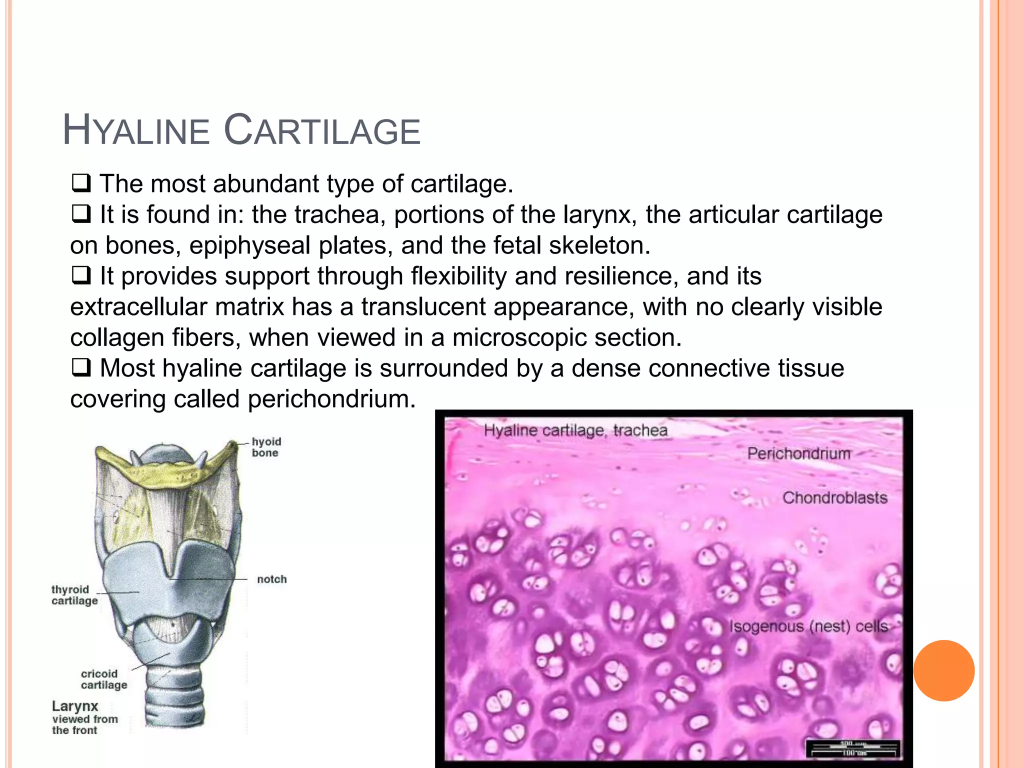
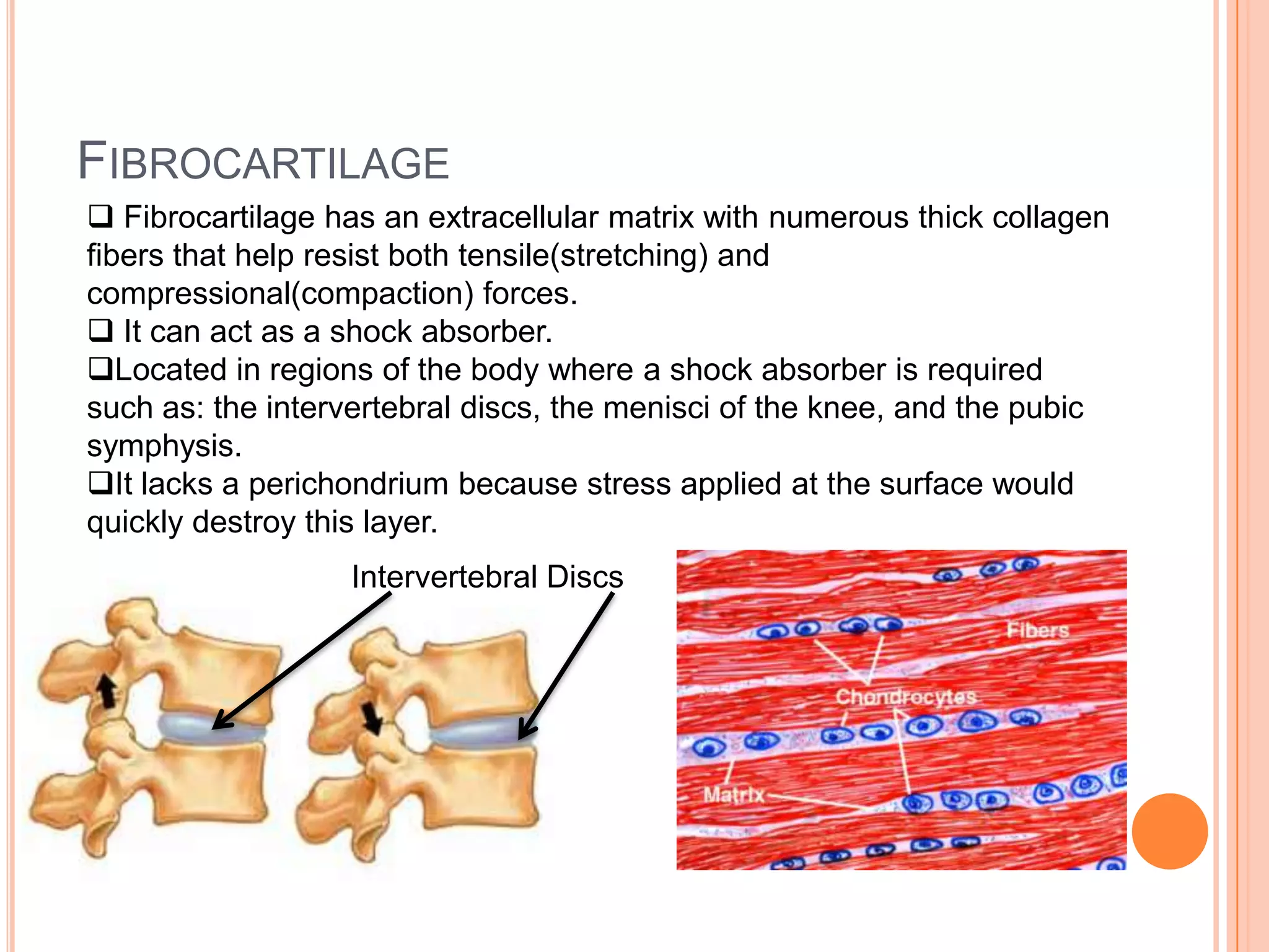
Cartilage & Bone Connective Tissue
Cartilage Connective TissueCartilage is a semi rigid connective tissue that is weaker thanbone, but more flexible and resilient.
There are three types of Cartilage: Hyaline Cartilage, Fibrocartilage, and Elastic Cartilage. Hyaline Cartilage The most abundant type of cartilage.
It is found in: the trachea, portions of the larynx, the articular cartilage on bones, epiphyseal plates, and the fetal skeleton.
It provides support through flexibility and resilience, and its extracellular matrix has a translucent appearance, with no clearly visible collagen fibers, when viewed in a microscopic section.
Most hyaline cartilage is surrounded by a dense connective tissue covering called perichondrium.FibrocartilageFibrocartilage has an extracellular matrix with numerous thick collagen fibers that help resist both tensile(stretching) and compressional(compaction) forces.
It can act as a shock absorber.
Located in regions of the body where a shock absorber is required such as: the intervertebral discs, the menisci of the knee, and the pubic symphysis.
It lacks a perichondrium because stress applied at the surface would quickly destroy this layer.Intervertebral Discs
Elastic CartilageElastic Cartilage contains highly branched elastic fibers with its extracellular matrix.
It’s typically found in regions requiring a highly flexible form of support.
It can be found in: the auricle of the ear, the external auditory canal, and the epiglottis.
Elastic cartilage is surrounded by a perichondrium. Auricle
Growth Patters of Cartilage Cartilage grows in two ways:
Interstitial Growth- Growth from within the cartilage itself.
Appositional Growth- Growth along the cartilage’s outside edge, or periphery.Bone Bones of the skeleton are complex, dynamic organs containing all tissue types.
The primary component is bone connective tissue, also called osseous connective tissue. They also contain connective tissue proper, cartilage connective tissue, smooth muscle tissue, fluid connective tissue, epithelial tissue, and nervous tissue.
The matrix of bone connective tissue is sturdy and rigid due to deposition of minerals in the matrix, a process called calcification.
Bones perform several basic functions: support and protection, movement, hemopoiesis, and storage of mineral and energy reserves.Bone Functions: Support & Protection Bone provides structural support and serve as a framework for the entire body.
They also protect many delicate tissues and organs from injury and trauma.

Cartilage & bone connective tissue

  • 1.
    Cartilage & BoneConnective Tissue
  • 2.
    Cartilage Connective TissueCartilageis a semi rigid connective tissue that is weaker thanbone, but more flexible and resilient.
  • 3.
    There are threetypes of Cartilage: Hyaline Cartilage, Fibrocartilage, and Elastic Cartilage. Hyaline Cartilage The most abundant type of cartilage.
  • 4.
    It is foundin: the trachea, portions of the larynx, the articular cartilage on bones, epiphyseal plates, and the fetal skeleton.
  • 5.
    It provides supportthrough flexibility and resilience, and its extracellular matrix has a translucent appearance, with no clearly visible collagen fibers, when viewed in a microscopic section.
  • 6.
    Most hyaline cartilageis surrounded by a dense connective tissue covering called perichondrium.FibrocartilageFibrocartilage has an extracellular matrix with numerous thick collagen fibers that help resist both tensile(stretching) and compressional(compaction) forces.
  • 7.
    It can actas a shock absorber.
  • 8.
    Located in regionsof the body where a shock absorber is required such as: the intervertebral discs, the menisci of the knee, and the pubic symphysis.
  • 9.
    It lacks aperichondrium because stress applied at the surface would quickly destroy this layer.Intervertebral Discs
  • 10.
    Elastic CartilageElastic Cartilagecontains highly branched elastic fibers with its extracellular matrix.
  • 11.
    It’s typically foundin regions requiring a highly flexible form of support.
  • 12.
    It can befound in: the auricle of the ear, the external auditory canal, and the epiglottis.
  • 13.
    Elastic cartilage issurrounded by a perichondrium. Auricle
  • 14.
    Growth Patters ofCartilage Cartilage grows in two ways:
  • 15.
    Interstitial Growth- Growthfrom within the cartilage itself.
  • 16.
    Appositional Growth- Growthalong the cartilage’s outside edge, or periphery.Bone Bones of the skeleton are complex, dynamic organs containing all tissue types.
  • 17.
    The primary componentis bone connective tissue, also called osseous connective tissue. They also contain connective tissue proper, cartilage connective tissue, smooth muscle tissue, fluid connective tissue, epithelial tissue, and nervous tissue.
  • 18.
    The matrix ofbone connective tissue is sturdy and rigid due to deposition of minerals in the matrix, a process called calcification.
  • 19.
    Bones perform severalbasic functions: support and protection, movement, hemopoiesis, and storage of mineral and energy reserves.Bone Functions: Support & Protection Bone provides structural support and serve as a framework for the entire body.
  • 20.
    They also protectmany delicate tissues and organs from injury and trauma.
  • 21.
    The rib cageprotects the heart and lungs, the cranial bones enclose and protect the brain, the vertebrae enclose the spinal cord, and the pelvis cradles some digestive, urinary, and reproductive organs.Bone Functions: Movement Individual groups of bones serve as attachment sites for skeletal muscles, other soft tissues, and some organs.
  • 22.
    Muscles attached tothe bones of the skeleton contract and exert a pull on the skeleton which then functions as a series of levers.
  • 23.
    The bones ofthe skeleton can alter the direction and magnitude of the forces generated by the skeletal muscles.
  • 24.
    Potential movements rangefrom powerful contractions needed for running and jumping to delicate, precise movements required to remove a splinter from the finger.Bone Movement: Hemopoiesis The process of blood cell production.
  • 25.
    Blood cells areproduced in a connective tissue called red bone marrow, which is located in some spongy bone.
  • 26.
    The locations ofred bone marrow differ between children and adults. In children it is located in the spongy bone, as children mature into adults, much of the red bone marrow degenerates and turns into a fatty tissue called bone marrow.
  • 27.
    Adults have redbone marrow only in selected portions or the axial skeleton, such as the flat bones of the skull, the vertebrae, the ribs, the sternum, and the ossa coxae. Adults also have red bone marrow in the proximal epiphyses of each humerus and femur.Bone Movement: Storage of Mineral and Energy Reserves More than 90% of the body’s reserves of the minerals calcium and phosphate are stored and released by the bone.
  • 28.
    Calcium is anessential mineral for such body functions as muscle contraction, blood clotting, and nerve impulse transmission.
  • 29.
    Phosphate is neededfor ATP utilization, among other things.
  • 30.
    When calcium orphosphate is needed by the body, some bone connective tissue is broken down, and the minerals are released into the bloodstream.
  • 31.
    Also potential energyis the form of lipids is stored in yellow bone marrow, which is located in the shafts of long bones.Classification and Anatomy of Bones There are four classes of bones as determined by shape: Long Bones, Short Bones, Flat Bones, and Irregular Bones.Long Bones Long Bones have greater length than width.
  • 32.
    They have anelongated, cylindrical shaft.
  • 33.
    This is themost common bone shape.
  • 34.
    Long Bones arefound in the upper limbs and lower limbs.
  • 35.
    They vary insize; the small bones in the fingers and toes are long bones, as are the larger tibia and fibula of the lower limb.
  • 36.
    The shaft ona long bone is also called the diaphysis.
  • 37.
    The end ofa long bone is called the epiphysis.Short Bones Short Bones have a length nearly equal to their width.
  • 38.
    The external surfacesof short bones are covered by compact bone, and their interior is composed of spongy bone.
  • 39.
    Some examples ofshort bones are: the carpals and tarsals.
  • 40.
    Sesamoid bones, whichare tiny, seed-shaped bones along the tendons of some muscles, are also classified as short bones.
  • 41.
    The patella isthe largest sesmoid bone.Carpals: Short Bones
  • 42.
    Flat Bones Haveflat, thin surfaces.
  • 43.
    These bones arecomposed of roughly parallel surfaces of compact bone with a layer of internally placed spongy bone.
  • 44.
    They provideextensive surfaces for muscle attachment and protect underlying soft tissues.
  • 45.
    Flat bones formthe roof of the skull, the scapulae, the sternum, and the ribs.Irregular Bones Irregular bones have elaborate, complex shapes and do not fit into any of the preceding categories.
  • 46.
    The vertebrae, ossacoxae, and several bones in the skull, such as the ethmoid and sphenoid bones are irregular bones.Compact Bone vs. Spongy Bone Compact Bone: Solid, relatively dense.
  • 47.
    Compact bone isusually forms the solid external walls of the bone.
  • 48.
    Spongy Bone:Appears porous, like a sponge.
  • 49.
    Forms an openlattice of narrow plates of bone, called tabeculae.
  • 50.
    Spongy bone islocated internally, primarily within the epiphyses.
  • 51.
    In aflat bone of the skull, spongy bone, called diploe, is sandwiched between two layers of compact bone.Bone Growth Interstitial Growth: Long Bones growth in length.
  • 52.
    Occurs withinthe epiphyseal plate.
  • 53.
    Appositional Growth: Growthin diameter or thickness.
  • 54.
    Occurs within theperiosteum.Bone MarkingsDistinctive bone markings characterize each bone in the body.
  • 55.
    Projections from thebone surface mark the point where tendons and ligaments attach.
  • 56.
    Sites of articulationbetween adjacent bones are smooth, flat areas.
  • 57.
    Depressions, grooves andtunnels through bones indicate sites where blood vessels and nerves either lie alongside or penetrate the bone.Aging of the Skeletal System Aging affects bone connective tissue in two ways:
  • 58.
    First, thetensile strength of bone decreases due to a reduced rate of protein synthesis, which in turn results in decreased ability to produce the organic portion of bone matrix.
  • 59.
    Second, boneloses calcium and other minerals. The bones of the skeleton become thinner and weaker, resulting in insufficient ossification, a condition called oseopenia.
  • 60.
    Osteoporosis – Bonemass becomes reduced enough to compromise normal function.