Downloaded 283 times


This document summarizes the structure and function of articular cartilage. It covers that articular cartilage is avascular and has a low capacity for healing after injury. It has several zones with different compositions that provide elasticity and distribute load across joints. Proteoglycans and collagen fibers give the tissue its resilience and ability to withstand pressure. Damage and degeneration of articular cartilage can occur from lack of use or overuse of joints.
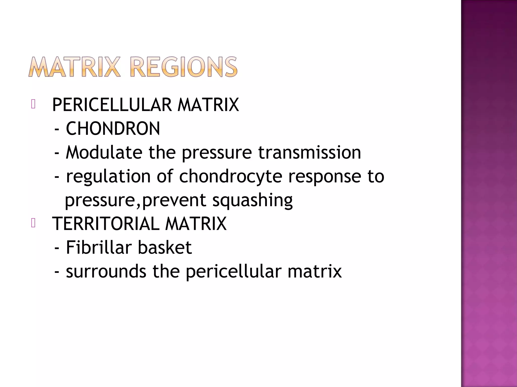
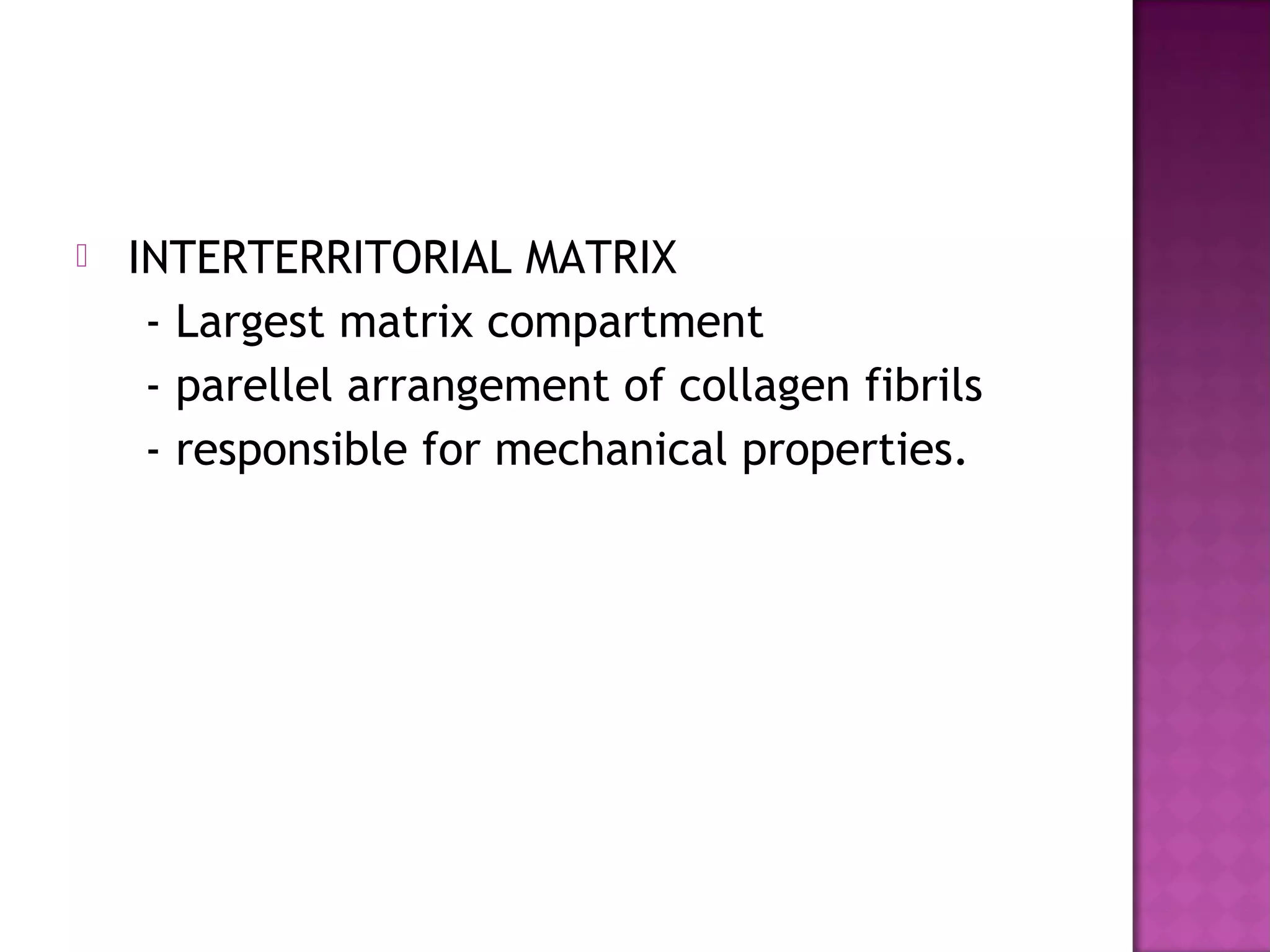
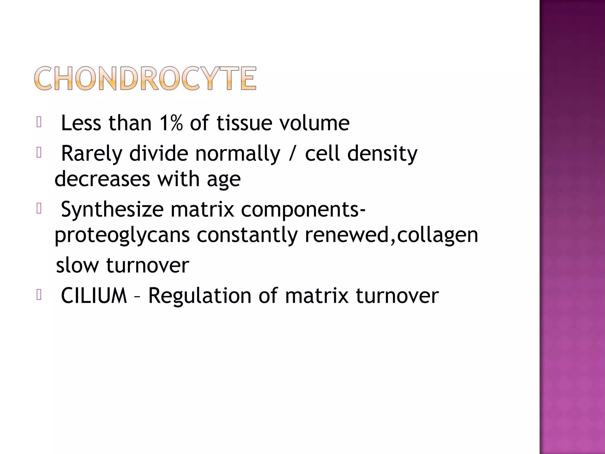
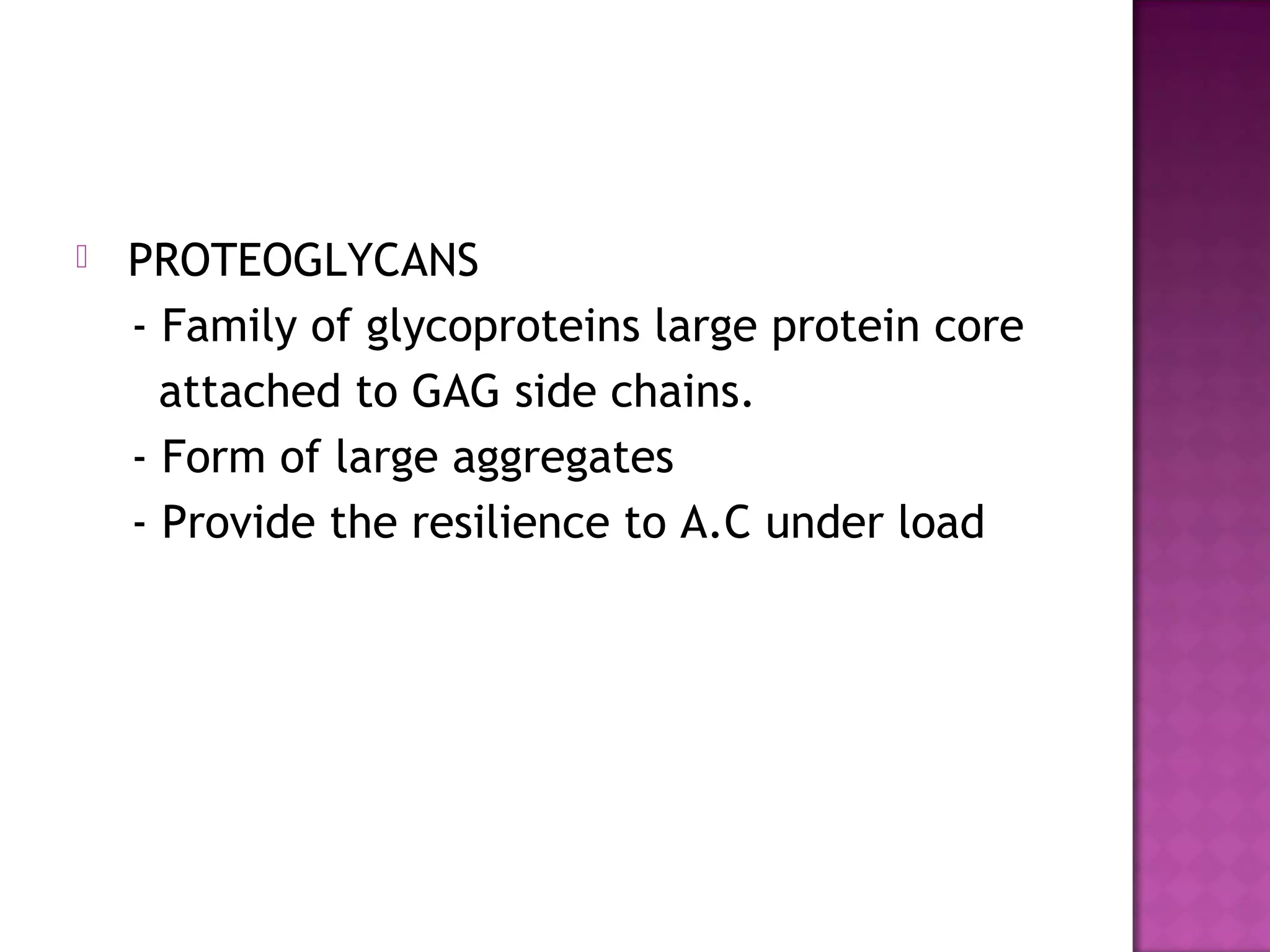
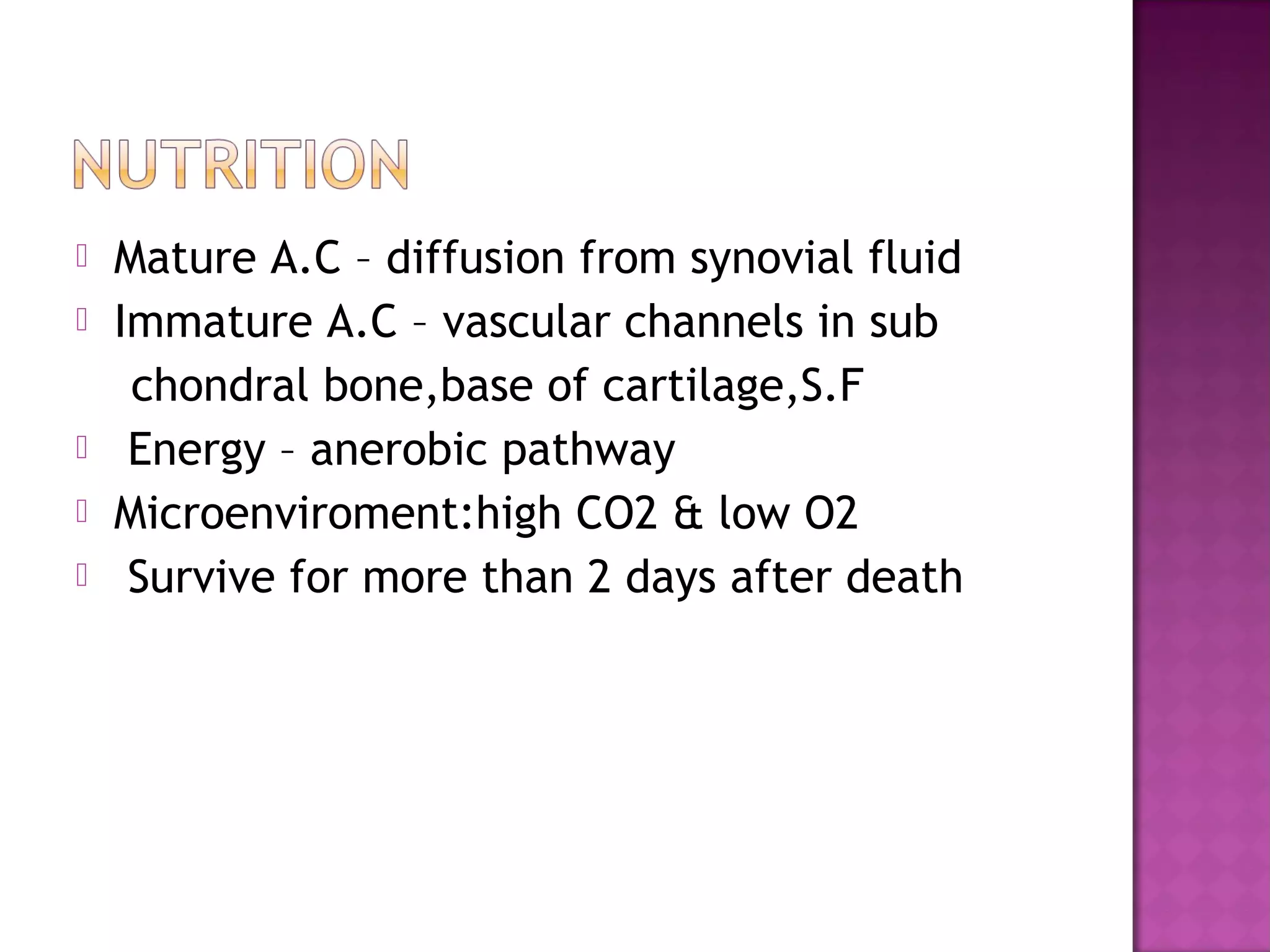
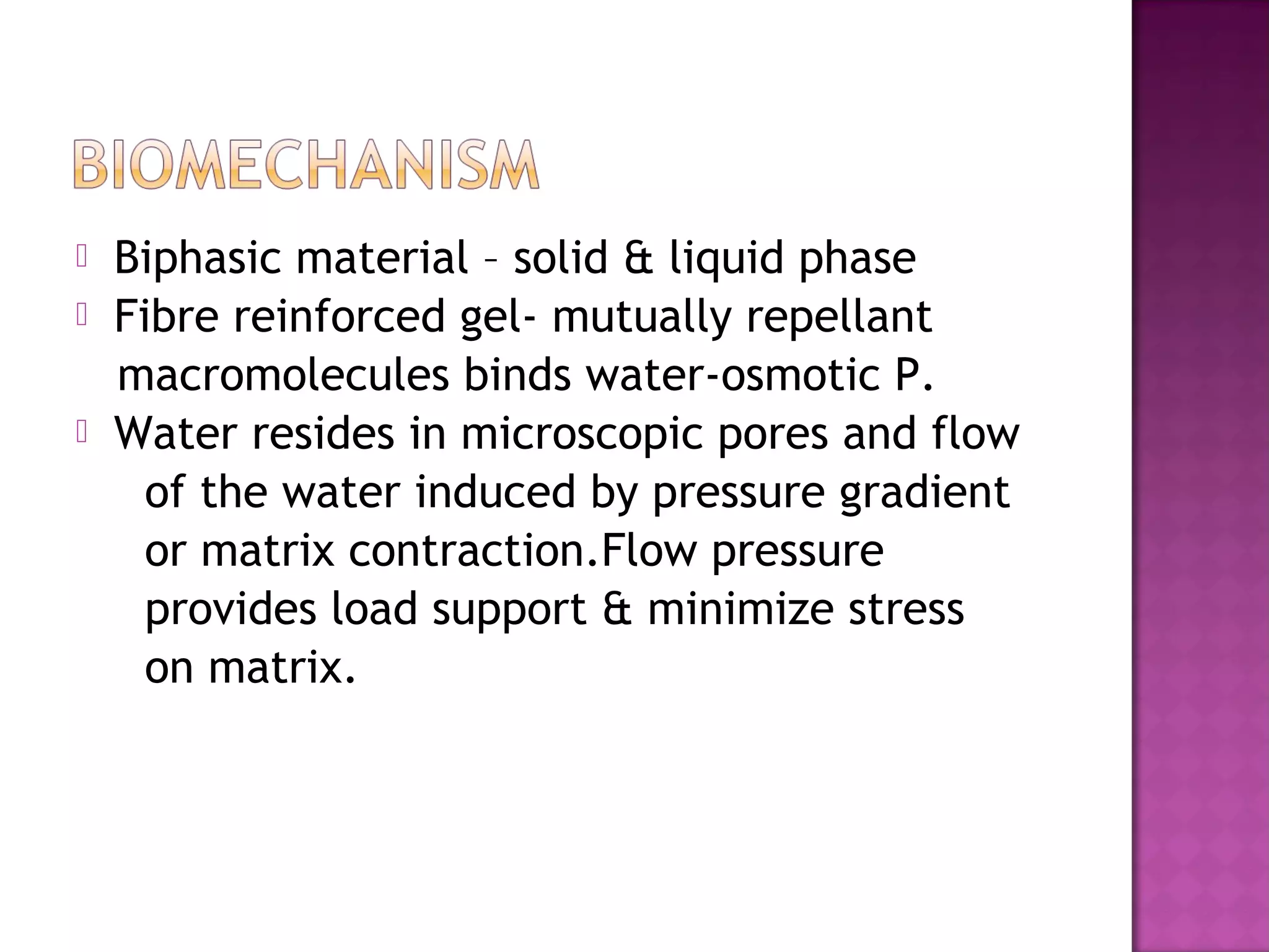
Describes hyaline cartilage features, composition, zones, and properties including collagen, proteoglycans and matrix structure.
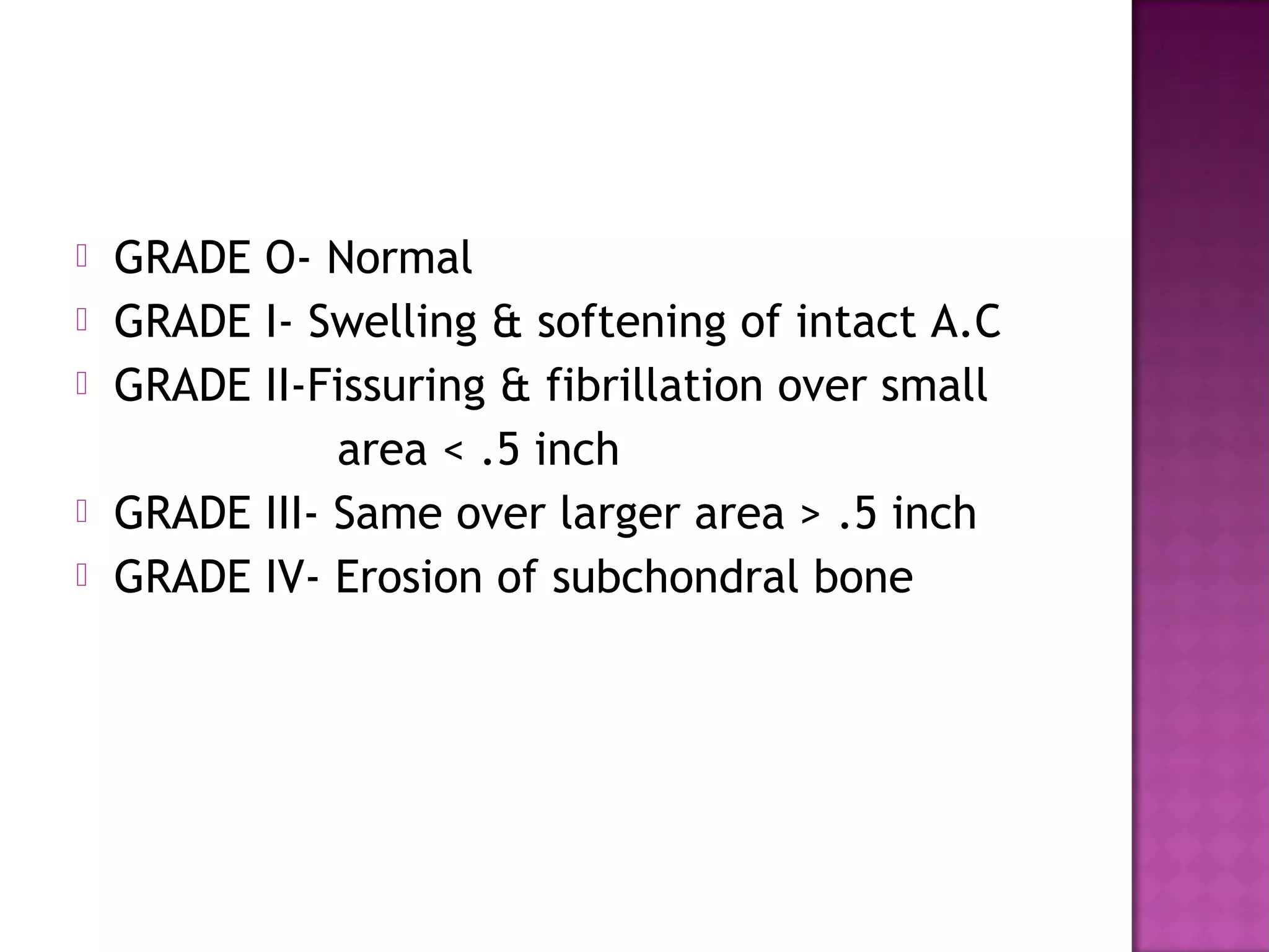
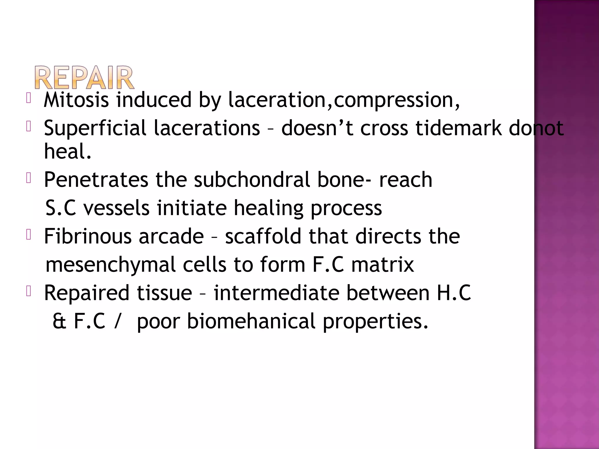
Explains aging effects, chondromalacia grades, and healing processes after cartilage injury.
Discusses joint loading, effects of immobilization, and surgical methods for cartilage repair and grafting.


