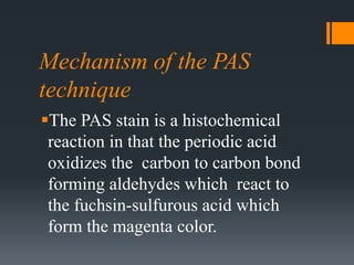
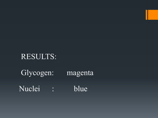
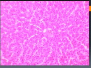
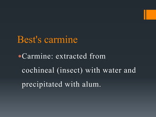
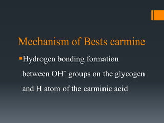
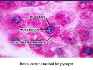
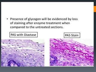
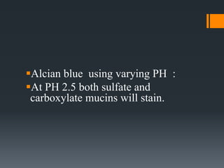
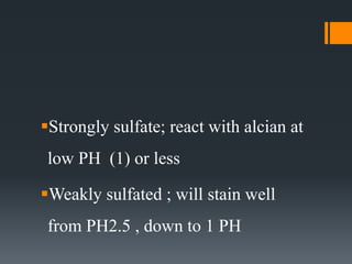
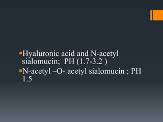
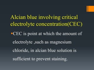
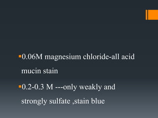
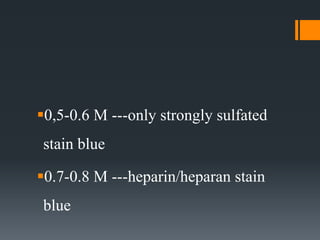
Carbohydrates
Mahmoud Ibrahim Osman
MSc in Histopathology and Cytology
Introduction
Carbohydrates- ( hydrated carbon)
are important organic compounds
that include sugars, starch, cellulose
and polymers that are mostly linked
to proteins.
The he role of carbohydrates in
cellular metabolism has been known
for many years, carbohydrates have
been more recently implicated in a
wide range of cellular functions
including:
protein folding, cell adhesion,
enzyme activity, and immune
recognition.
Classification of carbohydrates
Carbohydrates classified into two
main categories:
simple carbohydrates or those
molecules composed purely of
carbohydrates,
and glycoconjugates, those
molecules composed of
carbohydrates and other molecules
such as protein or lipid
Glycogen and Mucin are the two
main entities to be considered in
tissue carbohydrate demonstration
Polysaccharides
A polysaccharide is a large
macromolecule composed of
multiple monosaccharides joined by
covalent bonds referred to as
glycosidic linkages.
Glycogen
Glycogen is the only polysaccharide
found in animals that frequently is
evaluated by histochemical
techniques.
Simple polysaccharide with consists
of branched or straight chains D-
glucose units found intra cytoplasmic
serves as a major form of stored
energy reserves in humans.
In EM the glycogen occur in the
following forms;
Alpha: found as rosettes forms or
clusters of beta particles measured
60-250 nm in diameter.
Beta: occur as free particles or as a
part of a rosettes formation 20-40nm
Gamma: non-particulate found
between beta particles in the alpha
rosettes.
Normally glycogen found in:
Greatest amount in: liver, cardiac and
skeletal muscles.
Significant amount in: hair follicle,
endometrial gland
cervical and ectocervical epithelium
Few amount in: umbilical cord,
mesothelial cells, neutrophils,
megakaryocytes
Fixation and Section Preparation
Fixative containing alcohol or picric
acid are favorable for glycogen
demonstration eg; bouins and
Rossman solution, 80% alcohol .
Aqueous fixative eg; formalin give
adequate fixation.
Suza-zenker are contraindicated.
At EM: potassium permanganate and
glutraldehyde
Streaming artifacts (Polarization)
One of the difficulties associated
with the fixation of glycogen.
Definition: the tendency of glycogen
to stream through the cell with the
fixative toward one pole when it
fixed at RT.
. Overcoming:
1.Immediately fixation.
2.Fixation should be carried out at 4c
3.Using freeze drying technique
Glycogen staining
Langhan’s iodine method:
Glycogen gives mahogany brown
while other tissues stain yellow.
Impermanent- nonspecific
 H&E:
cells containing abundant glycogen
will stain deeply compared with those
containing little(weak red).
Periodic Acid-Schiff Reaction
(PAS)
The PAS technique is without
question the most versatile and
widely used technique for the
demonstration of carbohydrates or
glycoconjugates.
The PAS technique may aid in the
differential diagnosis of tumors
through the detection of mucins or
glycogen.
Mechanism of the PAS
technique
The PAS stain is a histochemical
reaction in that the periodic acid
oxidizes the carbon to carbon bond
forming aldehydes which react to
the fuchsin-sulfurous acid which
form the magenta color.
RESULTS:
Glycogen: magenta
Nuclei : blue
Best's carmine
Carmine: extracted from
cochineal (insect) with water and
precipitated with alum.
Mechanism of Bests carmine
Hydrogen bonding formation
between OHˉ groups on the glycogen
and H atom of the carminic acid
Hexamine sliver technique
Give similar result to PAS reaction.
Principle:
Following chromic acid oxidation, aldehydes
are formed from glycogen, these will reduce a
hexamine silver nitrate to black compounds.
Enzyme control
Use to specified the techniques applied in
glycogen demonstration:
Alpha amylase: extracted from hog
pancreas and Bacillus subtilis and
Aspergillus oryzea.both branched or
straight chain glycogen are digested, these
releases glucose and maltose.
Beta amylase: obtained from sweet
potato digest straight chain and release
maltose alone.
Diastase: commonly used as it is; easy,
stable, cheep, extracted from malt and
contain both alpha and beta amylase
Saliva: highly effective.
Pectinase: effective with abnormal
glycogen. Found in certain
glycogenesis, which is not digested
easy by amylase
Medical importance
Some glycogen storage diseases :
A-von Gierke disease (deficiency of
Glucose-6-phosphatase)
B-Pompe disease(deficiency of Acid
maltase)
Also demonstration of glycogen can
help in diagnosis of carcinomas of
Bladder,Kidney,Liver,ovary and
adenocarcinoma of
Pancrease,Lung,Seminoma
Mesothelioma
Mucin
High molecular weight glycolized
protein secreted by specialized
epithelial cells and connective tissue
cells
The carbohydrate content of a mucin
may account for up to 90% of its
molecular weight.
In contrast to the
glycosaminoglycan's, which are
strongly acidic polyanions, the
polysaccharide chains of the mucins
vary from neutral or weakly acidic to
strongly acidic.
mucoid carbohydrates can be
divided into three main types:
1.Mucopolysaccharides
2.Mucoprotein
3.Mucolipids
Mucoprotein
Predominantly protein and contains
more than 4% hexosamine , when
the polysaccharides over 20% known
as sialomucin ,
when the polysaccharides less than 4%
we called glycoprotein and classified
with mucoprotein and can not be
distinguished from them . PAS---positive
Glycolipids
Made up of fatty acids combined
with carbohydrates , usually
galactose the cerebrosides are the
most important glycolipids PAS---
positive
Functions of mucins
1.Lubricator function
2.Environment for ionic and molecular
diffusion
3.Cell to cell adhesion with specific cell
surface
Protective role (innate immunity) from
microbes and chemical and mechanical
agents throughout GIT, Respiratory
tract ,reproductive system .
Mucopolysaccharides(Mucin)
Polysaccharides-protein complexes
divided into:
- acid mucopolysaccharides
- and neutral mucopolysaccharides
Neutral mucopolysaccharides
carbohydrate made up of hexose
units usually acetylated such as
glucosamine combined with proteins
and calcium.
present in alimentary ,respiratory
tract , prostate gland ,PAS----positive
Acid mucopolysaccharides
composed of chains of glucosamine
and glucuronic acid and proteins
Divided into
1.Simple acid mucopolysaccharides;
Contain carboxylated glucose units
(glucuronic acid) e g (hyaluronic
acid )
2. Complex acid mucopolysaccharides:
In addition to glucuronic acid –sulfated
glucosamine units are includes e g chondroitin
which occur in cartilages and connective tissues
also heparin and heparan are others examples
Acid mucin : divided into sulfated
and carboxylated mucin
Sulfated mucin: divided into
strongly and weakly
Strongly sulfated mucin: divide into
strongly sulfated epithelial and strongly
sulfated connective tissue mucin
(proteoglycan )
Strongly sulfated CT mucin
Strongly sulfated CT mucin:
 Highly sulfated substances produced by
fibroblast ,endothelial, osteocytes ,
chondrocytes ,mast cells , react at low PH
with cationic dyes and PAS negative.
 The following types
1. Chondroitin sulfate A : found in
cartilage
2. C,S,B: found in aorta , heart valves ,
dermis of skin.
3. C,S,C : in cartilage ,umbilical cord ,
dermis of skin.
4- Heparin/heparan sulfate: heparan found
in aorta , cardiac CT, and heparin in mast
cells.
5-Keratan sulphate: present in
cornea, ageing cartilage
6-Hyaluronosulphate: found in
cornea
Strongly sulfated epithelial mucin :
 in bronchial glands and in minor
fraction in intestinal goblet cell ,
histologically react at low PH.
But differ from that of connective
tissue mucin in being PAS positive
Weakly sulfated epithelial mucin
(sulfomucin) : epithelial in type,
present in colonic goblet cell , react
at slightly higher PH
Carboxylated mucins :
Sialomucin: contain derivatives of
neuromeric acid (sialic ) epithelial in
origin, divide into:
a-Enzyme labile ( N-acetyl sialomucin):
digested by sialidase , present in
bronchial sub mucous gland , sub
mandibular salivary gland ,
goblet of small intestine ,PAS--
positive
b. Enzyme –resistant (N-acetyl -O-
acetyl neuromeric acid ) sialidase
resistant , found in mucosa of large
intestine –stomach-bronchus , PAS----
negative
2- Hyaluronic acid: occur in CT
formed by fibroblast ,important
constituent of synovial fluid also
found in umbilical cord ,
cardiac CT , dermis of skin , aorta ,
bone and cartilages
Diagnostic applications:
1/ To diagnose poorly differentiated
adenocarcinoma.
2/ Diagnostic significant in certain
situation :
* Hyaluronic acid –follicular
mucinosis/ -myxoid liposarcoma/-
mesothelioma
Neutral mucin- carcinoma of
stomach
Sialomucin (N-acetyl form)-
Transitional epithelium of
colon/rectum
Sialomucin (O-acetyl form )-ca of
colon/rectum
Demonstration of mucins
Aqueous fixatives (formalin) are
satisfactory.
Muco polysaccharide are better
preserved in alcoholic fixatives
Hyaluronic acid when occur free is better
fixed in 10% formal-alcohol or 10 %
formal sublimate.
Other fixatives 2% calcium acetate in
10%formalin
Mucin staining
1/ H&E:
 Mucin take up the eosin ,unless
Ehrlich Hematoxylin is used where
acid mucin stains blue.
2/ PAS: Mucin containing a reactive
hexose component will be PAS
positive, these include:
1. Neutral mucin
2. N acetyl sialomucin
1. Epithelial sulphated mucin
2. Mucoprotein
3. Glycolipid
3/ Alcian blue
Alcian blue is the highest molecular
weight (cationic dyes) formed
electrostatic bonds( salt linkage)
 with tissue polyanions bearing either
carboxyl or sulfate groups
 Alcian blue is a popular dye for
demonstrating acid mucin why?
1. Specificity
2. Strong coloration
1. In solubility of staining
2. Permanence of results
Alcian blue using varying PH :
At PH 2.5 both sulfate and
carboxylate mucins will stain.
Strongly sulfate; react with alcian at
low PH (1) or less
Weakly sulfated ; will stain well
from PH2.5 , down to 1 PH
Hyaluronic acid and N-acetyl
sialomucin; PH (1.7-3.2 )
N-acetyl –O- acetyl sialomucin ; PH
1.5
Alcian blue involving critical
electrolyte concentration(CEC)
CEC is point at which the amount of
electrolyte ,such as magnesium
chloride, in alcian blue solution is
sufficient to prevent staining.
Separate different types of acid
mucin, each type has CEC as
following:
0.06M magnesium chloride-all acid
mucin stain
0.2-0.3 M ---only weakly and
strongly sulfate ,stain blue
0,5-0.6 M ---only strongly sulfated
stain blue
0.7-0.8 M ---heparin/heparan stain
blue
4/ Dialyzed iron –prussian blue
tech.
This is another popular technique for
demonstration acid mucins.
At low PH colloidal iron will be
adsorbed onto tissue polyanions (
sulphate , carboxylate group)
and subsequently ,visualized by
conversion to ferric ferrocyanide
using (Perl's tech)
It is more sensitive ,but complex
5/ Southgate mucicarmine tech-
The rationale : not fully understood
,the aluminum/carmine compound
appear to be positively charged and
combined with negatively charged
acid mucosubstances.
Results :
Mucins :Red
Nuclei : Blue
6/Metachromatic staining
method
Acid mucosubstance can be
demonstrated by numbers of
metachromatic dyes.
The most useful dye is azure A
Sulfated and carboxylated mucins
are metachromatic while neutral
mucin is orthochromatic.
Sulfated mucin at PH3 and
carboxylated at PH5
Combined alcian blue PAS methods
Acid mucins and neutral mucins are
separated , also useful to demonstrate
any mucins . First all acid mucin
stained with alcian blue leaving only
neutral mucin to be demonstrated by
PAS reaction.
Alcian blue-Alcian yellow
Used to distinguished b/w sulphate and
carboxylate, involving initial staining
with low Ph. alcian blue, followed high
Ph. alcian yellow
Result :
sulphate mucin- blue
carboxylate mucin -yellow
Aldehyde fuchsin –Alcian blue
technique
Separate sulphate from carboxylate.
The rationale:
Depends on the greater affinity of
aldehyde fuchsin for sulphate mucin .
so that by first staining with this
solution sulphate stained purple
,rendering carboxylated mucin to be
stain by alcian blue (counterstaining
Blocking tech and Enzyme
controls
Improved specificity, by enzyme
action or by blocking staining
(chemical action )
Blocking
Methylation:
using hydrochloric acid in methyl alcohol for
blocking the staining reaction of carboxylated
mucin by esterification of carboxyl group and
sulphate group by desulphation.
Methylation and Saponification:
After Methylation ,saponification with
potassium hydroxide in ethyl alcohol ,
will restore the staining of carboxyl
groups but sulphate groups still blocked.
Sialidase digestion:
useful method for identifying a certain
type of sialomucin.
Other types of carbohydrates
Chitin:
This is hyaline substance wide
distributed in non human tissue for
example exoskeleton of insects.
In human only seen lining the wall of
hydatid cyst of lung or liver due to
infestation of larvae of dog tapeworm
Echinococcus granulosus.
PAS positive Diastase resistance
Starch
Can be found in tissue as
contaminant from surgical gloves
powder.
PAS positive with iodine pale blue-
Diastase labile
Cellulose
Seen in undigested food specially in
the section taken from GIT.
PAS positive brown color with
iodine

Carbohydrate

  • 1.
    Carbohydrates Mahmoud Ibrahim Osman MScin Histopathology and Cytology
  • 2.
    Introduction Carbohydrates- ( hydratedcarbon) are important organic compounds that include sugars, starch, cellulose and polymers that are mostly linked to proteins.
  • 3.
    The he roleof carbohydrates in cellular metabolism has been known for many years, carbohydrates have been more recently implicated in a wide range of cellular functions including:
  • 4.
    protein folding, celladhesion, enzyme activity, and immune recognition.
  • 5.
    Classification of carbohydrates Carbohydratesclassified into two main categories: simple carbohydrates or those molecules composed purely of carbohydrates,
  • 6.
    and glycoconjugates, those moleculescomposed of carbohydrates and other molecules such as protein or lipid
  • 7.
    Glycogen and Mucinare the two main entities to be considered in tissue carbohydrate demonstration
  • 8.
    Polysaccharides A polysaccharide isa large macromolecule composed of multiple monosaccharides joined by covalent bonds referred to as glycosidic linkages.
  • 9.
    Glycogen Glycogen is theonly polysaccharide found in animals that frequently is evaluated by histochemical techniques.
  • 10.
    Simple polysaccharide withconsists of branched or straight chains D- glucose units found intra cytoplasmic serves as a major form of stored energy reserves in humans.
  • 11.
    In EM theglycogen occur in the following forms; Alpha: found as rosettes forms or clusters of beta particles measured 60-250 nm in diameter.
  • 12.
    Beta: occur asfree particles or as a part of a rosettes formation 20-40nm Gamma: non-particulate found between beta particles in the alpha rosettes.
  • 13.
    Normally glycogen foundin: Greatest amount in: liver, cardiac and skeletal muscles. Significant amount in: hair follicle, endometrial gland
  • 14.
    cervical and ectocervicalepithelium Few amount in: umbilical cord, mesothelial cells, neutrophils, megakaryocytes
  • 15.
    Fixation and SectionPreparation Fixative containing alcohol or picric acid are favorable for glycogen demonstration eg; bouins and Rossman solution, 80% alcohol .
  • 16.
    Aqueous fixative eg;formalin give adequate fixation. Suza-zenker are contraindicated. At EM: potassium permanganate and glutraldehyde
  • 17.
    Streaming artifacts (Polarization) Oneof the difficulties associated with the fixation of glycogen. Definition: the tendency of glycogen to stream through the cell with the fixative toward one pole when it fixed at RT.
  • 18.
    . Overcoming: 1.Immediately fixation. 2.Fixationshould be carried out at 4c 3.Using freeze drying technique
  • 19.
    Glycogen staining Langhan’s iodinemethod: Glycogen gives mahogany brown while other tissues stain yellow. Impermanent- nonspecific
  • 20.
     H&E: cells containingabundant glycogen will stain deeply compared with those containing little(weak red).
  • 21.
    Periodic Acid-Schiff Reaction (PAS) ThePAS technique is without question the most versatile and widely used technique for the demonstration of carbohydrates or glycoconjugates.
  • 22.
    The PAS techniquemay aid in the differential diagnosis of tumors through the detection of mucins or glycogen.
  • 23.
    Mechanism of thePAS technique The PAS stain is a histochemical reaction in that the periodic acid oxidizes the carbon to carbon bond forming aldehydes which react to the fuchsin-sulfurous acid which form the magenta color.
  • 24.
  • 26.
    Best's carmine Carmine: extractedfrom cochineal (insect) with water and precipitated with alum.
  • 27.
    Mechanism of Bestscarmine Hydrogen bonding formation between OHˉ groups on the glycogen and H atom of the carminic acid
  • 29.
    Hexamine sliver technique Givesimilar result to PAS reaction. Principle: Following chromic acid oxidation, aldehydes are formed from glycogen, these will reduce a hexamine silver nitrate to black compounds.
  • 30.
    Enzyme control Use tospecified the techniques applied in glycogen demonstration: Alpha amylase: extracted from hog pancreas and Bacillus subtilis and Aspergillus oryzea.both branched or straight chain glycogen are digested, these releases glucose and maltose.
  • 31.
    Beta amylase: obtainedfrom sweet potato digest straight chain and release maltose alone. Diastase: commonly used as it is; easy, stable, cheep, extracted from malt and contain both alpha and beta amylase
  • 32.
    Saliva: highly effective. Pectinase:effective with abnormal glycogen. Found in certain glycogenesis, which is not digested easy by amylase
  • 34.
    Medical importance Some glycogenstorage diseases : A-von Gierke disease (deficiency of Glucose-6-phosphatase) B-Pompe disease(deficiency of Acid maltase)
  • 35.
    Also demonstration ofglycogen can help in diagnosis of carcinomas of Bladder,Kidney,Liver,ovary and adenocarcinoma of Pancrease,Lung,Seminoma Mesothelioma
  • 36.
    Mucin High molecular weightglycolized protein secreted by specialized epithelial cells and connective tissue cells
  • 37.
    The carbohydrate contentof a mucin may account for up to 90% of its molecular weight.
  • 38.
    In contrast tothe glycosaminoglycan's, which are strongly acidic polyanions, the polysaccharide chains of the mucins vary from neutral or weakly acidic to strongly acidic.
  • 39.
    mucoid carbohydrates canbe divided into three main types: 1.Mucopolysaccharides 2.Mucoprotein 3.Mucolipids
  • 40.
    Mucoprotein Predominantly protein andcontains more than 4% hexosamine , when the polysaccharides over 20% known as sialomucin ,
  • 41.
    when the polysaccharidesless than 4% we called glycoprotein and classified with mucoprotein and can not be distinguished from them . PAS---positive
  • 42.
    Glycolipids Made up offatty acids combined with carbohydrates , usually galactose the cerebrosides are the most important glycolipids PAS--- positive
  • 43.
    Functions of mucins 1.Lubricatorfunction 2.Environment for ionic and molecular diffusion 3.Cell to cell adhesion with specific cell surface
  • 44.
    Protective role (innateimmunity) from microbes and chemical and mechanical agents throughout GIT, Respiratory tract ,reproductive system .
  • 45.
    Mucopolysaccharides(Mucin) Polysaccharides-protein complexes divided into: -acid mucopolysaccharides - and neutral mucopolysaccharides
  • 46.
    Neutral mucopolysaccharides carbohydrate madeup of hexose units usually acetylated such as glucosamine combined with proteins and calcium.
  • 47.
    present in alimentary,respiratory tract , prostate gland ,PAS----positive
  • 48.
    Acid mucopolysaccharides composed ofchains of glucosamine and glucuronic acid and proteins Divided into
  • 49.
    1.Simple acid mucopolysaccharides; Containcarboxylated glucose units (glucuronic acid) e g (hyaluronic acid )
  • 50.
    2. Complex acidmucopolysaccharides: In addition to glucuronic acid –sulfated glucosamine units are includes e g chondroitin which occur in cartilages and connective tissues also heparin and heparan are others examples
  • 51.
    Acid mucin :divided into sulfated and carboxylated mucin Sulfated mucin: divided into strongly and weakly
  • 52.
    Strongly sulfated mucin:divide into strongly sulfated epithelial and strongly sulfated connective tissue mucin (proteoglycan )
  • 53.
    Strongly sulfated CTmucin Strongly sulfated CT mucin:  Highly sulfated substances produced by fibroblast ,endothelial, osteocytes , chondrocytes ,mast cells , react at low PH with cationic dyes and PAS negative.
  • 54.
     The followingtypes 1. Chondroitin sulfate A : found in cartilage 2. C,S,B: found in aorta , heart valves , dermis of skin.
  • 55.
    3. C,S,C :in cartilage ,umbilical cord , dermis of skin. 4- Heparin/heparan sulfate: heparan found in aorta , cardiac CT, and heparin in mast cells.
  • 56.
    5-Keratan sulphate: presentin cornea, ageing cartilage 6-Hyaluronosulphate: found in cornea
  • 57.
    Strongly sulfated epithelialmucin :  in bronchial glands and in minor fraction in intestinal goblet cell , histologically react at low PH.
  • 58.
    But differ fromthat of connective tissue mucin in being PAS positive
  • 59.
    Weakly sulfated epithelialmucin (sulfomucin) : epithelial in type, present in colonic goblet cell , react at slightly higher PH
  • 60.
    Carboxylated mucins : Sialomucin:contain derivatives of neuromeric acid (sialic ) epithelial in origin, divide into:
  • 61.
    a-Enzyme labile (N-acetyl sialomucin): digested by sialidase , present in bronchial sub mucous gland , sub mandibular salivary gland ,
  • 62.
    goblet of smallintestine ,PAS-- positive
  • 63.
    b. Enzyme –resistant(N-acetyl -O- acetyl neuromeric acid ) sialidase resistant , found in mucosa of large intestine –stomach-bronchus , PAS---- negative
  • 64.
    2- Hyaluronic acid:occur in CT formed by fibroblast ,important constituent of synovial fluid also found in umbilical cord ,
  • 65.
    cardiac CT ,dermis of skin , aorta , bone and cartilages
  • 66.
    Diagnostic applications: 1/ Todiagnose poorly differentiated adenocarcinoma. 2/ Diagnostic significant in certain situation :
  • 67.
    * Hyaluronic acid–follicular mucinosis/ -myxoid liposarcoma/- mesothelioma Neutral mucin- carcinoma of stomach
  • 68.
    Sialomucin (N-acetyl form)- Transitionalepithelium of colon/rectum Sialomucin (O-acetyl form )-ca of colon/rectum
  • 69.
    Demonstration of mucins Aqueousfixatives (formalin) are satisfactory. Muco polysaccharide are better preserved in alcoholic fixatives
  • 70.
    Hyaluronic acid whenoccur free is better fixed in 10% formal-alcohol or 10 % formal sublimate. Other fixatives 2% calcium acetate in 10%formalin
  • 71.
    Mucin staining 1/ H&E: Mucin take up the eosin ,unless Ehrlich Hematoxylin is used where acid mucin stains blue.
  • 72.
    2/ PAS: Mucincontaining a reactive hexose component will be PAS positive, these include: 1. Neutral mucin 2. N acetyl sialomucin
  • 73.
    1. Epithelial sulphatedmucin 2. Mucoprotein 3. Glycolipid
  • 74.
    3/ Alcian blue Alcianblue is the highest molecular weight (cationic dyes) formed electrostatic bonds( salt linkage)
  • 75.
     with tissuepolyanions bearing either carboxyl or sulfate groups
  • 76.
     Alcian blueis a popular dye for demonstrating acid mucin why? 1. Specificity 2. Strong coloration
  • 77.
    1. In solubilityof staining 2. Permanence of results
  • 78.
    Alcian blue usingvarying PH : At PH 2.5 both sulfate and carboxylate mucins will stain.
  • 79.
    Strongly sulfate; reactwith alcian at low PH (1) or less Weakly sulfated ; will stain well from PH2.5 , down to 1 PH
  • 80.
    Hyaluronic acid andN-acetyl sialomucin; PH (1.7-3.2 ) N-acetyl –O- acetyl sialomucin ; PH 1.5
  • 81.
    Alcian blue involvingcritical electrolyte concentration(CEC) CEC is point at which the amount of electrolyte ,such as magnesium chloride, in alcian blue solution is sufficient to prevent staining.
  • 82.
    Separate different typesof acid mucin, each type has CEC as following:
  • 83.
    0.06M magnesium chloride-allacid mucin stain 0.2-0.3 M ---only weakly and strongly sulfate ,stain blue
  • 84.
    0,5-0.6 M ---onlystrongly sulfated stain blue 0.7-0.8 M ---heparin/heparan stain blue
  • 85.
    4/ Dialyzed iron–prussian blue tech. This is another popular technique for demonstration acid mucins. At low PH colloidal iron will be adsorbed onto tissue polyanions ( sulphate , carboxylate group)
  • 86.
    and subsequently ,visualizedby conversion to ferric ferrocyanide using (Perl's tech) It is more sensitive ,but complex
  • 87.
    5/ Southgate mucicarminetech- The rationale : not fully understood ,the aluminum/carmine compound appear to be positively charged and combined with negatively charged acid mucosubstances.
  • 88.
  • 89.
    6/Metachromatic staining method Acid mucosubstancecan be demonstrated by numbers of metachromatic dyes. The most useful dye is azure A
  • 90.
    Sulfated and carboxylatedmucins are metachromatic while neutral mucin is orthochromatic. Sulfated mucin at PH3 and carboxylated at PH5
  • 91.
    Combined alcian bluePAS methods Acid mucins and neutral mucins are separated , also useful to demonstrate any mucins . First all acid mucin stained with alcian blue leaving only neutral mucin to be demonstrated by PAS reaction.
  • 92.
    Alcian blue-Alcian yellow Usedto distinguished b/w sulphate and carboxylate, involving initial staining with low Ph. alcian blue, followed high Ph. alcian yellow Result : sulphate mucin- blue carboxylate mucin -yellow
  • 93.
    Aldehyde fuchsin –Alcianblue technique Separate sulphate from carboxylate. The rationale: Depends on the greater affinity of aldehyde fuchsin for sulphate mucin .
  • 94.
    so that byfirst staining with this solution sulphate stained purple ,rendering carboxylated mucin to be stain by alcian blue (counterstaining
  • 95.
    Blocking tech andEnzyme controls Improved specificity, by enzyme action or by blocking staining (chemical action )
  • 96.
    Blocking Methylation: using hydrochloric acidin methyl alcohol for blocking the staining reaction of carboxylated mucin by esterification of carboxyl group and sulphate group by desulphation.
  • 97.
    Methylation and Saponification: AfterMethylation ,saponification with potassium hydroxide in ethyl alcohol , will restore the staining of carboxyl groups but sulphate groups still blocked.
  • 98.
    Sialidase digestion: useful methodfor identifying a certain type of sialomucin.
  • 99.
    Other types ofcarbohydrates Chitin: This is hyaline substance wide distributed in non human tissue for example exoskeleton of insects.
  • 100.
    In human onlyseen lining the wall of hydatid cyst of lung or liver due to infestation of larvae of dog tapeworm Echinococcus granulosus. PAS positive Diastase resistance
  • 101.
    Starch Can be foundin tissue as contaminant from surgical gloves powder. PAS positive with iodine pale blue- Diastase labile
  • 102.
    Cellulose Seen in undigestedfood specially in the section taken from GIT. PAS positive brown color with iodine