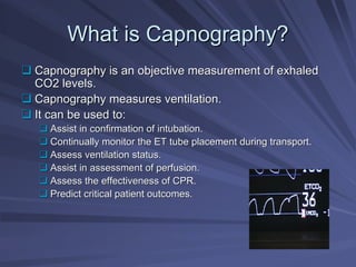
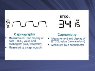
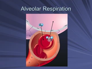
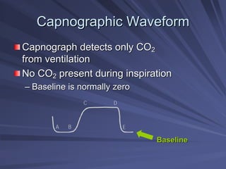
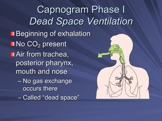
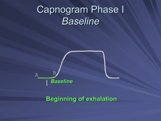
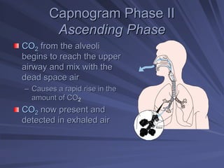
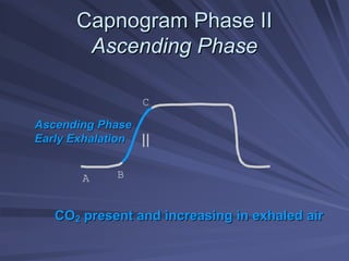
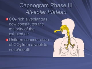
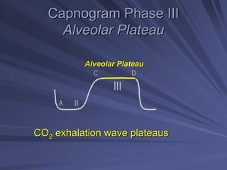
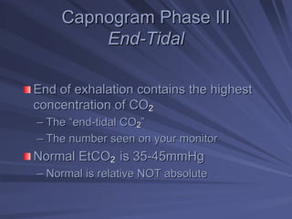
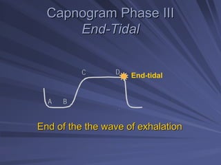
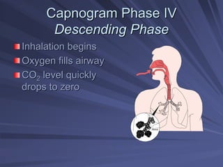
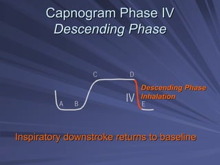
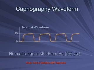
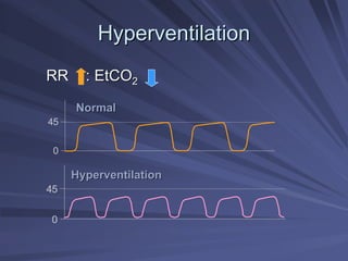
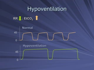
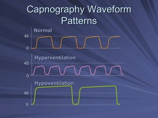
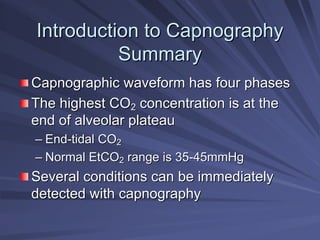
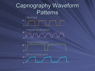
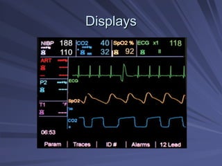
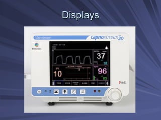
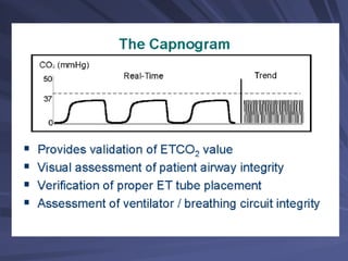
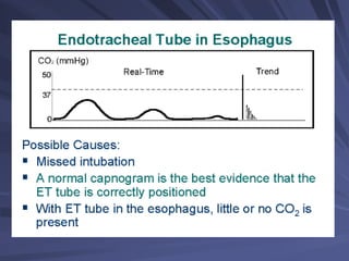
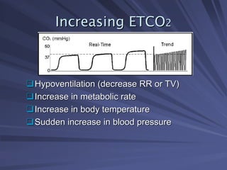
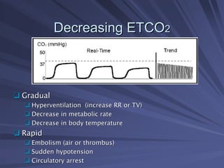
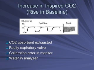
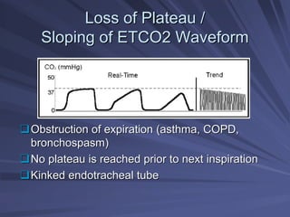
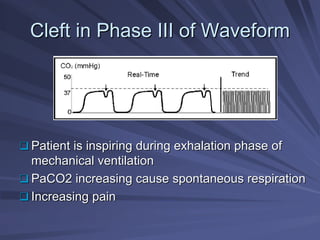
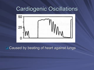
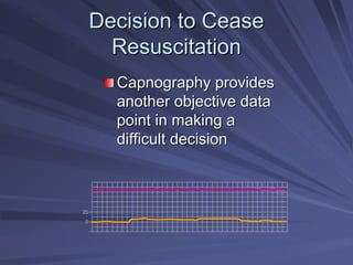
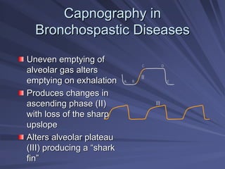
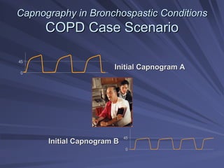
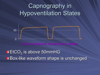
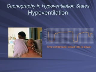
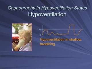
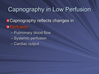
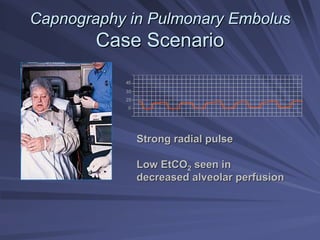
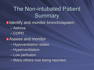
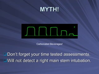
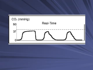
2. What is Capnography?
What is Capnography?
â°
â° Capnography is an objective measurement of exhaled
Capnography is an objective measurement of exhaled
CO2 levels.
CO2 levels.
â°
â° Capnography measures ventilation.
Capnography measures ventilation.
â°
â° It can be used to:
It can be used to:
â°
â° Assist in confirmation of intubation.
Assist in confirmation of intubation.
â°
â° Continually monitor the ET tube placement during transport.
Continually monitor the ET tube placement during transport.
â°
â° Assess ventilation status.
Assess ventilation status.
â°
â° Assist in assessment of perfusion.
Assist in assessment of perfusion.
â°
â° Assess the effectiveness of CPR.
Assess the effectiveness of CPR.
â°
â° Predict critical patient outcomes.
Predict critical patient outcomes.
3. CAPNOGRAPHY
CAPNOGRAPHY
â°
â° Term capnography comes from the Greek
Term capnography comes from the Greek
work
work KAPNOS,
KAPNOS, meaning smoke.
meaning smoke.
â°
â° Anesthesia context: inspired and expired
Anesthesia context: inspired and expired
gases sampled at the Y connector, mask
or nasal cannula.
or nasal cannula.
â°
â° Gives insight into alterations in ventilation,
Gives insight into alterations in ventilation,
cardiac output, distribution of pulmonary
blood flow and metabolic activity.
6. Oxygenation
Oxygenation â
â How we get oxygen to the
How we get oxygen to the
tissues
tissues
Ventilation (the movement of air)
Ventilation (the movement of air) â
â How
How
we get rid of carbon dioxide.
9. Capnography
Capnography vs
vs Pulse Oximetry
Pulse Oximetry
Capnography provides an immediate
picture of patient condition. Pulse oximetry
is delayed. Hold your breath.
Capnography will show immediate apnea,
while pulse oximetry will show a high
saturation for several minutes.
10. Capnography
Capnography vs
vs Pulse Oximetry
Pulse Oximetry
Good pulse
Good pulseâŚ
âŚ.good Spo2
.good Spo2âŚ
âŚ.No Capnography!
.No Capnography!
11. Introduction to Capnography
Introduction to Capnography
Summary
Summary
Oxygenation and ventilation
Pulse oximetry
Pulse oximetry
â
â Measures O
Measures O2
2 saturation in blood
saturation in blood
â
â Slow to indicate change in ventilation
Slow to indicate change in ventilation
Capnography
Capnography
â
â Measures CO
Measures CO2
2 in the
in the the
the airway
airway
â
â Provides a breath
Provides a breath-
-to
to-
-breath
breath
status of ventilation
13. Capnogram Phase I
Capnogram Phase I
Dead Space Ventilation
Beginning of exhalation
No CO
No CO2
2 present
present
Air from trachea,
Air from trachea,
posterior pharynx,
posterior pharynx,
mouth and nose
mouth and nose
â
â No gas exchange
No gas exchange
occurs there
occurs there
â
â Called
Called â
âdead space
dead spaceâ
â
14. Capnogram Phase I
Capnogram Phase I
Baseline
Baseline
Beginning of exhalation
A B
I Baseline
Baseline
15. Capnogram Phase II
Capnogram Phase II
Ascending Phase
Ascending Phase
CO
CO2
2 from the alveoli
from the alveoli
begins to reach the upper
airway and mix with the
dead space air
dead space air
â
â Causes a rapid rise in the
Causes a rapid rise in the
amount of CO
amount of CO2
2
CO
CO2
2 now present and
now present and
detected in exhaled air
Alveoli
16. Capnogram Phase II
Capnogram Phase II
Ascending Phase
Ascending Phase
CO
CO2
2 present and increasing in exhaled air
present and increasing in exhaled air
II
A B
C
Ascending Phase
Ascending Phase
Early Exhalation
Early Exhalation
17. Capnogram Phase III
Capnogram Phase III
Alveolar Plateau
Alveolar Plateau
CO
CO2
2 rich alveolar gas
rich alveolar gas
now constitutes the
now constitutes the
majority of the
majority of the
exhaled air
exhaled air
Uniform concentration
of CO
of CO2
2 from alveoli to
from alveoli to
nose/mouth
nose/mouth
18. Capnogram Phase III
Capnogram Phase III
Alveolar Plateau
Alveolar Plateau
CO
CO2
2 exhalation wave plateaus
exhalation wave plateaus
A B
C D
III
Alveolar Plateau
Alveolar Plateau
19. Capnogram Phase III
Capnogram Phase III
End
End-
-Tidal
Tidal
End of exhalation contains the highest
concentration of CO
concentration of CO2
2
â
â The
The â
âend
end-
-tidal CO
tidal CO2
2â
â
â
â The number seen on your monitor
The number seen on your monitor
Normal EtCO
Normal EtCO2
2 is 35
is 35-
-45mmHg
45mmHg
â
â Normal is relative NOT absolute
Normal is relative NOT absolute
20. Capnogram Phase III
Capnogram Phase III
End
End-
-Tidal
Tidal
End of the
End of the the
the wave of exhalation
wave of exhalation
A B
C D End-tidal
21. Capnogram Phase IV
Capnogram Phase IV
Descending Phase
Descending Phase
Inhalation begins
Inhalation begins
Oxygen fills airway
Oxygen fills airway
CO
CO2
2 level quickly
level quickly
drops to zero
drops to zero
Alveoli
22. Capnogram Phase IV
Capnogram Phase IV
Descending Phase
Descending Phase
Inspiratory
Inspiratory downstroke
downstroke returns to baseline
returns to baseline
A B
C D
E
IV
Descending Phase
Descending Phase
Inhalation
Inhalation
23. Capnography Waveform
Capnography Waveform
Normal range is 35
Normal range is 35-
-45mm Hg (5%
45mm Hg (5% vol
vol)
)
Normal Waveform
Normal Waveform
45
0
Note: This is Relative NOT absolute
24. Capnography Waveform
Capnography Waveform
Question
Question
How would your capnogram
change if you intentionally started
to breathe at a rate of 30?
â
â Frequency
Frequency
â
â Duration
Duration
â
â Height
Height
â
â Shape
Shape
26. Capnography Waveform
Capnography Waveform
Question
Question
How would your capnogram change
if you intentionally decreased your
respiratory rate to 8?
â
â Frequency
Frequency
â
â Duration
Duration
â
â Height
Height
â
â Shape
Shape
29. Introduction to Capnography
Introduction to Capnography
Summary
Summary
Capnographic
Capnographic waveform has four phases
waveform has four phases
The highest CO
The highest CO2
2 concentration is at the
concentration is at the
end of alveolar plateau
â
â End
End-
-tidal CO
tidal CO2
2
â
â Normal EtCO
Normal EtCO2
2 range is 35
range is 35-
-45mmHg
45mmHg
Several conditions can be immediately
detected with capnography
31. 2 Techniques for Monitoring ETCO2
2 Techniques for Monitoring ETCO2
â°
â°2 methods for obtaining gas sample of analysis
2 methods for obtaining gas sample of analysis
â°
â°Mainstream
Mainstream
â°
â°Sidestream
Sidestream
â°
â°Mainstream (Flow
Mainstream (Flow-
-through or In
through or In-
-line)
line)
â°
â°Adapter placed in the breathing circuit
Adapter placed in the breathing circuit
â°
â°No gas is removed from the airway
No gas is removed from the airway
â°
â°Adds bulk to the breathing system
Adds bulk to the breathing system
â°
â°Electronics are vulnerable to mechanical damage
Electronics are vulnerable to mechanical damage
33. Sidestream Analyzer
Sidestream Analyzer
â°
â°Sidestream (aspiration)
Sidestream (aspiration)
â°
â°Aspirate gas from an airway sampling site and
Aspirate gas from an airway sampling site and
transport the gas sample through a tube to a
remote CO2 analyzer
remote CO2 analyzer
â°
â°Provides ability to analyze multiple gases
Provides ability to analyze multiple gases
â°
â°Can use in non
Can use in non-
-intubated patients
intubated patients
â°
â°Potential for disconnect or leak giving false
Potential for disconnect or leak giving false
readings
readings
â°
â°Withdrawals 50 to 500ml/min of gas from
Withdrawals 50 to 500ml/min of gas from
breathing circuit (most common is 150
breathing circuit (most common is 150-
-
200ml/min)
200ml/min)
â°
â°Water vapor from circuit condenses on its way to
Water vapor from circuit condenses on its way to
monitor
monitor
â°
â°A water trap is usually interposed between the sample
A water trap is usually interposed between the sample
line and analyzer to protect optical equipment
42. Colorimetric Etco2 Sensor
Colorimetric Etco2 Sensor
Remember!!
Remember!!
â
âGood as Gold
Good as Goldâ
â
â
âYellow is Sunshine
Yellow is Sunshineâ
â
â
âYellow is YES
Yellow is YES!
!â
â
â
âPurple is Poop
Purple is Poopâ
â
43. Location of Sensor
Location of Sensor
â°
â°The location of CO
The location of CO2
2 sensor greatly affects
sensor greatly affects
the measurement
the measurement
â°
â°Measurement made further from the
Measurement made further from the
alveolus can become mixed with fresh gas
causing a dilution of CO
causing a dilution of CO2
2 values and
values and
rounding of capnogram
44. How ETCO
How ETCO2
2 Works
Works
â°
â°ETCO
ETCO2
2 monitoring determines the CO
monitoring determines the CO2
2
concentration of exhaled gas
â°
â°Photo detector measures the amount of infrared
Photo detector measures the amount of infrared
light absorbed by airway gas during inspiration
and expiration
and expiration
â°
â°CO2 molecules absorb specific wavelengths of
CO2 molecules absorb specific wavelengths of
infrared light energy
â°
â°Light absorption increases directly with CO
Light absorption increases directly with CO2
2
concentration
concentration
â°
â°A monitor converts this data to a CO
A monitor converts this data to a CO2
2 value and
value and
a corresponding waveform (capnogram)
45. Capnography
Capnography
â°
â°Expressed in numerical value in mm Hg.
Expressed in numerical value in mm Hg.
â°
â°Normal value between of 35
Normal value between of 35 â
â 45 mm Hg.
45 mm Hg.
â°
â°For all age groups.
For all age groups.
46. Capnography
Capnography
â°
â°If the number is > 45, the CO2 is high.
If the number is > 45, the CO2 is high.
â°
â°Hypoventilation
Hypoventilation
â°
â°Respiratory Acidosis
Respiratory Acidosis
â°
â°If the number is < 35, the CO2 is low.
If the number is < 35, the CO2 is low.
â°
â°Hyperventilation
Hyperventilation
â°
â°Respiratory Alkalosis.
Respiratory Alkalosis.
47. Interpreting Capnography
Interpreting Capnography
â°
â°Capnography (Numerical)
Capnography (Numerical)
â°
â°Capnogram (Visual)
Capnogram (Visual)
â°
â°ROSC
ROSC
â
âA 2005 study comparing field intubations that
A 2005 study comparing field intubations that
used continuous capnography to confirm
intubations vs. non
intubations vs. non-
-use showed zero
use showed zero
unrecognized misplaced intubations in the
monitoring group vs. 23% misplaced tubes in the
unmonitored group.
unmonitored group.â
â
â°
â° Annals of Emergency Medicine, May 2005
Annals of Emergency Medicine, May 2005
48. Confirm ET Tube Placement
Confirm ET Tube Placement
Capnography provides
â
â Objective confirmation of correct
Objective confirmation of correct
tube placement
tube placement
â
â Documentation of correct placement
Documentation of correct placement
50. Confirm ET Tube Placement
Confirm ET Tube Placement
ET tube placement in esophagus may
briefly detect CO
briefly detect CO2
2
â
â Following carbonated beverage ingestion
Following carbonated beverage ingestion
â
â When gastric distention was produced by
When gastric distention was produced by
mouth to mouth ventilation
Residual CO
Residual CO2
2 will be washed out after
will be washed out after
6 positive pressure breaths
51. Detect ET Tube Displacement
Detect ET Tube Displacement
Traditional methods of monitoring
tube position
tube position
â
â Periodic auscultation of breath sounds
Periodic auscultation of breath sounds
â
â Gastric distention
Gastric distention
â
â Worsening of patient
Worsening of patientâ
âs color
s color
Late sign of tube displacement
These methods are subjective and
unreliable
unreliableâ
âand delayed
and delayed
55. Increasing ETCO
Increasing ETCO2
2
â°
â°Hypoventilation (decrease RR or TV)
Hypoventilation (decrease RR or TV)
â°
â°Increase in metabolic rate
Increase in metabolic rate
â°
â°Increase in body temperature
Increase in body temperature
â°
â°Sudden increase in blood pressure
Sudden increase in blood pressure
56. Decreasing ETCO
Decreasing ETCO2
2
â°
â° Gradual
Gradual
â°
â° Hyperventilation (increase RR or TV)
Hyperventilation (increase RR or TV)
â°
â° Decrease in metabolic rate
Decrease in metabolic rate
â°
â° Decrease in body temperature
Decrease in body temperature
â°
â° Rapid
Rapid
â°
â° Embolism (air or thrombus)
Embolism (air or thrombus)
â°
â° Sudden hypotension
Sudden hypotension
â°
â° Circulatory arrest
Circulatory arrest
57. Increase in Inspired CO2
Increase in Inspired CO2
(Rise in Baseline)
(Rise in Baseline)
â°
â° CO2 absorbent exhausted
CO2 absorbent exhausted
â°
â° Faulty expiratory valve
Faulty expiratory valve
â°
â° Calibration error in monitor
Calibration error in monitor
â°
â° Water in analyzer
Water in analyzer
58. Loss of Plateau /
Loss of Plateau /
Sloping of ETCO2 Waveform
â°
â°Obstruction of expiration (asthma, COPD,
Obstruction of expiration (asthma, COPD,
bronchospasm)
bronchospasm)
â°
â°No plateau is reached prior to next inspiration
No plateau is reached prior to next inspiration
â°
â°Kinked endotracheal tube
Kinked endotracheal tube
59. Cleft in Phase III of Waveform
Cleft in Phase III of Waveform
â°
â° Patient is inspiring during exhalation phase of
Patient is inspiring during exhalation phase of
mechanical ventilation
â°
â° PaCO2 increasing cause spontaneous respiration
PaCO2 increasing cause spontaneous respiration
â°
â° Increasing pain
Increasing pain
61. Decision to Cease
Decision to Cease
Resuscitation
Resuscitation
Capnography provides
another objective data
point in making a
point in making a
difficult decision
difficult decision
0
25
63. The Non
The Non-
-intubated Patient
intubated Patient
CC:
CC: â
âtrouble breathing
trouble breathingâ
â
Identifying the problem and
underlying pathogenesis
Assessing the patient
Assessing the patientâ
âs status
s status
Anticipating sudden changes
64. The Non
The Non-
-intubated Patient
intubated Patient
Capnography Applications
Identify and monitor bronchospasm
â
â Asthma
Asthma
â
â COPD
COPD
Assess and monitor
Assess and monitor
â
â Hypoventilation states
Hypoventilation states
â
â Hyperventilation
Hyperventilation
â
â Low
Low-
-perfusion states
perfusion states
65. The Non
The Non-
-intubated Patient
intubated Patient
Capnography Applications
Capnography reflects changes in
â
â Ventilation
Ventilation -
- movement of gases in and out of
movement of gases in and out of
the lungs
the lungs
â
â Diffusion
Diffusion -
- exchange of gases between the
exchange of gases between the
air
air-
-filled alveoli and the pulmonary circulation
filled alveoli and the pulmonary circulation
â
â Perfusion
Perfusion -
- circulation of blood through the
circulation of blood through the
arterial and venous systems
68. Capnography in
Capnography in
Bronchospastic
Bronchospastic Conditions
Conditions
Air trapped due to
Air trapped due to
irregularities in airways
Uneven emptying of
Uneven emptying of
alveolar gas
alveolar gas
â
â Dilutes exhaled CO
Dilutes exhaled CO2
2
â
â Slower rise in
Slower rise in CO
CO2
2
concentration during
exhalation
exhalation
Alveoli
69. Capnography in
Capnography in
Bronchospastic
Bronchospastic Diseases
Diseases
Uneven emptying of
Uneven emptying of
alveolar gas alters
alveolar gas alters
emptying on exhalation
Produces changes in
Produces changes in
ascending phase (II)
with loss of the sharp
upslope
upslope
Alters alveolar plateau
(III) producing a
(III) producing a â
âshark
shark
fin
finâ
â
A B
C D
E
II
III
70. Capnography in
Capnography in Bronchospastic
Bronchospastic Conditions
Conditions
Capnogram of Asthma
Capnogram of Asthma
Source: Krauss B., et al. 2003. FEV1 in Restrictive Lung Disease Does Not Predict
the Shape of the Capnogram. Oral presentation. Annual Meeting, American Thoracic
Society, May, Seattle, WA
Changes in CO
Changes in CO2
2 seen with increasing bronchospasm
seen with increasing bronchospasm
Bronchospasm
Normal
71. Capnography in
Capnography in Bronchospastic
Bronchospastic Conditions
Conditions
Asthma Case Scenario
16 year old female
16 year old female
C/O
C/O â
âhaving difficulty breathing
having difficulty breathingâ
â
Visible distress
Visible distress
History of asthma, physical exertion,
History of asthma, physical exertion, â
âa cold
a coldâ
â
Patient has used her
Patient has used her â
âpuffer
pufferâ
â 8 times over the
8 times over the
last two hours
last two hours
Pulse 126, BP 148/86, RR 34
Wheezing noted on expiration
72. Capnography in
Capnography in Bronchospastic
Bronchospastic Conditions
Conditions
Asthma
Asthma Case Scenario
Case Scenario
Initial
Initial
After therapy
After therapy
73. Capnography in
Capnography in Bronchospastic
Bronchospastic Conditions
Conditions
Pathology of COPD
Pathology of COPD
Progressive
Progressive
Partially reversible
Airways obstructed
Airways obstructed
â
â Hyperplasia of mucous glands
Hyperplasia of mucous glands
and smooth muscle
and smooth muscle
â
â Excess mucous production
Excess mucous production
â
â Some hyper
Some hyper-
-responsiveness
responsiveness
Hyperplasia: An abnormal increase in the number of cells in an o
Hyperplasia: An abnormal increase in the number of cells in an organ or a
rgan or a
tissue with consequent enlargement.
74. Capnography in
Capnography in Bronchospastic
Bronchospastic Conditions
Conditions
Pathology of COPD
Pathology of COPD
Small airways
Small airways
â
â Main sites of airway obstruction
Main sites of airway obstruction
â
â Inflammation
Inflammation
â
â Fibrosis and narrowing
Fibrosis and narrowing
â
â Chronic damage to alveoli
Chronic damage to alveoli
â
â Hyper
Hyper-
-expansion due to
expansion due to
air trapping
air trapping
â
â Impaired gas exchange
Impaired gas exchange
Alveoli
75. Capnography in
Capnography in Bronchospastic
Bronchospastic Conditions
Conditions
Capnography in COPD
Capnography in COPD
Arterial CO
Arterial CO2
2 in COPD
in COPD
â
â PaCO
PaCO2
2 increases as disease progresses
increases as disease progresses
â
â Requires frequent arterial punctures for
Requires frequent arterial punctures for ABGs
ABGs
Correlating
Correlating capnograph
capnograph to patient status
to patient status
â
â Ascending phase and plateau are altered by
Ascending phase and plateau are altered by
uneven emptying of gases
â
â PaC02 is the (
PaC02 is the (P)artial
P)artial pressure of (
pressure of (a)rterial
a)rterial (C02) in the human body. In
(C02) in the human body. In
other words, it is a calculation of the amount of carbon dioxide
other words, it is a calculation of the amount of carbon dioxide present
present
in the artery of a person. The normal level is 35
in the artery of a person. The normal level is 35-
-45 mmHg. An amount
45 mmHg. An amount
greater than 45 is dangerous, even life
greater than 45 is dangerous, even life-
-threatening.
threatening.
76. Capnography in
Capnography in Bronchospastic
Bronchospastic Conditions
Conditions
COPD Case Scenario
COPD Case Scenario
72 year old male
72 year old male
C/O difficulty breathing
History of CAD, CHF, smoking
and COPD
and COPD
Productive cough, recent
respiratory infection
Pulse 90, BP 158/82 RR 27
77. Capnography in
Capnography in Bronchospastic
Bronchospastic Conditions
Conditions
COPD Case Scenario
COPD Case Scenario
45
0
45
0
Initial Capnogram A
Initial Capnogram A
Initial Capnogram B
Initial Capnogram B
78. Capnography in CHF
Capnography in CHF
Case Scenario
Case Scenario
88 year old male
88 year old male
C/O: Short of breath
H/O: MI X 2, on oxygen at 2 L/m
Pulse 66, BP 114/76/p, RR 36 labored and
shallow, skin cool and diaphoretic,
2+ pedal edema
2+ pedal edema
Initial SpO
Initial SpO2
2 69%; EtCO
69%; EtCO2
2 17mmHG
17mmHG
79. Capnography in CHF
Capnography in CHF
Case Scenario
Case Scenario
Placed on non
Placed on non-
-rebreather
rebreather mask with 100%
mask with 100%
oxygen at 15 L/m; IV Fentanyl and SL
Nitroglycerin as per local protocol
Ten minutes after treatment:
SpO
SpO2
2 69% 99%
69% 99%
EtCO
EtCO2
2 17mmHG 35
17mmHG 35 mmHG
mmHG
4 5
3 5
0
2 5
Time condensed
Time condensed
to show changes
to show changes
80. Capnography in
Capnography in
Hypoventilation States
Altered mental status
â
â Sedation
Sedation
â
â Alcohol intoxication
Alcohol intoxication
â
â Drug Ingestion
Drug Ingestion
â
â Stroke
Stroke
â
â CNS infections
CNS infections
â
â Head injury
Head injury
Abnormal breathing
Abnormal breathing
CO
CO2
2 retention
retention
â
â EtCO
EtCO2
2 >50mmHg
>50mmHg
81. Capnography in
Capnography in
Hypoventilation States
EtCO
EtCO2
2 is above 50mmHG
is above 50mmHG
Box
Box-
-like waveform shape is unchanged
like waveform shape is unchanged
45
0
Time condensed; actual rate is slower
82. Capnography in Hypoventilation States
Capnography in Hypoventilation States
Case Scenario
Case Scenario
Observer called 911
Observer called 911
76 year old male sleeping and
unresponsive on sidewalk,
unresponsive on sidewalk, â
âgash
gash
on his head
on his headâ
â
Known history of hypertension,
EtOH
EtOH intoxication
intoxication
Pulse 100, BP 188/82, RR 10, SpO
Pulse 100, BP 188/82, RR 10, SpO2
2 96%
96%
on room air
on room air
83. Capnography in Hypoventilation States
Capnography in Hypoventilation States
Hypoventilation
Hypoventilation
45
35
0
25
55
65
Time condensed; actual rate is slower
84. Capnography in Hypoventilation States
Capnography in Hypoventilation States
Hypoventilation
Hypoventilation
45
0
Hypoventilation in shallow
breathing
breathing
85. Capnography in Low Perfusion
Capnography in Low Perfusion
Capnography reflects changes in
Perfusion
Perfusion
â
â Pulmonary blood flow
Pulmonary blood flow
â
â Systemic perfusion
Systemic perfusion
â
â Cardiac output
Cardiac output
86. Capnography in Low Perfusion
Capnography in Low Perfusion
Case Scenario
Case Scenario
57 year old male
57 year old male
Motor vehicle crash with injury to chest
History of atrial fib, anticoagulant
Unresponsive
Unresponsive
Pulse 100 irregular, BP 88/p
Intubated on scene
Intubated on scene
87. Capnography in Low Perfusion
Capnography in Low Perfusion
Case Scenario
Case Scenario
45
35
0
25
Low EtCO
Low EtCO2
2 seen in
seen in
low cardiac output
low cardiac output
Ventilation controlled
88. Capnography in Pulmonary Embolus
Capnography in Pulmonary Embolus
Case Scenario
Case Scenario
72 year old female
72 year old female
CC: Sharp chest pain, short of breath
History: Legs swollen and pain in right
calf following flight from Alaska
Pulse 108 and regular, RR 22, BP
158/88 SpO
158/88 SpO2
2 95%
95%
89. Capnography in Pulmonary Embolus
Capnography in Pulmonary Embolus
Case Scenario
Case Scenario
45
35
0
25
Strong radial pulse
Strong radial pulse
Low EtCO
Low EtCO2
2 seen in
seen in
decreased alveolar perfusion
90. Capnography in Seizing Patients
Capnography in Seizing Patients
Only accurate and reliable modality for
assessment of
assessment of ventilatory
ventilatory status
status
Helps to distinguish between
Central apnea
Central apnea
Ineffective ventilations
Effective ventilations
91. Capnography in DKA
Capnography in DKA
The more
The more acidotic
acidotic the patient, the lower the
the patient, the lower the
HCO3 and the higher the respiratory rate
and lower the EtCO2
and lower the EtCO2
Helps is distinguishing DKA
Helps is distinguishing DKA vs
vs HHNK
HHNK
92. Capnography use with a Head
Capnography use with a Head
Injured Patient
Injured Patient
Helps to avoid hyperventilation
Target value of 35mmHG is recommended
93. The Non
The Non-
-intubated Patient
intubated Patient
Summary
Summary
Identify and monitor bronchospasm
â
â Asthma
Asthma
â
â COPD
COPD
Assess and monitor
Assess and monitor
â
â Hypoventilation states
Hypoventilation states
â
â Hyperventilation
Hyperventilation
â
â Low perfusion
Low perfusion
â
â Many others now being reported
Many others now being reported
94.
95. Capnography will detect a tracheal
intubation, every time.
Capnography Fact or Myth?
96. MYTH!
MYTH!
â°
â°Don
Donâ
ât forget your time tested assessments.
t forget your time tested assessments.
â°
â°Will not detect a right main stem intubation.
Will not detect a right main stem intubation.
Carbonated Beverages!
97. I saw the tube go through the cords,
and my capnometer reads zero. I must
have missed.
Capnography Fact or Myth?
98. MYTH!
MYTH!
â°
â°NEVER forget your time tested
NEVER forget your time tested
assessments!
assessments!
â°
â°What if the body is not making CO2?
What if the body is not making CO2?
â°
â°Cellular Death
Cellular Death
â°
â°Extreme Hypothermia
Extreme Hypothermia
â°
â°Extended down time without CPR
Extended down time without CPR
99. Capnography Fact or Myth?
Capnography Fact or Myth?
Capnography is just another confusing
thing I donât need to know about and
doesnât need to be on the ambulance!
100. MYTH!
MYTH!
â°
â°Airway Management poses the 2
Airway Management poses the 2nd
nd
highest risk of liability for EMS.
â°
â°What is the riskiest?
What is the riskiest?
â°
â°Its easy to use!
Its easy to use!
â°
â°Just look for the little boxes!
Just look for the little boxes!
â°
â°Much better indicator than pulse
Much better indicator than pulse
oximetry.
oximetry.
â°
â°Pulse oximetry is oxygenation
Pulse oximetry is oxygenationâŚ
âŚ.ETC02 is
.ETC02 is
ventilation!
ventilation!
108. Review
Review
Capnography is used as long as EMS
feels it provides benefit for the patient.
110. Review
Review
The device is harmless and causes no
pain or discomfort to the patient.
111. Case Study
Case Study
You have one of your fellow crew
members ventilating a cardiac arrest
victim.
victim.
Capnography has been applied after the
tube (ET or King) has been inserted and a
EtCo2 of 19 has been found.
What should you do next?
112. Case Study
Case Study
Your crew would want to slow the
respiratory rate to bring the EtCo2 up to a
normal level of 35
normal level of 35 â
â 45.
45.
Continue to monitor the patients Spo2 and
EtCo2 readings during transport.
113. Question?
Question?
You have one of your crew ventilating a
Traumatic Head Injury patient and the initial
EtCo2 reading that you obtain is 49. This
reading indicates that you will need to:
â
â A) Slow the respiratory rate
A) Slow the respiratory rate
â
â B) Increase the respiratory rate
B) Increase the respiratory rate
â
â C) Does not matter what this is. We need 100% on
C) Does not matter what this is. We need 100% on
the Sp02
the Sp02
â
â D) Increase the flow of 02 into the BVM
D) Increase the flow of 02 into the BVM
114. You have one of your crew ventilating a
You have one of your crew ventilating a
Traumatic Head Injury patient and the initial
EtCo2 reading that you obtain is 49. This
reading indicates that you will need to:
â
â A) Slow the respiratory rate
A) Slow the respiratory rate
â
â B) Increase the respiratory rate
B) Increase the respiratory rate
â
â C) Does not matter what this is. We need 100% on
C) Does not matter what this is. We need 100% on
the Sp02
the Sp02
â
â D) Increase the flow of 02 into the BVM
D) Increase the flow of 02 into the BVM
115. Case Study
Case Study
You are treating a full arrest and have
hooked up your capnography to your
advanced airway. You have a reading of 0
and no wave form. Is this reason to
remove your airway?
remove your airway?
117. Case Study
Case Study
Listen to lung sounds
Look for symmetrical chest rise
Vapor in the tube
Vapor in the tube
Change in patient color
Absent abdominal sounds
Re
Re-
-visualize placement of tube
visualize placement of tube
EIDD
EIDD
118. Case Study
Case Study
6 year old has overdosed on parents pain
medications. Agonal respirations of 7 were
noted on your arrival. You have inserted
an appropriate sized King Tube and you
see the following wave form.
119.
120. Leaking or inadequate inflation of
Leaking or inadequate inflation of
airway cuff
airway cuff
121. Is this color good or bad?
Is this color good or bad?
123. Sources
Sources
â°
â° Paramedic Care: Principles and Practice
Paramedic Care: Principles and Practice. Bledsoe,
. Bledsoe,
Bryan E. Brady Publishers. 2003.
â°
â° Emergency Care
Emergency Care. 10
. 10th
th Edition.
Edition. Limmer
Limmer, Daniel. Brady
, Daniel. Brady
Publishers. 2005.
Publishers. 2005.
â°
â° Capnography for Paramedics
Capnography for Paramedics.
.
http://
http://emscapnography.blogspot.com
emscapnography.blogspot.com
â°
â° Capnography in EMS
Capnography in EMS. Kraus, Baruch
. Kraus, Baruch EdM
EdM.
.
JEMS. January 2003.
JEMS. January 2003.
â°
â° Medtronic ERS
Medtronic ERS
124. Special Thanks
Special Thanks
Staci Rivas, CRNA, MSN
KU Nurse Anesthesia Department
Jeff Lesniak, EMT
Jeff Lesniak, EMT-
-P
P
Woodstock Fire Rescue District
&
&
Medtronics Corporation
for the use of their materials in this
presentation
presentation
125. Now let
Now letâ
âs play!!
s play!!
Everyone needs to see how
their specific departments
EtCo2 monitor works
EtCo2 monitor works