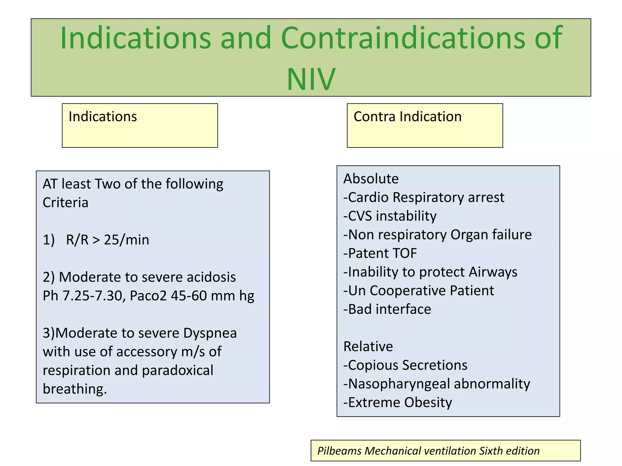
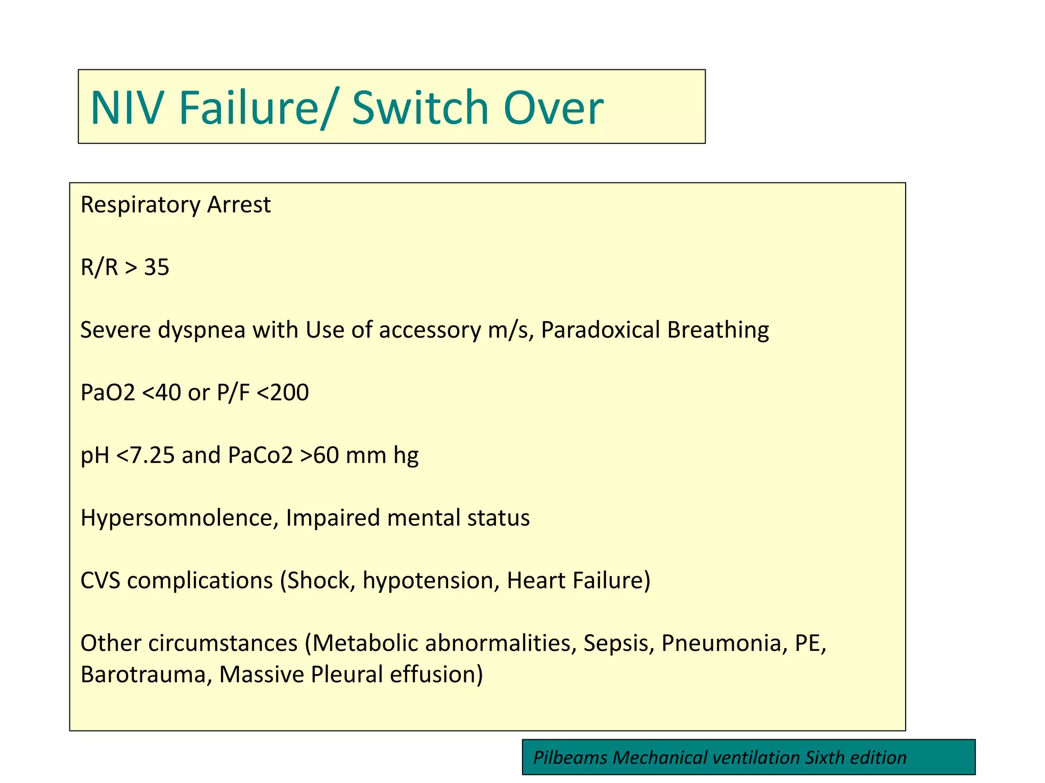
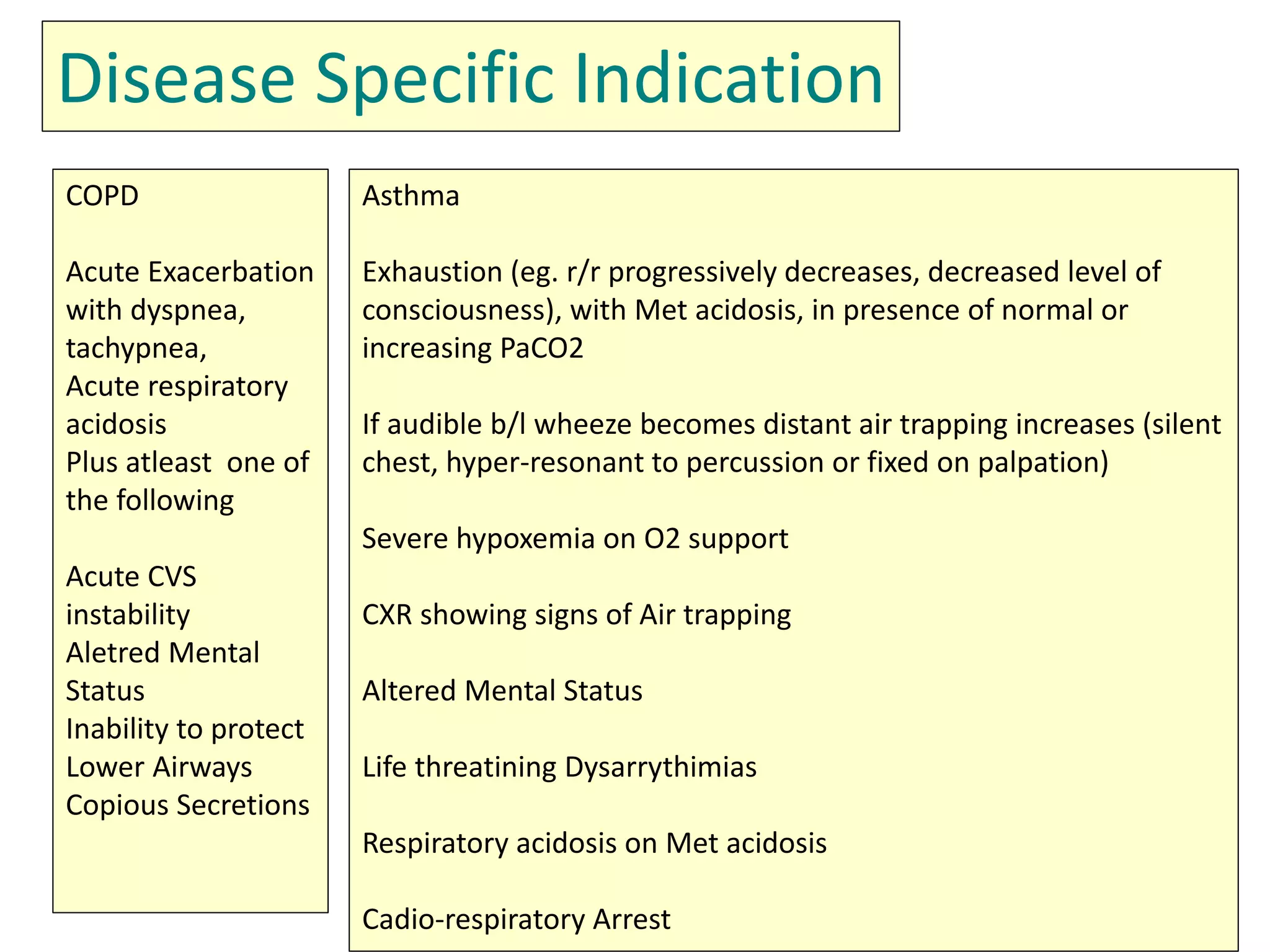
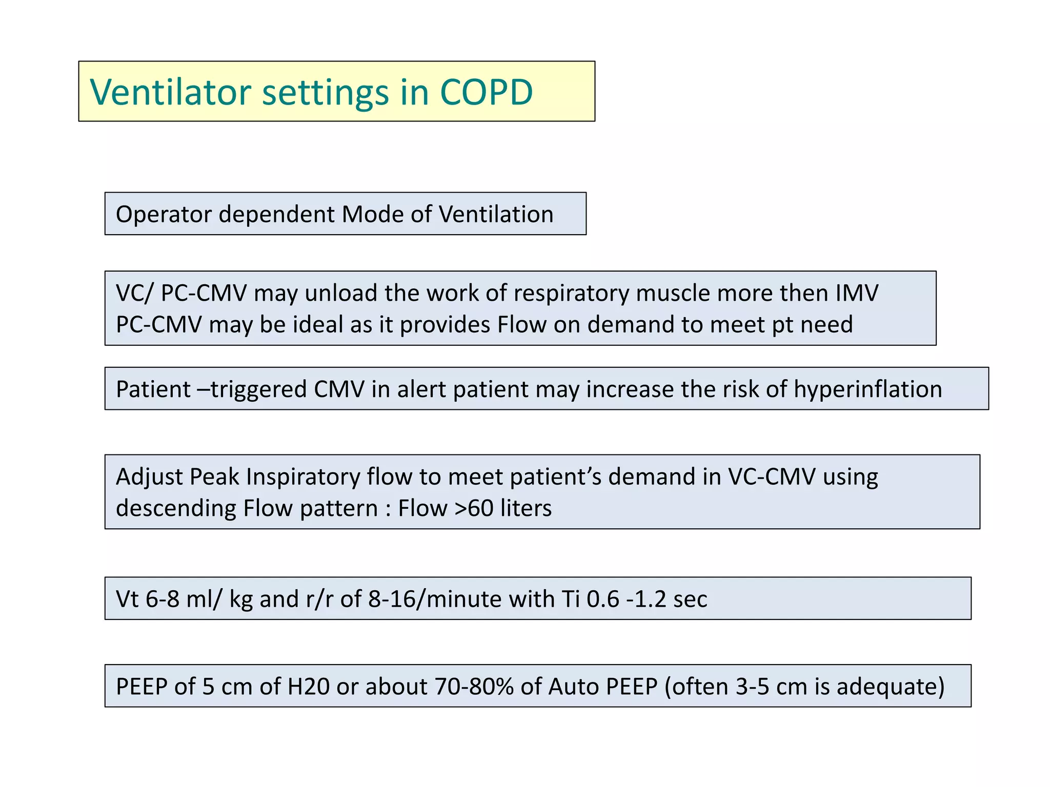
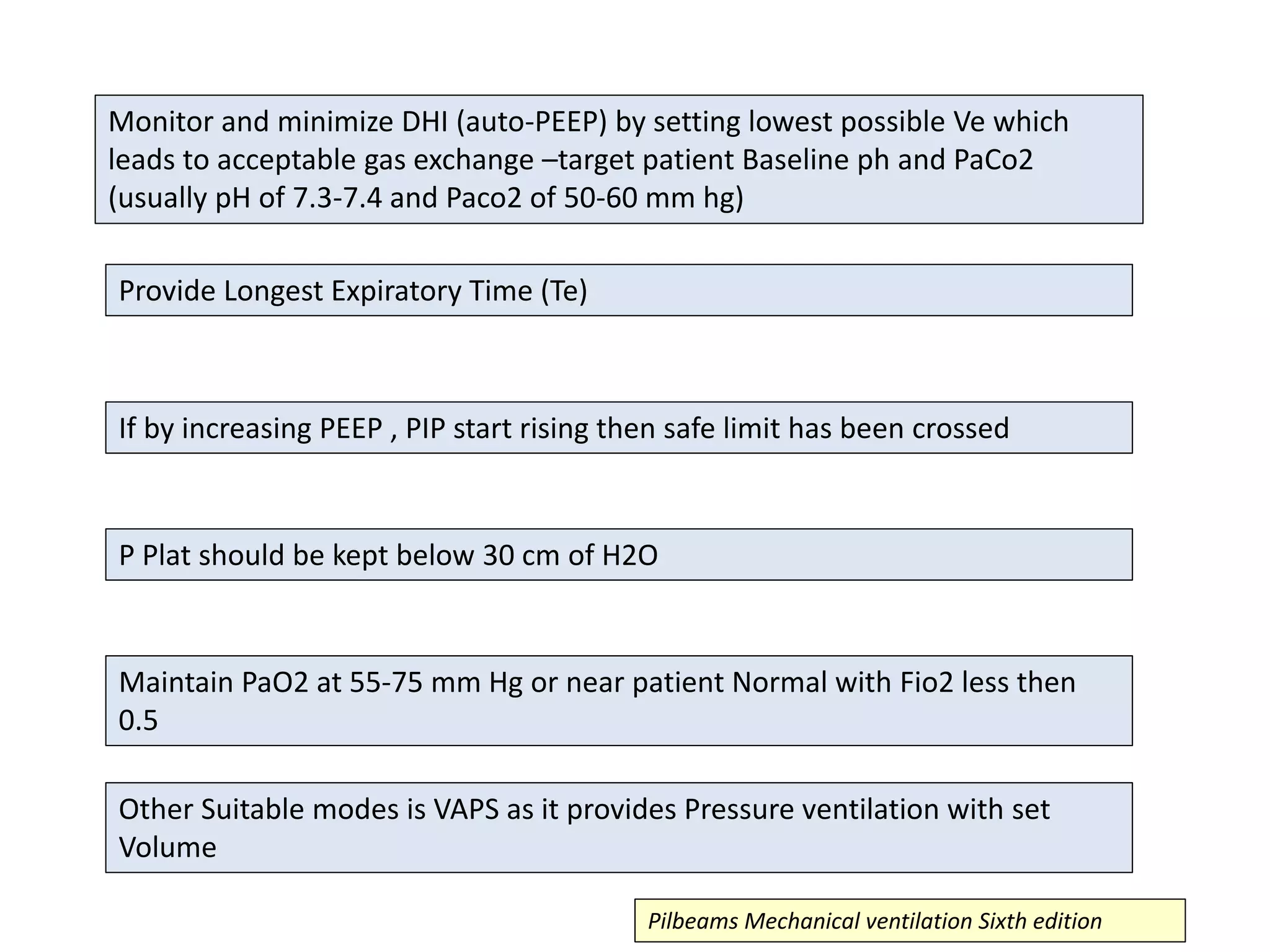
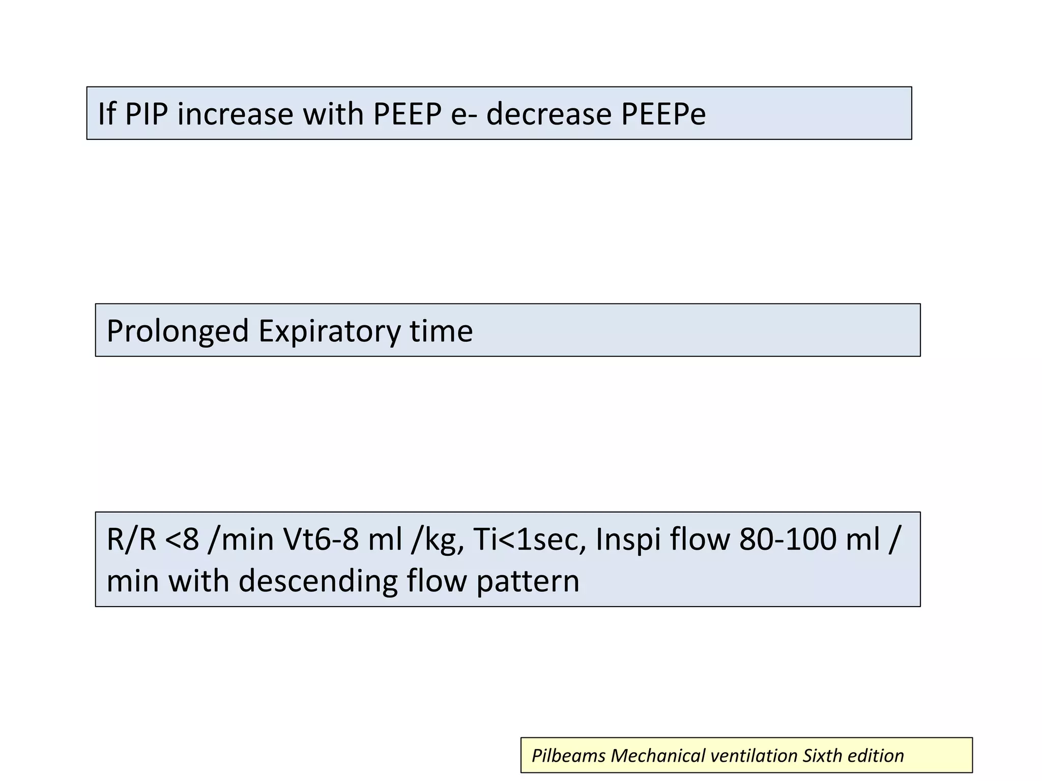
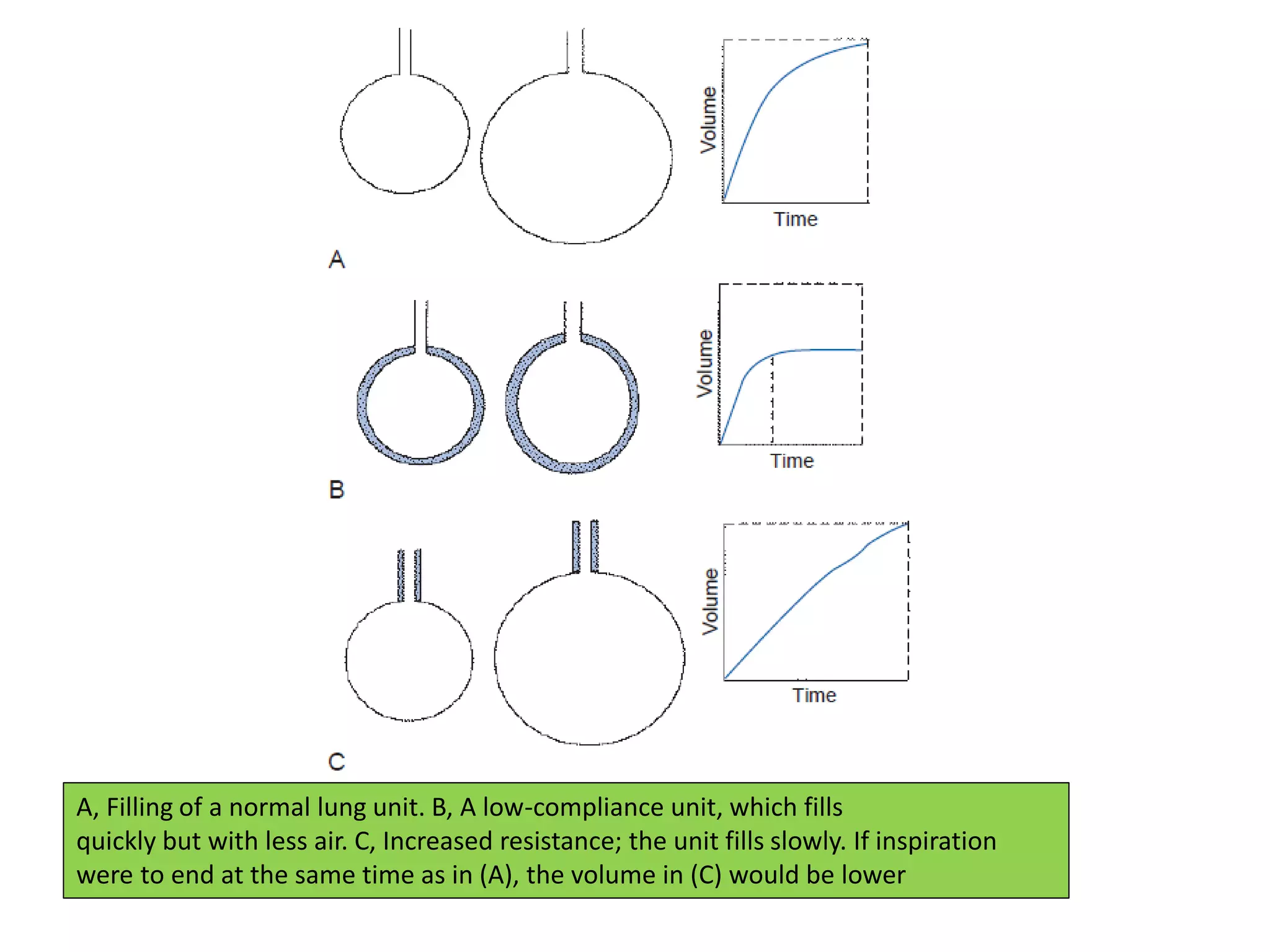
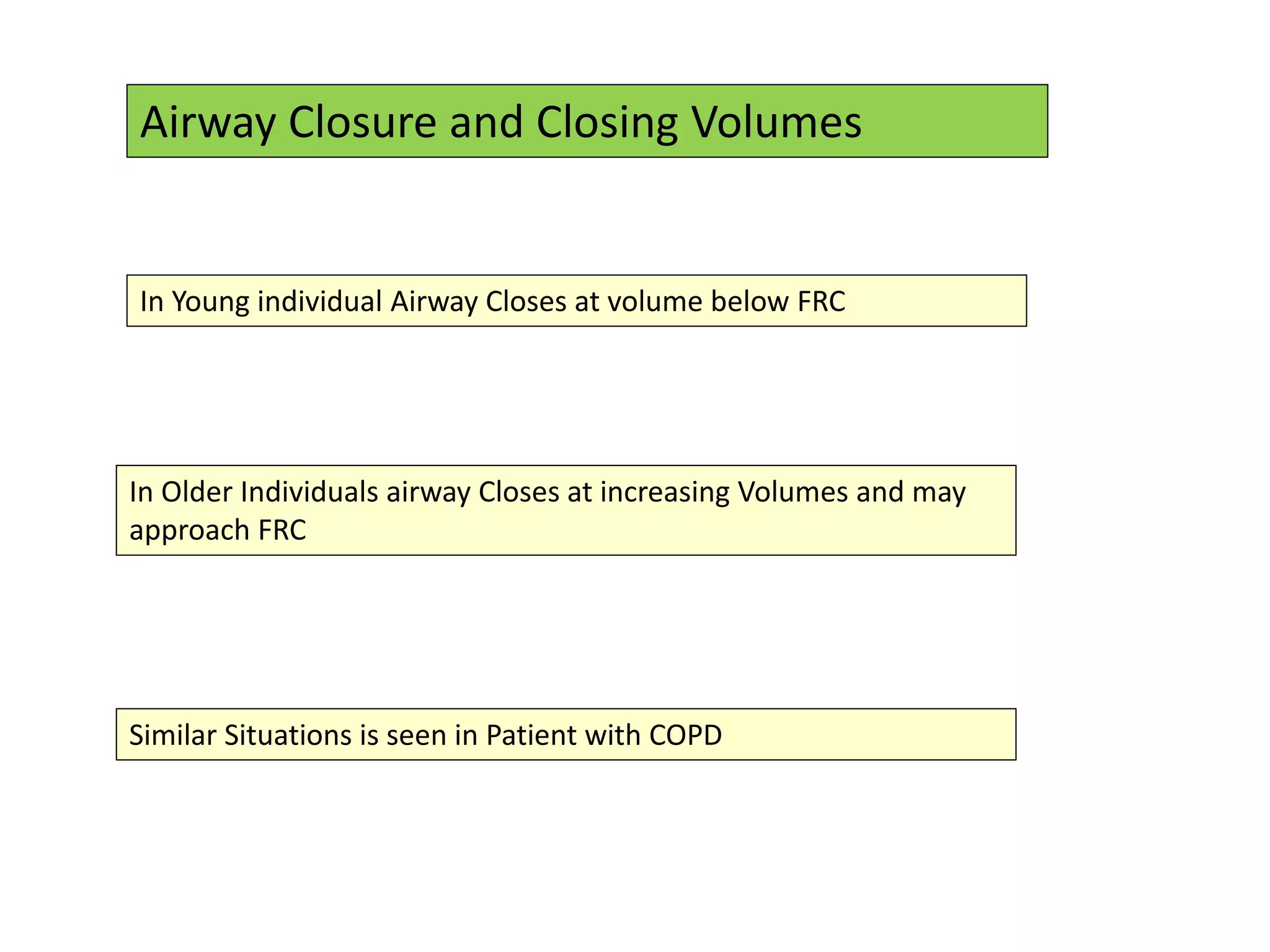
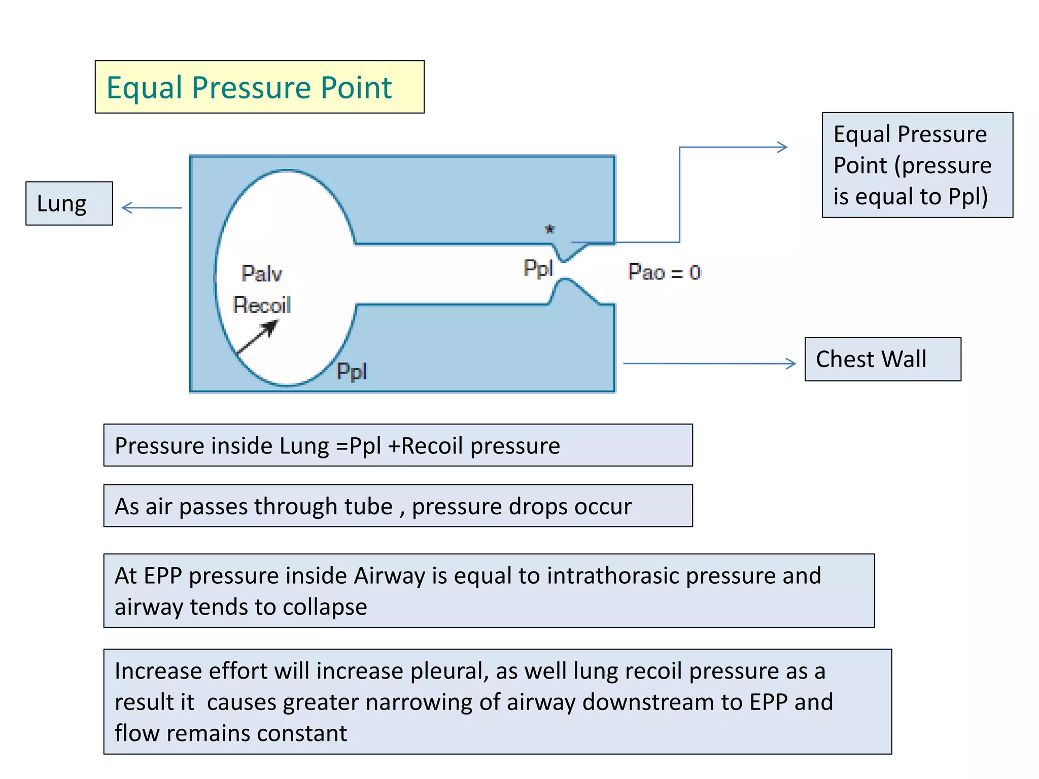
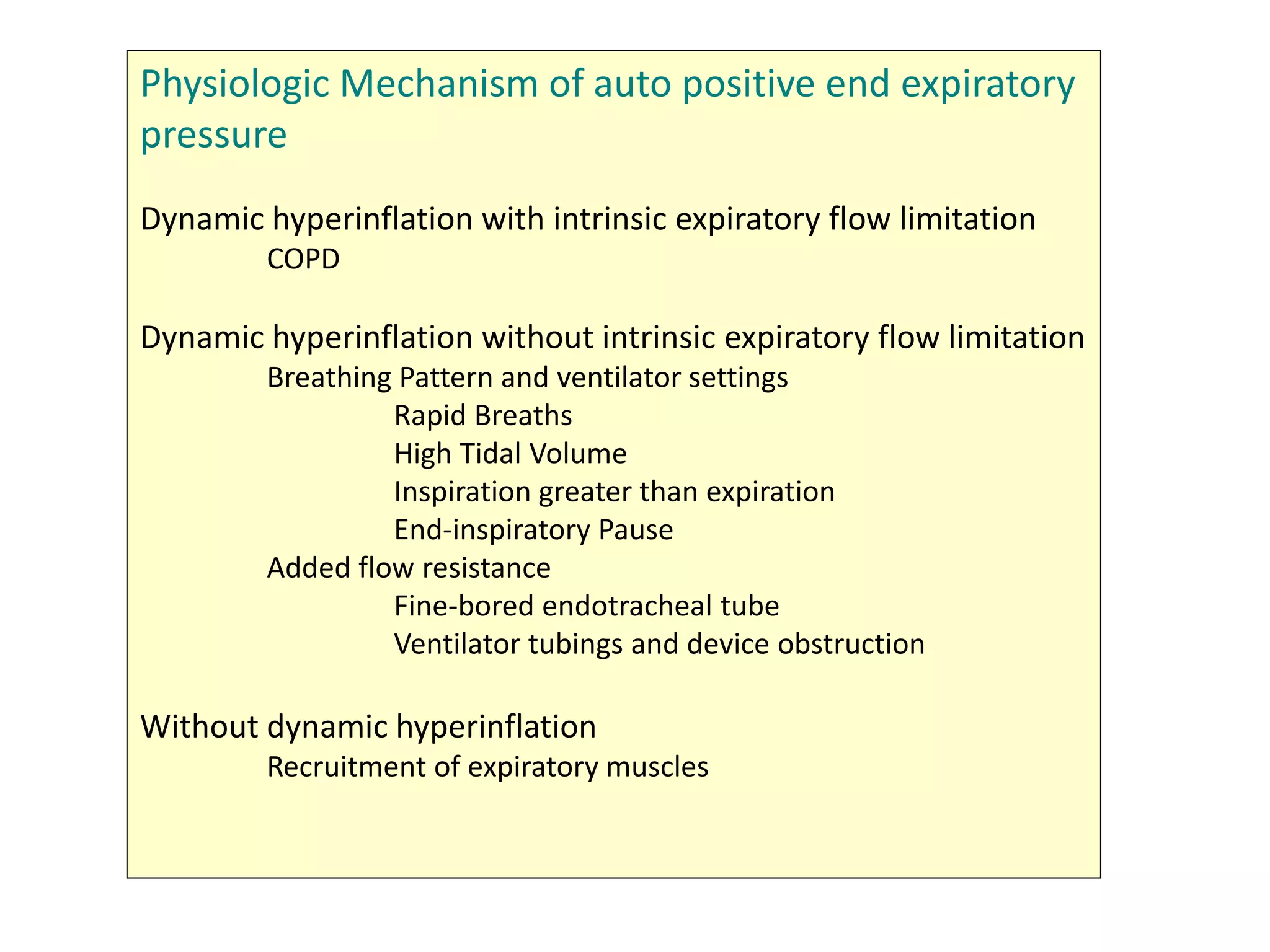
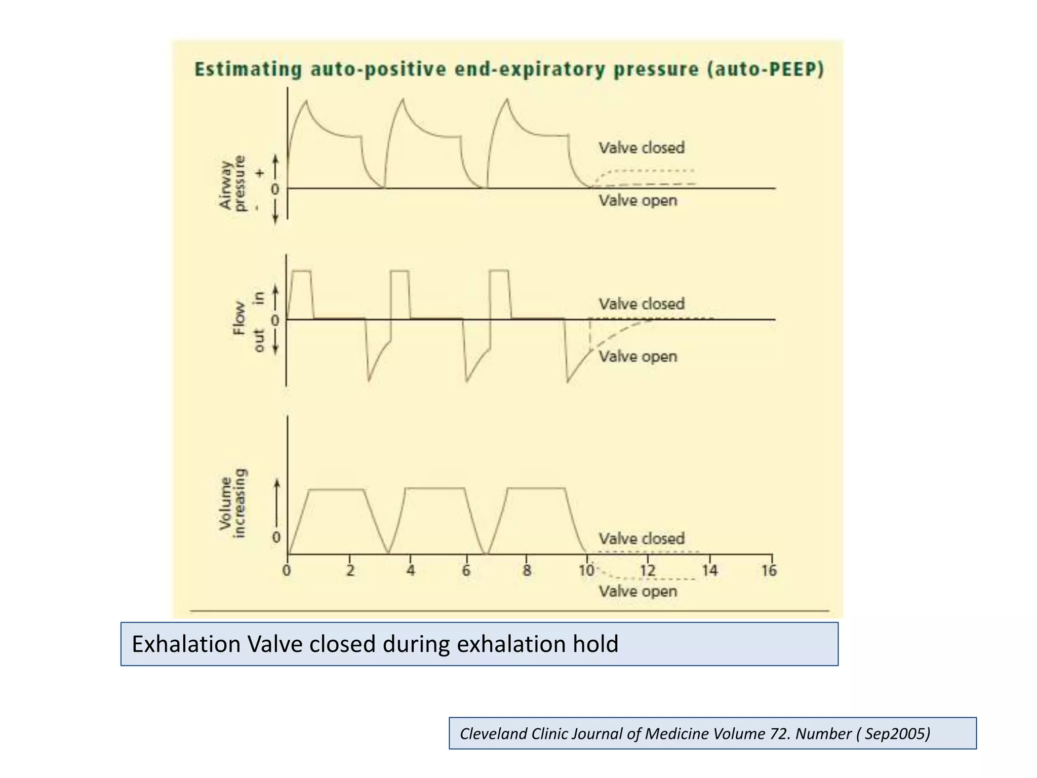
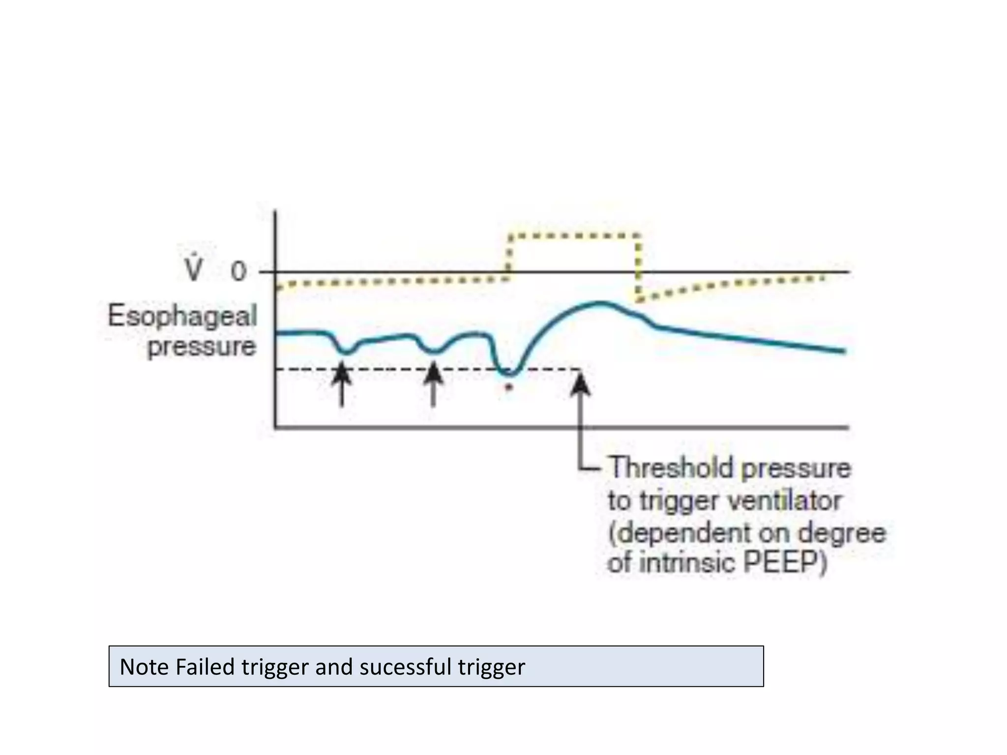
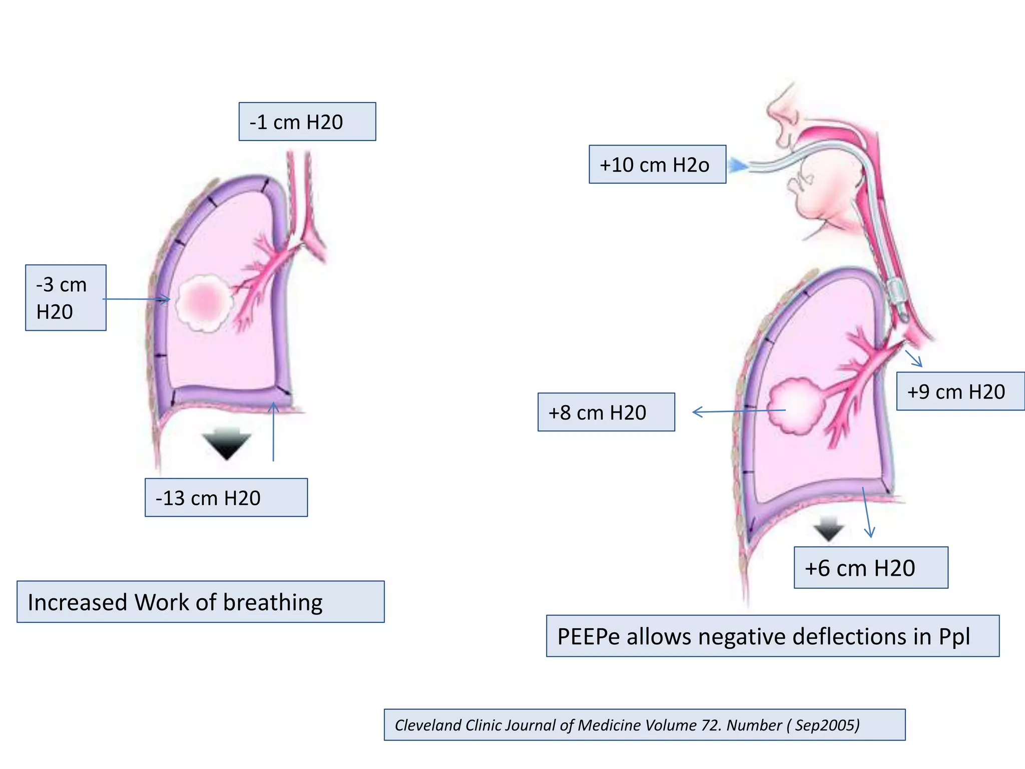
Introduction to ArtificiaI Intelligence in Higher Education
Ventilation in obstructive airway disease
1. Ventilation In Obstructive Airway
Disease
Dr Girish Agrawal
Consultant
Pulmonary, Critical Care and Sleep Medicine
Ramkrishna Care Hospital
Raipur C.G.
2. NIV Invasive
Ventilation
Criteria for Intubation
Full Spontaneous Breathing
NO YES
Failure
Success
Success Success
Acute Respiratory Failure Requiring Ventilatory Support
3. Indications and Contraindications of
NIV
Indications
AT least Two of the following
Criteria
1) R/R > 25/min
2) Moderate to severe acidosis
Ph 7.25-7.30, Paco2 45-60 mm hg
3)Moderate to severe Dyspnea
with use of accessory m/s of
respiration and paradoxical
breathing.
Absolute
-Cardio Respiratory arrest
-CVS instability
-Non respiratory Organ failure
-Patent TOF
-Inability to protect Airways
-Un Cooperative Patient
-Bad interface
Relative
-Copious Secretions
-Nasopharyngeal abnormality
-Extreme Obesity
Contra Indication
Pilbeams Mechanical ventilation Sixth edition
4. NIV Failure/ Switch Over
Respiratory Arrest
R/R > 35
Severe dyspnea with Use of accessory m/s, Paradoxical Breathing
PaO2 <40 or P/F <200
pH <7.25 and PaCo2 >60 mm hg
Hypersomnolence, Impaired mental status
CVS complications (Shock, hypotension, Heart Failure)
Other circumstances (Metabolic abnormalities, Sepsis, Pneumonia, PE,
Barotrauma, Massive Pleural effusion)
Pilbeams Mechanical ventilation Sixth edition
5. Disease Specific Indication
COPD
Acute Exacerbation
with dyspnea,
tachypnea,
Acute respiratory
acidosis
Plus atleast one of
the following
Acute CVS
instability
Aletred Mental
Status
Inability to protect
Lower Airways
Copious Secretions
Asthma
Exhaustion (eg. r/r progressively decreases, decreased level of
consciousness), with Met acidosis, in presence of normal or
increasing PaCO2
If audible b/l wheeze becomes distant air trapping increases (silent
chest, hyper-resonant to percussion or fixed on palpation)
Severe hypoxemia on O2 support
CXR showing signs of Air trapping
Altered Mental Status
Life threatining Dysarrythimias
Respiratory acidosis on Met acidosis
Cadio-respiratory Arrest
6. Objectives
Physiological objective
To Support Gas Exchange
Increase Lung Volume
Reduce work of Breathing
Clinical Objectives
Reverse acute respiratory failure
Reverse respiratory distress and muscle fatigue
Reverse hypoxemia
Prevent/reverse atelectasis and ,maintain FRC
Reduce systemic and myocardial oxygen
consumption
To minimize associated complication
Pilbeams Mechanical ventilation Sixth edition
7. Ventilator settings in COPD
Operator dependent Mode of Ventilation
VC/ PC-CMV may unload the work of respiratory muscle more then IMV
PC-CMV may be ideal as it provides Flow on demand to meet pt need
Patient –triggered CMV in alert patient may increase the risk of hyperinflation
Adjust Peak Inspiratory flow to meet patient’s demand in VC-CMV using
descending Flow pattern : Flow >60 liters
Vt 6-8 ml/ kg and r/r of 8-16/minute with Ti 0.6 -1.2 sec
PEEP of 5 cm of H20 or about 70-80% of Auto PEEP (often 3-5 cm is adequate)
8. Monitor and minimize DHI (auto-PEEP) by setting lowest possible Ve which
leads to acceptable gas exchange –target patient Baseline ph and PaCo2
(usually pH of 7.3-7.4 and Paco2 of 50-60 mm hg)
Provide Longest Expiratory Time (Te)
If by increasing PEEP , PIP start rising then safe limit has been crossed
P Plat should be kept below 30 cm of H2O
Maintain PaO2 at 55-75 mm Hg or near patient Normal with Fio2 less then
0.5
Other Suitable modes is VAPS as it provides Pressure ventilation with set
Volume
Pilbeams Mechanical ventilation Sixth edition
9. Ventilator settings in Bronchial Asthma
PC-VC/CMV are acceptable, easier to control Pressure with PC-CMV
Plat < 30, Maintain Minimal PIP
Target Pao2 60-100 mm hg (fio2>0.5)
Permissive Hypercapnia (PaCo2 45-80 mm hg) , pH >7.20
Bicarbonate can be used as and when required to maintain pH
Paralytics if required then for first 24 hour only
PEEP only to counter PEEPi (as already has high FRC)
10. If PIP increase with PEEP e- decrease PEEPe
Prolonged Expiratory time
R/R <8 /min Vt6-8 ml /kg, Ti<1sec, Inspi flow 80-100 ml /
min with descending flow pattern
Pilbeams Mechanical ventilation Sixth edition
13. Weibel ER/ west JB: Respiratory Physiology 9th ed
Anato
mical
Dead
Space
Dist-
5 mm
Note Extemely Rapid Increase in total cross
sectional are of airways in Respiratory Zone
As a result gas Flow changes from Conductive to
diffusive mode and spatial co relation of Flow
14. In other words
All the alveoli are not equally ventilated, even in normal lung
Tc of a region of Lung is product of its resistance and compliance
Reasons are Gravitational and non gravitational influence on
gas distribution
Time Constant (Tc) is Non – Gravitational concept, besides
series in-equality.
15. • It is specific for particular region of lung
• i.e diff for diff region or diff lung unit
• i.e each lung unit inflates and deflates at different time constant
• A unit with large Tc is still incompletely filled before expiration starts i.e
poor ventilation
• i.e at higher respiratory rate , lesser time for ventilation
• A unit with small TC , filss rapidly, and may receive higher proportion of gas
from anatomic dead space, hence poor ventilation
• Smaller lung unit (small Tc) recieves greater gas flow by diffusion due to
asymmetry of structure
16. A, Filling of a normal lung unit. B, A low-compliance unit, which fills
quickly but with less air. C, Increased resistance; the unit fills slowly. If inspiration
were to end at the same time as in (A), the volume in (C) would be lower
18. Airway Closure and Closing Volumes
In Young individual Airway Closes at volume below FRC
In Older Individuals airway Closes at increasing Volumes and may
approach FRC
Similar Situations is seen in Patient with COPD
19. Equal Pressure Point
Pressure inside Lung =Ppl +Recoil pressure
At EPP pressure inside Airway is equal to intrathorasic pressure and
airway tends to collapse
As air passes through tube , pressure drops occur
Increase effort will increase pleural, as well lung recoil pressure as a
result it causes greater narrowing of airway downstream to EPP and
flow remains constant
Chest Wall
Lung
Equal Pressure
Point (pressure
is equal to Ppl)
20. Dynamic Hyperinflation
Worsening of Smooth muscle cell contraction, Airway Wall edema and luminal
obstruction with mucus are the reason for Asthma Exacerbation
This trapping of air is called Dynamic Hyper inflation
Narrowing of the airways (smooth muscle contraction) occurs disproportionately
in medium and large bronchi
As a result patient breaths at higher FRC’s , have low Tidal Volume and thus Low
FVC
EPP is reached much distally- leading to air trapping
21. Levy BD, Kitch B, Fanta CH: Medical and ventilatory management
of status asthmaticus. Intensive Care Med 1998, 24:105-117.
22. 10 cm
H2o
10 cm H20
Obstructed Airway
0 cm H2O
Alveolar Pressure
Pleural Pressure
Ppl=Palv
Cleveland Clinic Journal of Medicine Volume 72. Number ( Sep2005)
23. Auto- PEEP (PEEPi)
Presence of Positive Pressure in alveoli at the end of Expiration that is
greater than atmospheric pressure without application of external
PEEP
Dynamic hyperinflation ,without Intrinsic expiratory flow limitation
Dynamic hyperinflation ,with Intrinsic expiratory flow limitation
Exaggerated expiratory Activity without dynamic hyperinflation
It can be measured by applying Pause at the end of Expiration
Three Types
24. Physiologic Mechanism of auto positive end expiratory
pressure
Dynamic hyperinflation with intrinsic expiratory flow limitation
COPD
Dynamic hyperinflation without intrinsic expiratory flow limitation
Breathing Pattern and ventilator settings
Rapid Breaths
High Tidal Volume
Inspiration greater than expiration
End-inspiratory Pause
Added flow resistance
Fine-bored endotracheal tube
Ventilator tubings and device obstruction
Without dynamic hyperinflation
Recruitment of expiratory muscles
25. How to recognize Auto-PEEP
• Exhalation that continues until the next breath starts, as seen on
graphic display of expiratory flow vs time in a patient on a ventilator
that is set to deliver a certain number of breaths per minute
• A delay between the start of inspiratory effort and the drop in airway
pressure or the start of machine-delivered flow in a patient on a
ventilator that is set to deliver breaths on demand
• Failure of peak airway pressure to change when external PEEP is
applied
• In paralyzed or heavily sedated patients, reduction of plateau
pressure after prolonged exhalation.
26. Exhalation Valve closed during exhalation hold
Cleveland Clinic Journal of Medicine Volume 72. Number ( Sep2005)
27. Decrease trigger Sensitivity (to generate negative pressure that is
great enough to trigger the ventilator )
Hemodynamic Compromise (increased Intra-thorasic pressure –
decrease venous return)
Hyperinflation- Volutrauma
Consequences
Increased work of breathing , leading to fatigue
30. Treatment of auto-positive end-expiratory pressure
Change ventilator settings
Increase expiratory time
Decrease respiratory rate
Decrease tidal volume
Reduce ventilatory demand
Reduce anxiety, pain, fever, shivering
Reduce dead space
Give sedatives and paralytics
Reduce flow resistance
Use large-bore endotracheal tube
Suction frequently
Give bronchodilators
Counterbalance expiratory flow limitation
External positive end-expiratory pressure
31. -1 cm H20
-3 cm
H20
-13 cm H20
Increased Work of breathing
+10 cm H2o
+8 cm H20
+9 cm H20
+6 cm H20
PEEPe allows negative deflections in Ppl
Cleveland Clinic Journal of Medicine Volume 72. Number ( Sep2005)
32. GOTTFRIED SB: THE ROLE OF PEEP IN MECHANICALLY VENTILATED COPD PATIENT. IN MARINI JJ, ROUSSOS C (EDITORS):
VENTILATORY FAILURE. NEW YORK, SPRINGER-VERLAG,1991:392–418:
Editor's Notes
Spatial Flow : neighbouring region will have similar flow then region located apart
Similarly there is bernoulli effect
It says that as gas flows through a tube , lateral pressure exerted by particles are less then the pressure driving flow by an amount proprotional to velocity of the gas.
Thus as gas velocity increases there is tendency for tubes to collapse.
Bernoulli effect is prominent at high volumes and flow , where as EPP is important at low volumes and flow
Top, expiratory flow limitation within a
lung. The alveolar pressure at the end of passive
expiration (auto-PEEP) in a dynamically hyperinflated
patient exceeds the critical pressure at which
dynamic airway compression occurs. External PEEP,
applied at the airway opening, will not worsen auto-
PEEP if it does not exceed the critical pressure.
Bottom, analogous circumstances governing
hydrostatic pressure above and below a waterfall.
The amount of flow over the waterfall remains
constant until the level of water in the stream below
(the airway pressure) reaches the height of waterfall
(the critical pressure) but not the stream above (the
alveolar pressure).