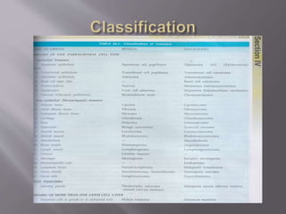
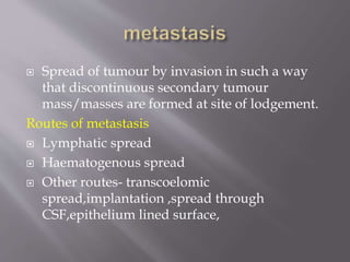
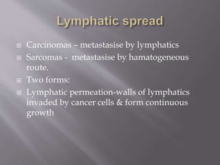
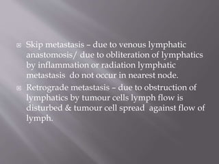
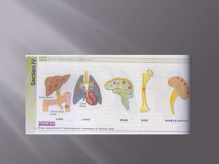
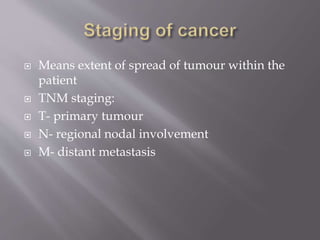
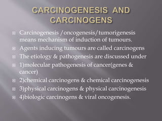
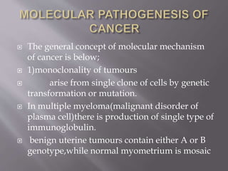
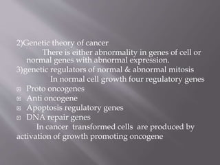
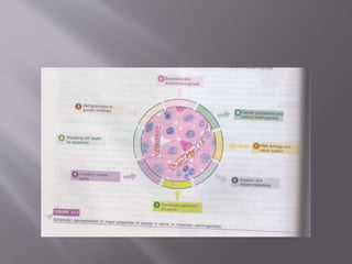
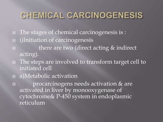
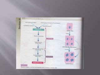
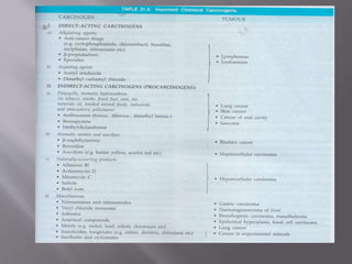
Melbia Shiny
First MDS
 Definition
 classification
 Difference between benign & malignant
 Metastasis
 Carcinogens & carcinogenesis
 A mass of tissue formed as a result of abnormal
,excessive , uncoordinated , autonomous , &
purposeless proliferation of cells.
 Spread of tumour by invasion in such a way
that discontinuous secondary tumour
mass/masses are formed at site of lodgement.
Routes of metastasis
 Lymphatic spread
 Haematogenous spread
 Other routes- transcoelomic
spread,implantation ,spread through
CSF,epithelium lined surface,
 Carcinomas – metastasise by lymphatics
 Sarcomas - metastasise by hamatogeneous
route.
 Two forms:
 Lymphatic permeation-walls of lymphatics
invaded by cancer cells & form continuous
growth
 Lymphatic emboli - malignant cell detach to
form tumour emboli & are carried along lymph
to next lymph node & get lodged in
subcapsular sinus.
 Regional nodal metastasis seen in:
 Carcinoma breast – to axillary lymph nodes
 Carcinoma thyroid – to lateral cervical lymph
node
 Skip metastasis – due to venous lymphatic
anastomosis/ due to obliteration of lymphatics
by inflammation or radiation lymphatic
metastasis do not occur in nearest node.
 Retrograde metastasis – due to obstruction of
lymphatics by tumour cells lymph flow is
disturbed & tumour cell spread against flow of
lymph.
 Virchow’s lymph node – nodal metastasis to
supraclavicular lymph node from cancer of
abdominal organs.
 Common sites are – liver ,lung ,brain,
bone,kidney, & adrenals
 These provide good soil for growth of good
seed - Seed soil theory
 Do not allow metastasis to grow – heart ,
spleen , skeletal muscle.
 Arterial spread is less likely because of thick
wall & elastic tissue.
 Systemic vein drain into vena cava from limbs
,head ,neck & pelvis.- metastasis to lungs
 Portal vein drain from bowel , spleen, &
pancreas – secondaries in lungs
 Retrograde spread due to venous obstruction
,seen in vertebral metastases in cancer of
thyroid & prostrate.
Transcoelomic spread
 Cancers invade serosal wall of coelomic cavity
so tumour fragments break & are carried in
coelomic fluid & get implanted in body cavity.
 Peritoneal cavity mostly involved
 Carcinoma of stomach into ovary
 Carcinoma of ovary into peritoneal cavity
 Carcinoma of bronchus & breast into pleura &
peritoneum
 Along epithelium lined surfaces
 Unusual because intact epithelium are resistant
to penetration by tumour cell
 Fallopian tube from endometrium to ovaries &
vice versa
 Through bronchus into alveoli
 Through ureters from kidney into lower
urinary tract
 Spread via CSF
 Malignant tumours of leptomeninges spread
by releasing tumour cells into CSF to produce
metastasis
 Implantation
 Due to implantation by needles , sutures, or
transfer from lower lip to upper lip.
steps a involved at cell molecular level:
 1)aggressive clonal proliferation and angiogenesis
rapidly proliferating clone of cancer cells
based on tumour heterogenicity.Tumour
angiogenesis (new blood vessels are formed from
pre existing ones) plays a important role,which
provide nourishment to growing tumours.The
morphologic features are:
i)microvascular density –the new capillaries add
to vascular density of tumour.
 ii)central necrosis –if tumour outgrows its
blood supply tumour angiogenesis fail & core
undergoes ischaemic necrosis.
 2)Tumour cell loosening
 Normally cell adhesion is due to
CAMs,E(epithelial)-cadherin.in carcinomas
there is loss or inactivation of E-cadherin &
other CAMs.
 3)tumour cell ECM interaction
 Loosened cancer cells are attached to
ECM proteins laminin & fibronectin.
 4)degradation of ECM
 Tumour cells overexpress proteases and
matrix degrading enzymes ,cathepsind
metalloproteinases(collagenase,gelatinase).
(dissolution of ECM).
5)entry of tumour cells into capillary lumen
This is by these mechanism:
 Autocrine motility factor (AMF)-this stimulate
receptor mediated motility of tumours cells
 Cleavage products of matrix components-these
promote chemotaxis,growth promotion &
angiogenesis.
 6)thrombus formation
 The protruding tumour cells are covered
with circulating blood and form
thrombus.Thrombus provide nourishment and
protect from immune attack.
 7)extravasation of tumour cells
 Tumour cells block these vascular
channels and attach to vascular endothelium.
 8)survival and growth of metastatic deposit
 The extravasated malignant cell on
lodgement grow further under growth
factors(PDGF, FGF, TGF-B & VEGF.
 Grading is defined as macroscopic &
microscopic degree of differentiation of tumour
 Based on :
 Degree of anaplasia
 Rate ofgrowth
 Borders grading
 Grade I – well differentiated (<25% anaplastic)
 Grade II – moderately differentiated (25-50%
anaplastic cells)
 Grade III – moderately differentiated(50-75%
anaplastic cells)
 Grade IV – poorly differentiated / anaplastic
(>75% anaplastic cells)
 Means extent of spread of tumour within the
patient
 TNM staging:
 T- primary tumour
 N- regional nodal involvement
 M- distant metastasis
 Carcinogenesis /oncogenesis/tumorigenesis
means mechanism of induction of tumours.
 Agents inducing tumours are called carcinogens
 The etiology & pathogenesis are discussed under
 1)molecular pathogenesis of cancer(genes &
cancer)
 2)chemical carcinogens & chemical carcinogenesis
 3)physical carcinogens & physical carcinogenesis
 4)biologic carcinogens & viral oncogenesis.
 The general concept of molecular mechanism
of cancer is below;
 1)monoclonality of tumours
 arise from single clone of cells by genetic
transformation or mutation.
 In multiple myeloma(malignant disorder of
plasma cell)there is production of single type of
immunoglobulin.
 benign uterine tumours contain either A or B
genotype,while normal myometrium is mosaic
2)Genetic theory of cancer
There is either abnormality in genes of cell or
normal genes with abnormal expression.
3)genetic regulators of normal & abnormal mitosis
In normal cell growth four regulatory genes
 Proto oncogenes
 Anti oncogene
 Apoptosis regulatory genes
 DNA repair genes
In cancer transformed cells are produced by
activation of growth promoting oncogene
 Inactivation of cancer suppressor gene
 Abnormal apoptosis regulatory genes
 Failure of DNA repair
4)multi step processof cancer growth &
progression
i)excessive & autonomous growth –growth promoting
oncogenes.
ii)refractoriness to growth inhibition- growth suppressing
anti oncogenes.
iii)escaping cell death by apoptosis – genes regulating
apoptosis and cancer.
iv)avoiding cellular aging – telomeres & telomerase.
v)continued perfusion of cancer – cancer angiogenesis.
vi)invasion & distant metastasis- cancer dissemination
vii)DNA damage & repair system- mutator gene& cancer
viii)cancer progression & tumour heterogeneity.- clonal
aggressiveness.
 3)Gene regulating apoptosis & cancer:
 In cancer cells apoptosis is interfered due to
mutation in the gene.eg are
 BCL2 gene seen in B cell lymphoma removes
the apoptosis –inhibitary control on cancer
cells.
 CD95 receptors are depleted in hepatocellular
carcinoma.
 4)Telomereses & telomerase in cancer
 Telomerase is RNA enzyme that help in
repair of damage to DNA and maintain normal
telomere length in successive cell division.
 After repetitive mitosis telomeres are lost in
normal cell & cease mitosis.Cancer cell have
upregulated telomerase enzyme ,hence avoid
aging & mitosis does not slow down.
 5)Tumour angiogenesis
 The stimulus is provided by
 Promoters of tumour angiogenesis- vascular
endothelial growth factors(VEGF)& basic
fibroblast growth factors(bFGF)
 Antiangiogenesis factors – thrombospondin
1,angiostatin ,endostatin & vasculostatin.
6)Mutated genes & cancer
The example mutated gene present are ;
Hereditary nonpolyposis colon cancer(lynch syndrome)
Ataxia telangiectasia(ATM)
Xeroderma pigmentosum
Bloom syndrome
Hereditary breast cancer
 7)Clonal aggressiveness
 With passage of time tumour cells
acquire more heterogenecity & leads to
tumour progression.

 The stages of chemical carcinogenesis is :
 i)Initiation of carcinogenesis
 there are two (direct acting & indirect
acting).
 The steps are involved to transform target cell to
initiated cell
 a)Metabolic activation
 procarcinogens needs activation & are
activated in liver by monooxygenase of
cytochrome& P-450 system in endoplasmic
reticulum
 b)Reactive electrophiles
 these bind to electron rich portions such
as DNA, RNA, other proteins.
 c)Target molecules
 the primary target is DNA producing
mutagenesis
 d)Initiated cell
 the changes produced in DNA become
fixed only if the altered cell undergoes one cell
cycle
 ii)Promotion of carcinogenesis
 they are not mutagenic
 need repeated dose exposure for long time
 produce slow response
 the change may be reversible
 applied after exposure to initiators
 is not effective alone
 there is a clonal expansion of mutated gene
 eg- hormones , phorbol esters
 They are of two types;
 Radiation & nonradiation
 i)Radiation carcinogenesis
 UV & ionizing radiation
 the UV radiation produce pyrimidine dimmers
in DNA which remains unrepaired in
exceesively exposed individuals.
 Ionizing radiation are X rays ,alpha
,beta,gamma rays radioactive isotopes ,protons
& neutrons can cause cancer.
 ii)Non radiation carcinogenesis
 these include mechanical injury from
stones in gallbladder, urinary tract, healed
scars from burns /trauma,implant materials
like plastic ,glass, or foreign bodies.
 BIOLOGIC CARCINOGENESIS
 THEY ARE
 Parasites- schistosoma haematobium cause
carcinoma of urinary tract
 Fungus – aspergillus flavus cause
hepatocellular carcinoma
 Bacteria – helicobacter pylori cause gastric
lymphoma
 Oncogenic viruses is transmitted by
Vertical transmission-from parent to offspring
 Horizontal transmission- one to another by
direct contact
 Two types of oncogenic viruses are DNA
oncogenic viruses & RNA oncogenic viruses
1)Papova virus
Human papilloma virus
Papilloma virus
Polyoma virus
2)Herpes virus
EB virus
Human herpesvirus 8
3)Adeno virus
4)Pox virus
5)Hepadnavirus
HB virus
 1)Mode of DNA viral oncogenesis
Replication
 The DNA virus invade the host cell & is
incorporated into host nucleus & T antigen is
expressed.
 Virions are formed in cell nucleus.The new virions
released are accompanied by host cell lysis.
Integration
 Integration of viral genome into host
cell genome occurs which requires essential
presence of functional T antigen.
 2)Mode of RNA viral oncogenesis
 a)RNA virus invade the host cell ,the viral
envelope fuses with plasma membrane of host
cell,viral RNA genome as well as reverse
transcriptase are released into cytosol
 b)Reverse transcriptase act as template to
synthesise single strand of matching viral DNA
which is copied to form complementary DNA
resulting in double stranded viral
DNA(proviruses)
 c)The provirus is integrated into host cell genome
 d)Finally brings about replication of viral
components.
 1)histological methods
 2)cytological methods
 Exfoliative cytology
 FNAC
 3)histochemistry & cytochemistry
 4)immunohistochemistry
 5)electron microscopy
 6)tumour markers
 7)modern aids
 Flow cytometry
 In situ hybridization
 Molecular diagnostic techniques
 DNA micro assay analysis of tumours

Thank you

Neoplasia

  • 1.
  • 2.
     Definition  classification Difference between benign & malignant  Metastasis  Carcinogens & carcinogenesis
  • 3.
     A massof tissue formed as a result of abnormal ,excessive , uncoordinated , autonomous , & purposeless proliferation of cells.
  • 6.
     Spread oftumour by invasion in such a way that discontinuous secondary tumour mass/masses are formed at site of lodgement. Routes of metastasis  Lymphatic spread  Haematogenous spread  Other routes- transcoelomic spread,implantation ,spread through CSF,epithelium lined surface,
  • 7.
     Carcinomas –metastasise by lymphatics  Sarcomas - metastasise by hamatogeneous route.  Two forms:  Lymphatic permeation-walls of lymphatics invaded by cancer cells & form continuous growth
  • 8.
     Lymphatic emboli- malignant cell detach to form tumour emboli & are carried along lymph to next lymph node & get lodged in subcapsular sinus.  Regional nodal metastasis seen in:  Carcinoma breast – to axillary lymph nodes  Carcinoma thyroid – to lateral cervical lymph node
  • 10.
     Skip metastasis– due to venous lymphatic anastomosis/ due to obliteration of lymphatics by inflammation or radiation lymphatic metastasis do not occur in nearest node.  Retrograde metastasis – due to obstruction of lymphatics by tumour cells lymph flow is disturbed & tumour cell spread against flow of lymph.
  • 11.
     Virchow’s lymphnode – nodal metastasis to supraclavicular lymph node from cancer of abdominal organs.
  • 12.
     Common sitesare – liver ,lung ,brain, bone,kidney, & adrenals  These provide good soil for growth of good seed - Seed soil theory  Do not allow metastasis to grow – heart , spleen , skeletal muscle.  Arterial spread is less likely because of thick wall & elastic tissue.
  • 13.
     Systemic veindrain into vena cava from limbs ,head ,neck & pelvis.- metastasis to lungs  Portal vein drain from bowel , spleen, & pancreas – secondaries in lungs  Retrograde spread due to venous obstruction ,seen in vertebral metastases in cancer of thyroid & prostrate.
  • 15.
    Transcoelomic spread  Cancersinvade serosal wall of coelomic cavity so tumour fragments break & are carried in coelomic fluid & get implanted in body cavity.  Peritoneal cavity mostly involved  Carcinoma of stomach into ovary  Carcinoma of ovary into peritoneal cavity  Carcinoma of bronchus & breast into pleura & peritoneum
  • 16.
     Along epitheliumlined surfaces  Unusual because intact epithelium are resistant to penetration by tumour cell  Fallopian tube from endometrium to ovaries & vice versa  Through bronchus into alveoli  Through ureters from kidney into lower urinary tract
  • 17.
     Spread viaCSF  Malignant tumours of leptomeninges spread by releasing tumour cells into CSF to produce metastasis  Implantation  Due to implantation by needles , sutures, or transfer from lower lip to upper lip.
  • 19.
    steps a involvedat cell molecular level:  1)aggressive clonal proliferation and angiogenesis rapidly proliferating clone of cancer cells based on tumour heterogenicity.Tumour angiogenesis (new blood vessels are formed from pre existing ones) plays a important role,which provide nourishment to growing tumours.The morphologic features are: i)microvascular density –the new capillaries add to vascular density of tumour.  ii)central necrosis –if tumour outgrows its blood supply tumour angiogenesis fail & core undergoes ischaemic necrosis.
  • 20.
     2)Tumour cellloosening  Normally cell adhesion is due to CAMs,E(epithelial)-cadherin.in carcinomas there is loss or inactivation of E-cadherin & other CAMs.  3)tumour cell ECM interaction  Loosened cancer cells are attached to ECM proteins laminin & fibronectin.
  • 21.
     4)degradation ofECM  Tumour cells overexpress proteases and matrix degrading enzymes ,cathepsind metalloproteinases(collagenase,gelatinase). (dissolution of ECM). 5)entry of tumour cells into capillary lumen This is by these mechanism:  Autocrine motility factor (AMF)-this stimulate receptor mediated motility of tumours cells  Cleavage products of matrix components-these promote chemotaxis,growth promotion & angiogenesis.
  • 22.
     6)thrombus formation The protruding tumour cells are covered with circulating blood and form thrombus.Thrombus provide nourishment and protect from immune attack.  7)extravasation of tumour cells  Tumour cells block these vascular channels and attach to vascular endothelium.  8)survival and growth of metastatic deposit  The extravasated malignant cell on lodgement grow further under growth factors(PDGF, FGF, TGF-B & VEGF.
  • 23.
     Grading isdefined as macroscopic & microscopic degree of differentiation of tumour  Based on :  Degree of anaplasia  Rate ofgrowth
  • 24.
     Borders grading Grade I – well differentiated (<25% anaplastic)  Grade II – moderately differentiated (25-50% anaplastic cells)  Grade III – moderately differentiated(50-75% anaplastic cells)  Grade IV – poorly differentiated / anaplastic (>75% anaplastic cells)
  • 25.
     Means extentof spread of tumour within the patient  TNM staging:  T- primary tumour  N- regional nodal involvement  M- distant metastasis
  • 27.
     Carcinogenesis /oncogenesis/tumorigenesis meansmechanism of induction of tumours.  Agents inducing tumours are called carcinogens  The etiology & pathogenesis are discussed under  1)molecular pathogenesis of cancer(genes & cancer)  2)chemical carcinogens & chemical carcinogenesis  3)physical carcinogens & physical carcinogenesis  4)biologic carcinogens & viral oncogenesis.
  • 28.
     The generalconcept of molecular mechanism of cancer is below;  1)monoclonality of tumours  arise from single clone of cells by genetic transformation or mutation.  In multiple myeloma(malignant disorder of plasma cell)there is production of single type of immunoglobulin.  benign uterine tumours contain either A or B genotype,while normal myometrium is mosaic
  • 30.
    2)Genetic theory ofcancer There is either abnormality in genes of cell or normal genes with abnormal expression. 3)genetic regulators of normal & abnormal mitosis In normal cell growth four regulatory genes  Proto oncogenes  Anti oncogene  Apoptosis regulatory genes  DNA repair genes In cancer transformed cells are produced by activation of growth promoting oncogene
  • 31.
     Inactivation ofcancer suppressor gene  Abnormal apoptosis regulatory genes  Failure of DNA repair 4)multi step processof cancer growth & progression
  • 32.
    i)excessive & autonomousgrowth –growth promoting oncogenes. ii)refractoriness to growth inhibition- growth suppressing anti oncogenes. iii)escaping cell death by apoptosis – genes regulating apoptosis and cancer. iv)avoiding cellular aging – telomeres & telomerase. v)continued perfusion of cancer – cancer angiogenesis. vi)invasion & distant metastasis- cancer dissemination vii)DNA damage & repair system- mutator gene& cancer viii)cancer progression & tumour heterogeneity.- clonal aggressiveness.
  • 36.
     3)Gene regulatingapoptosis & cancer:  In cancer cells apoptosis is interfered due to mutation in the gene.eg are  BCL2 gene seen in B cell lymphoma removes the apoptosis –inhibitary control on cancer cells.  CD95 receptors are depleted in hepatocellular carcinoma.
  • 37.
     4)Telomereses &telomerase in cancer  Telomerase is RNA enzyme that help in repair of damage to DNA and maintain normal telomere length in successive cell division.  After repetitive mitosis telomeres are lost in normal cell & cease mitosis.Cancer cell have upregulated telomerase enzyme ,hence avoid aging & mitosis does not slow down.
  • 38.
     5)Tumour angiogenesis The stimulus is provided by  Promoters of tumour angiogenesis- vascular endothelial growth factors(VEGF)& basic fibroblast growth factors(bFGF)  Antiangiogenesis factors – thrombospondin 1,angiostatin ,endostatin & vasculostatin.
  • 39.
    6)Mutated genes &cancer The example mutated gene present are ; Hereditary nonpolyposis colon cancer(lynch syndrome) Ataxia telangiectasia(ATM) Xeroderma pigmentosum Bloom syndrome Hereditary breast cancer
  • 40.
     7)Clonal aggressiveness With passage of time tumour cells acquire more heterogenecity & leads to tumour progression. 
  • 41.
     The stagesof chemical carcinogenesis is :  i)Initiation of carcinogenesis  there are two (direct acting & indirect acting).  The steps are involved to transform target cell to initiated cell  a)Metabolic activation  procarcinogens needs activation & are activated in liver by monooxygenase of cytochrome& P-450 system in endoplasmic reticulum
  • 42.
     b)Reactive electrophiles these bind to electron rich portions such as DNA, RNA, other proteins.  c)Target molecules  the primary target is DNA producing mutagenesis  d)Initiated cell  the changes produced in DNA become fixed only if the altered cell undergoes one cell cycle
  • 43.
     ii)Promotion ofcarcinogenesis  they are not mutagenic  need repeated dose exposure for long time  produce slow response  the change may be reversible  applied after exposure to initiators  is not effective alone  there is a clonal expansion of mutated gene  eg- hormones , phorbol esters
  • 46.
     They areof two types;  Radiation & nonradiation  i)Radiation carcinogenesis  UV & ionizing radiation  the UV radiation produce pyrimidine dimmers in DNA which remains unrepaired in exceesively exposed individuals.  Ionizing radiation are X rays ,alpha ,beta,gamma rays radioactive isotopes ,protons & neutrons can cause cancer.
  • 47.
     ii)Non radiationcarcinogenesis  these include mechanical injury from stones in gallbladder, urinary tract, healed scars from burns /trauma,implant materials like plastic ,glass, or foreign bodies.
  • 48.
     BIOLOGIC CARCINOGENESIS THEY ARE  Parasites- schistosoma haematobium cause carcinoma of urinary tract  Fungus – aspergillus flavus cause hepatocellular carcinoma  Bacteria – helicobacter pylori cause gastric lymphoma
  • 49.
     Oncogenic virusesis transmitted by Vertical transmission-from parent to offspring  Horizontal transmission- one to another by direct contact  Two types of oncogenic viruses are DNA oncogenic viruses & RNA oncogenic viruses
  • 51.
    1)Papova virus Human papillomavirus Papilloma virus Polyoma virus 2)Herpes virus EB virus Human herpesvirus 8 3)Adeno virus 4)Pox virus 5)Hepadnavirus HB virus
  • 52.
     1)Mode ofDNA viral oncogenesis Replication  The DNA virus invade the host cell & is incorporated into host nucleus & T antigen is expressed.  Virions are formed in cell nucleus.The new virions released are accompanied by host cell lysis. Integration  Integration of viral genome into host cell genome occurs which requires essential presence of functional T antigen.
  • 53.
     2)Mode ofRNA viral oncogenesis  a)RNA virus invade the host cell ,the viral envelope fuses with plasma membrane of host cell,viral RNA genome as well as reverse transcriptase are released into cytosol  b)Reverse transcriptase act as template to synthesise single strand of matching viral DNA which is copied to form complementary DNA resulting in double stranded viral DNA(proviruses)  c)The provirus is integrated into host cell genome  d)Finally brings about replication of viral components.
  • 55.
     1)histological methods 2)cytological methods  Exfoliative cytology  FNAC  3)histochemistry & cytochemistry  4)immunohistochemistry  5)electron microscopy  6)tumour markers  7)modern aids  Flow cytometry  In situ hybridization  Molecular diagnostic techniques  DNA micro assay analysis of tumours 
  • 56.