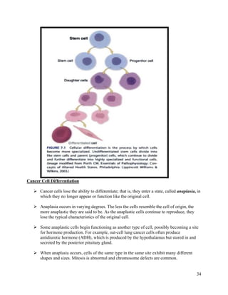
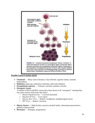
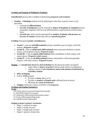
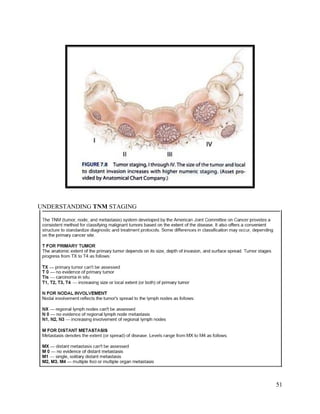
Cancer results from abnormal cell growth and differentiation. The four main steps in carcinogenesis are initiation, promotion, progression, and metastasis. Initiation involves DNA damage from factors like chemicals, radiation, or viruses. Promotion enhances the growth of mutated cells through exposure to factors like hormones or food additives. Progression refers to further growth and spread within the tissue. Metastasis is the spread of cancer to other organs. Genetic mutations in oncogenes, tumor suppressor genes, and DNA repair genes can drive uncontrolled cell growth. Cancer manifests through local effects on tissues and systemic effects like fatigue, weight loss, and abnormal hormone production. Tumor markers are substances released by cancer cells or in response to cancer that can
Neoplasia - Characteristics & Classification of Neoplasm Chhavi Singh
This power point presentation take a detail note on neoplasm (cancer), types of neoplasm, stages of neoplasm, various kinds of carcinogens. This presentation also take interest in the classification and characteristics of the tumor & difference between the normal cells and tumor cells.
Presentation for the cytology (cell biology) course on cancer/tumour. A document with more information can be found for better presenting and understanding of the material.
Neoplasia: Is the abnormal growth and proliferation of abnormal cells or abnormal amounts of cells due to a benign or malignant process. There can be benign tumors, or neoplasms, and malignant ones.
Cancer
Cancer is one of the most common diseases in the developed world:
1 in 4 deaths are due to cancer
1 in 17 deaths are due to lung cancer
Lung cancer is the most common cancer in men
Breast cancer is the most common cancer in women
There are over 100 different forms of cancer
Cancer
The division of normal cells is precisely controlled. New cells are only formed for growth or to replace dead ones.
Cancerous cells divide repeatedly out of control even though they are not needed, they crowd out other normal cells and function abnormally. They can also destroy the correct functioning of major organs.
What causes cancer?
Cancer arises from the mutation of a normal gene.
Mutated genes that cause cancer are called oncogenes.
It is thought that several mutations need to occur to give rise to cancer
Cells that are old or not functioning properly normally self destruct and are replaced by new cells.
However, cancerous cells do not self destruct and continue to divide rapidly producing millions of new cancerous cells.
A factor which brings about a mutation is called a mutagen.
A mutagen is mutagenic.
Any agent that causes cancer is called a carcinogen and is described as carcinogenic.
So some mutagens are carcinogenic.
Carcinogens
Ionising radiation – X Rays, UV light
Chemicals – tar from cigarettes
Virus infection – papilloma virus can be responsible for cervical cancer.
Hereditary predisposition – Some families are more susceptible to getting certain cancers. Remember you can’t inherit cancer its just that you maybe more susceptible to getting it.
Benign or malignant?
Benign tumours do not spread from their site of origin, but can crowd out (squash) surrounding cells eg brain tumour, warts.
Malignant tumours can spread from the original site and cause secondary tumours. This is called metastasis. They interfere with neighbouring cells and can block blood vessels, the gut, glands, lungs etc.
Why are secondary tumours so bad?
Both types of tumour can tire the body out as they both need a huge amount of nutrients to sustain the rapid growth and division of the cells.
The Development of Cancer
Within every nucleus of every one of the human body's 30 trillion cells exists DNA, the substance that contains the information needed to make and control every cell within the body. Here is a close-up view of a tiny fragment of DNA.
1. DNA of a normal cell
This piece of DNA is an exact copy of the DNA from which it came. When the parent cell divided to create two cells, the cell's DNA also divided, creating two identical copies of the original DNA.
2. Mutation of DNA
Here is the same section of DNA but from another cell. If you can imagine that DNA is a twisted ladder, then each rung of the ladder is a pair of joined molecules, or a base pair. With this section of DNA, one of the base pairs is different from the original. This DNA has suffered a mutation, either through mis-copying (when its parent cell divided), or through the damaging effects
MBBS 2nd Year Pathology - Neoplasia : IntroductionNida Us Sahr
Chapter 7 (Neoplasia) from Robbins and Cotran Pathologic Basis of Disease (9th Edition) for MBBS 2nd Year.
After going through this presentation, it will be easy to understand Neoplasia from Robbins.
Cancer basically starts with uncontrolled growth level of cells and goes beyond the blood lymph or healthy tissues to create tumor in their targeted organ(s).
A tumor marker is a substance found in your blood, urine, or body tissue. The term "tumor markers" may refer to proteins that are made by both healthy
....
Multicellular organisms coordinate their functions for the proper survival of the individual. Cells send, receive and interpret an elaborate set of extracellular signals that serve as social controls. Resting, growing dividing, differentiating and dying all depend on these signals.
Mutations tend to disrupt these social controls. Mutations give single cells a selective advantage of more prolific growth and division as compared to other cells.
Cancer develops when an individual mutant clone of cells begins prospering at the expense of its neighbours.
So, Cancer is a genetic disease characterized by uncontrolled division of abnormal cells leading to a malignant and invasive growth or tumor.
TUMORS may be benign or malignant.
BENIGN TUMORS: These, such as a common skin wart, remains confined to their original location, neither invade surrounding normal tissue nor spread to distant body sites.
MALIGNANT TUMORS: These are capable of both invading surrounding normal tissue and spreading throughout the body via the circulatory or lymphatic systems (Metastasis).
Announcement about my previous presentations - Thank youAreej Abu Hanieh
ANNOUNCEMENT Thank you for all of you, my followers who sent me messages with a lot of love and appreciations, I finally graduated after 6 years of studying in Birzeit University , In doctor of Pharmacy department I hope all of you benefited from all the presentations posted before Thank you a new PharmD GraduatedAreej ^^
Recomendações da OMS sobre cuidados maternos e neonatais para uma experiência pós-natal positiva.
Em consonância com os ODS – Objetivos do Desenvolvimento Sustentável e a Estratégia Global para a Saúde das Mulheres, Crianças e Adolescentes, e aplicando uma abordagem baseada nos direitos humanos, os esforços de cuidados pós-natais devem expandir-se para além da cobertura e da simples sobrevivência, de modo a incluir cuidados de qualidade.
Estas diretrizes visam melhorar a qualidade dos cuidados pós-natais essenciais e de rotina prestados às mulheres e aos recém-nascidos, com o objetivo final de melhorar a saúde e o bem-estar materno e neonatal.
Uma “experiência pós-natal positiva” é um resultado importante para todas as mulheres que dão à luz e para os seus recém-nascidos, estabelecendo as bases para a melhoria da saúde e do bem-estar a curto e longo prazo. Uma experiência pós-natal positiva é definida como aquela em que as mulheres, pessoas que gestam, os recém-nascidos, os casais, os pais, os cuidadores e as famílias recebem informação consistente, garantia e apoio de profissionais de saúde motivados; e onde um sistema de saúde flexível e com recursos reconheça as necessidades das mulheres e dos bebês e respeite o seu contexto cultural.
Estas diretrizes consolidadas apresentam algumas recomendações novas e já bem fundamentadas sobre cuidados pós-natais de rotina para mulheres e neonatos que recebem cuidados no pós-parto em unidades de saúde ou na comunidade, independentemente dos recursos disponíveis.
É fornecido um conjunto abrangente de recomendações para cuidados durante o período puerperal, com ênfase nos cuidados essenciais que todas as mulheres e recém-nascidos devem receber, e com a devida atenção à qualidade dos cuidados; isto é, a entrega e a experiência do cuidado recebido. Estas diretrizes atualizam e ampliam as recomendações da OMS de 2014 sobre cuidados pós-natais da mãe e do recém-nascido e complementam as atuais diretrizes da OMS sobre a gestão de complicações pós-natais.
O estabelecimento da amamentação e o manejo das principais intercorrências é contemplada.
Recomendamos muito.
Vamos discutir essas recomendações no nosso curso de pós-graduação em Aleitamento no Instituto Ciclos.
Esta publicação só está disponível em inglês até o momento.
Prof. Marcus Renato de Carvalho
www.agostodourado.com
Flu Vaccine Alert in Bangalore Karnatakaaddon Scans
As flu season approaches, health officials in Bangalore, Karnataka, are urging residents to get their flu vaccinations. The seasonal flu, while common, can lead to severe health complications, particularly for vulnerable populations such as young children, the elderly, and those with underlying health conditions.
Dr. Vidisha Kumari, a leading epidemiologist in Bangalore, emphasizes the importance of getting vaccinated. "The flu vaccine is our best defense against the influenza virus. It not only protects individuals but also helps prevent the spread of the virus in our communities," he says.
This year, the flu season is expected to coincide with a potential increase in other respiratory illnesses. The Karnataka Health Department has launched an awareness campaign highlighting the significance of flu vaccinations. They have set up multiple vaccination centers across Bangalore, making it convenient for residents to receive their shots.
To encourage widespread vaccination, the government is also collaborating with local schools, workplaces, and community centers to facilitate vaccination drives. Special attention is being given to ensuring that the vaccine is accessible to all, including marginalized communities who may have limited access to healthcare.
Residents are reminded that the flu vaccine is safe and effective. Common side effects are mild and may include soreness at the injection site, mild fever, or muscle aches. These side effects are generally short-lived and far less severe than the flu itself.
Healthcare providers are also stressing the importance of continuing COVID-19 precautions. Wearing masks, practicing good hand hygiene, and maintaining social distancing are still crucial, especially in crowded places.
Protect yourself and your loved ones by getting vaccinated. Together, we can help keep Bangalore healthy and safe this flu season. For more information on vaccination centers and schedules, residents can visit the Karnataka Health Department’s official website or follow their social media pages.
Stay informed, stay safe, and get your flu shot today!
- Video recording of this lecture in English language: https://youtu.be/lK81BzxMqdo
- Video recording of this lecture in Arabic language: https://youtu.be/Ve4P0COk9OI
- Link to download the book free: https://nephrotube.blogspot.com/p/nephrotube-nephrology-books.html
- Link to NephroTube website: www.NephroTube.com
- Link to NephroTube social media accounts: https://nephrotube.blogspot.com/p/join-nephrotube-on-social-media.html
New Drug Discovery and Development .....NEHA GUPTA
The "New Drug Discovery and Development" process involves the identification, design, testing, and manufacturing of novel pharmaceutical compounds with the aim of introducing new and improved treatments for various medical conditions. This comprehensive endeavor encompasses various stages, including target identification, preclinical studies, clinical trials, regulatory approval, and post-market surveillance. It involves multidisciplinary collaboration among scientists, researchers, clinicians, regulatory experts, and pharmaceutical companies to bring innovative therapies to market and address unmet medical needs.
NVBDCP.pptx Nation vector borne disease control programSapna Thakur
NVBDCP was launched in 2003-2004 . Vector-Borne Disease: Disease that results from an infection transmitted to humans and other animals by blood-feeding arthropods, such as mosquitoes, ticks, and fleas. Examples of vector-borne diseases include Dengue fever, West Nile Virus, Lyme disease, and malaria.
Title: Sense of Smell
Presenter: Dr. Faiza, Assistant Professor of Physiology
Qualifications:
MBBS (Best Graduate, AIMC Lahore)
FCPS Physiology
ICMT, CHPE, DHPE (STMU)
MPH (GC University, Faisalabad)
MBA (Virtual University of Pakistan)
Learning Objectives:
Describe the primary categories of smells and the concept of odor blindness.
Explain the structure and location of the olfactory membrane and mucosa, including the types and roles of cells involved in olfaction.
Describe the pathway and mechanisms of olfactory signal transmission from the olfactory receptors to the brain.
Illustrate the biochemical cascade triggered by odorant binding to olfactory receptors, including the role of G-proteins and second messengers in generating an action potential.
Identify different types of olfactory disorders such as anosmia, hyposmia, hyperosmia, and dysosmia, including their potential causes.
Key Topics:
Olfactory Genes:
3% of the human genome accounts for olfactory genes.
400 genes for odorant receptors.
Olfactory Membrane:
Located in the superior part of the nasal cavity.
Medially: Folds downward along the superior septum.
Laterally: Folds over the superior turbinate and upper surface of the middle turbinate.
Total surface area: 5-10 square centimeters.
Olfactory Mucosa:
Olfactory Cells: Bipolar nerve cells derived from the CNS (100 million), with 4-25 olfactory cilia per cell.
Sustentacular Cells: Produce mucus and maintain ionic and molecular environment.
Basal Cells: Replace worn-out olfactory cells with an average lifespan of 1-2 months.
Bowman’s Gland: Secretes mucus.
Stimulation of Olfactory Cells:
Odorant dissolves in mucus and attaches to receptors on olfactory cilia.
Involves a cascade effect through G-proteins and second messengers, leading to depolarization and action potential generation in the olfactory nerve.
Quality of a Good Odorant:
Small (3-20 Carbon atoms), volatile, water-soluble, and lipid-soluble.
Facilitated by odorant-binding proteins in mucus.
Membrane Potential and Action Potential:
Resting membrane potential: -55mV.
Action potential frequency in the olfactory nerve increases with odorant strength.
Adaptation Towards the Sense of Smell:
Rapid adaptation within the first second, with further slow adaptation.
Psychological adaptation greater than receptor adaptation, involving feedback inhibition from the central nervous system.
Primary Sensations of Smell:
Camphoraceous, Musky, Floral, Pepperminty, Ethereal, Pungent, Putrid.
Odor Detection Threshold:
Examples: Hydrogen sulfide (0.0005 ppm), Methyl-mercaptan (0.002 ppm).
Some toxic substances are odorless at lethal concentrations.
Characteristics of Smell:
Odor blindness for single substances due to lack of appropriate receptor protein.
Behavioral and emotional influences of smell.
Transmission of Olfactory Signals:
From olfactory cells to glomeruli in the olfactory bulb, involving lateral inhibition.
Primitive, less old, and new olfactory systems with different path
Knee anatomy and clinical tests 2024.pdfvimalpl1234
This includes all relevant anatomy and clinical tests compiled from standard textbooks, Campbell,netter etc..It is comprehensive and best suited for orthopaedicians and orthopaedic residents.
Report Back from SGO 2024: What’s the Latest in Cervical Cancer?bkling
Are you curious about what’s new in cervical cancer research or unsure what the findings mean? Join Dr. Emily Ko, a gynecologic oncologist at Penn Medicine, to learn about the latest updates from the Society of Gynecologic Oncology (SGO) 2024 Annual Meeting on Women’s Cancer. Dr. Ko will discuss what the research presented at the conference means for you and answer your questions about the new developments.
Ozempic: Preoperative Management of Patients on GLP-1 Receptor Agonists Saeid Safari
Preoperative Management of Patients on GLP-1 Receptor Agonists like Ozempic and Semiglutide
ASA GUIDELINE
NYSORA Guideline
2 Case Reports of Gastric Ultrasound
micro teaching on communication m.sc nursing.pdfAnurag Sharma
Microteaching is a unique model of practice teaching. It is a viable instrument for the. desired change in the teaching behavior or the behavior potential which, in specified types of real. classroom situations, tends to facilitate the achievement of specified types of objectives.
Couples presenting to the infertility clinic- Do they really have infertility...Sujoy Dasgupta
Dr Sujoy Dasgupta presented the study on "Couples presenting to the infertility clinic- Do they really have infertility? – The unexplored stories of non-consummation" in the 13th Congress of the Asia Pacific Initiative on Reproduction (ASPIRE 2024) at Manila on 24 May, 2024.
Tom Selleck Health: A Comprehensive Look at the Iconic Actor’s Wellness Journeygreendigital
Tom Selleck, an enduring figure in Hollywood. has captivated audiences for decades with his rugged charm, iconic moustache. and memorable roles in television and film. From his breakout role as Thomas Magnum in Magnum P.I. to his current portrayal of Frank Reagan in Blue Bloods. Selleck's career has spanned over 50 years. But beyond his professional achievements. fans have often been curious about Tom Selleck Health. especially as he has aged in the public eye.
Follow us on: Pinterest
Introduction
Many have been interested in Tom Selleck health. not only because of his enduring presence on screen but also because of the challenges. and lifestyle choices he has faced and made over the years. This article delves into the various aspects of Tom Selleck health. exploring his fitness regimen, diet, mental health. and the challenges he has encountered as he ages. We'll look at how he maintains his well-being. the health issues he has faced, and his approach to ageing .
Early Life and Career
Childhood and Athletic Beginnings
Tom Selleck was born on January 29, 1945, in Detroit, Michigan, and grew up in Sherman Oaks, California. From an early age, he was involved in sports, particularly basketball. which played a significant role in his physical development. His athletic pursuits continued into college. where he attended the University of Southern California (USC) on a basketball scholarship. This early involvement in sports laid a strong foundation for his physical health and disciplined lifestyle.
Transition to Acting
Selleck's transition from an athlete to an actor came with its physical demands. His first significant role in "Magnum P.I." required him to perform various stunts and maintain a fit appearance. This role, which he played from 1980 to 1988. necessitated a rigorous fitness routine to meet the show's demands. setting the stage for his long-term commitment to health and wellness.
Fitness Regimen
Workout Routine
Tom Selleck health and fitness regimen has evolved. adapting to his changing roles and age. During his "Magnum, P.I." days. Selleck's workouts were intense and focused on building and maintaining muscle mass. His routine included weightlifting, cardiovascular exercises. and specific training for the stunts he performed on the show.
Selleck adjusted his fitness routine as he aged to suit his body's needs. Today, his workouts focus on maintaining flexibility, strength, and cardiovascular health. He incorporates low-impact exercises such as swimming, walking, and light weightlifting. This balanced approach helps him stay fit without putting undue strain on his joints and muscles.
Importance of Flexibility and Mobility
In recent years, Selleck has emphasized the importance of flexibility and mobility in his fitness regimen. Understanding the natural decline in muscle mass and joint flexibility with age. he includes stretching and yoga in his routine. These practices help prevent injuries, improve posture, and maintain mobilit
Lung Cancer: Artificial Intelligence, Synergetics, Complex System Analysis, S...Oleg Kshivets
RESULTS: Overall life span (LS) was 2252.1±1742.5 days and cumulative 5-year survival (5YS) reached 73.2%, 10 years – 64.8%, 20 years – 42.5%. 513 LCP lived more than 5 years (LS=3124.6±1525.6 days), 148 LCP – more than 10 years (LS=5054.4±1504.1 days).199 LCP died because of LC (LS=562.7±374.5 days). 5YS of LCP after bi/lobectomies was significantly superior in comparison with LCP after pneumonectomies (78.1% vs.63.7%, P=0.00001 by log-rank test). AT significantly improved 5YS (66.3% vs. 34.8%) (P=0.00000 by log-rank test) only for LCP with N1-2. Cox modeling displayed that 5YS of LCP significantly depended on: phase transition (PT) early-invasive LC in terms of synergetics, PT N0—N12, cell ratio factors (ratio between cancer cells- CC and blood cells subpopulations), G1-3, histology, glucose, AT, blood cell circuit, prothrombin index, heparin tolerance, recalcification time (P=0.000-0.038). Neural networks, genetic algorithm selection and bootstrap simulation revealed relationships between 5YS and PT early-invasive LC (rank=1), PT N0—N12 (rank=2), thrombocytes/CC (3), erythrocytes/CC (4), eosinophils/CC (5), healthy cells/CC (6), lymphocytes/CC (7), segmented neutrophils/CC (8), stick neutrophils/CC (9), monocytes/CC (10); leucocytes/CC (11). Correct prediction of 5YS was 100% by neural networks computing (area under ROC curve=1.0; error=0.0).
CONCLUSIONS: 5YS of LCP after radical procedures significantly depended on: 1) PT early-invasive cancer; 2) PT N0--N12; 3) cell ratio factors; 4) blood cell circuit; 5) biochemical factors; 6) hemostasis system; 7) AT; 8) LC characteristics; 9) LC cell dynamics; 10) surgery type: lobectomy/pneumonectomy; 11) anthropometric data. Optimal diagnosis and treatment strategies for LC are: 1) screening and early detection of LC; 2) availability of experienced thoracic surgeons because of complexity of radical procedures; 3) aggressive en block surgery and adequate lymph node dissection for completeness; 4) precise prediction; 5) adjuvant chemoimmunoradiotherapy for LCP with unfavorable prognosis.
Prix Galien International 2024 Forum ProgramLevi Shapiro
June 20, 2024, Prix Galien International and Jerusalem Ethics Forum in ROME. Detailed agenda including panels:
- ADVANCES IN CARDIOLOGY: A NEW PARADIGM IS COMING
- WOMEN’S HEALTH: FERTILITY PRESERVATION
- WHAT’S NEW IN THE TREATMENT OF INFECTIOUS,
ONCOLOGICAL AND INFLAMMATORY SKIN DISEASES?
- ARTIFICIAL INTELLIGENCE AND ETHICS
- GENE THERAPY
- BEYOND BORDERS: GLOBAL INITIATIVES FOR DEMOCRATIZING LIFE SCIENCE TECHNOLOGIES AND PROMOTING ACCESS TO HEALTHCARE
- ETHICAL CHALLENGES IN LIFE SCIENCES
- Prix Galien International Awards Ceremony
1. 30
Lecture 2: Cancer
Cancer: Introduction
• Cancer is a disease that results from abnormal growth and differentiation of tissues.
Cancer is the second leading cause of death in the United States after cardiovascular
disease.
• The most common sites for cancer development are the prostate, breast, lung and colon.
• Although cancer can arise at any age, the incidence of cancer increases proportionally
with increasing age.
2. 31
PATHOPHYSIOLOGIC CHANGES
Normal Cell: Cell Growth
• Tissue growth and repair involve cell proliferation and differentiation.
1) Cell Proliferation ()ﺗﻜﺎﺛﺮ
• Cell proliferation is the process by which cells divide and reproduce. The process of cell
growth and division is called the cell cycle.
• Cell division provides the body with the means for replacing cells that have a limited life
span such as skin and blood cells, increasing tissue mass during periods of growth, and
providing for tissue repair and wound healing.
• In normal tissue, cell proliferation is regulated so that the number of cells actively
dividing is equivalent to the number dying or being shed.
• These controls are absent in cancer cells, and cell production exceeds cell loss.
• Consequently, cancer cells enter the cell cycle more frequently and at different rates.
Cell Cycle
The mitotic phase (M) actually includes both mitosis and cytokinesis. This is when the
nucleus and then the cytoplasm divide.
The other three phases (G1, S, and G2) are generally grouped together as Interphase
1) Growth Phase 1 (G1): during this phase, the cell grows rapidly, while performing routine
metabolic processes. It also makes proteins needed for DNA replication and copies some
3. 32
of its organelles in preparation for cell division. A cell typically spends most of its life in
this phase. This phase is sometimes referred to as Gap 1.
2) Synthesis Phase (S): during this phase, the cell’s DNA is copied in the process of DNA
replication.
3) Growth Phase 2 (G2): during this phase, the cell makes final preparations to divide. For
example, it makes additional proteins and organelles. This phase is sometimes referred
to as Gap 2.
Control of the Cell Cycle
Checkpoints in the cell cycle ensure that the cell is ready to proceed before it moves on to the
next phase of the cycle.
The G1 checkpoint, just before entry into S phase, makes the key decision of whether the
cell should divide.
The S checkpoint determines if the DNA has been replicated properly.
The mitotic spindle checkpoint occurs at the point in metaphase where all the
chromosomes should have aligned at the mitotic plate.
4. 33
Cell Cycle Summary
The cell cycle is a repeating series of events that cells go through. It includes growth,
DNA synthesis, and cell division. There are two growth phases, and cell division includes
mitosis.
The cell cycle is controlled by regulatory proteins at three key checkpoints in the
cycle. The proteins signal the cell to either start or delay the next phase of the cycle.
Cancer is a disease that occurs when the cell cycle is no longer regulated. Cancer cells
grow rapidly and may form a mass of abnormal cells called a tumor.
2) Cell differentiation ()ﺍﻟﺘﻤﻴﻴﺰ
Cell differentiation is the process whereby proliferating cells are transformed into
different and more specialized cell types. It determines the structure, function, and life
span of a cell. For example, a red blood cell is programmed to develop into a concave
disk that functions as a vehicle for oxygen transport and lives approximately 120 days.
There are three types of cells:
o The well differentiated neurons and cells of skeletal and cardiac muscle that are
unable to divide and reproduce;
o The parent, or progenitor cells, that continue to divide and reproduce, such as
blood cells, skin cells, and liver cells.
o The undifferentiated stem cells that can be triggered to enter the cell cycle and
produce large numbers of progenitor cells when the need arises.
As a cell line becomes more differentiated, it becomes more highly specialized in its
function and less able to divide.
5. 34
Cancer Cell Differentiation
Cancer cells lose the ability to differentiate; that is, they enter a state, called anaplasia, in
which they no longer appear or function like the original cell.
Anaplasia occurs in varying degrees. The less the cells resemble the cell of origin, the
more anaplastic they are said to be. As the anaplastic cells continue to reproduce, they
lose the typical characteristics of the original cell.
Some anaplastic cells begin functioning as another type of cell, possibly becoming a site
for hormone production. For example, oat-cell lung cancer cells often produce
antidiuretic hormone (ADH), which is produced by the hypothalamus but stored in and
secreted by the posterior pituitary gland.
When anaplasia occurs, cells of the same type in the same site exhibit many different
shapes and sizes. Mitosis is abnormal and chromosome defects are common.
6. 35
Cancer terminology
• Tumor or neoplasm
Cancers result from a process of altered cell differentiation and growth. The
resulting tissue is called neoplasia.
The term neoplasm comes from a Greek word meaning new formation.
• Benign neoplasm
Tumor cells that tend to be clustered in a single mass and are not malignant.
Benign tumors usually will not cause death unless they interfere with vital
function.
• Malignant neoplasm
Tumors that have the ability to metastasize or break loose and spread to other
areas of the body. If untreated, such tumors can cause great suffering and death.
7. 36
Metastasis
Metastatic tumors are those in which the cancer cells have traveled from the original or
primary site to a second or more distant site.
Most commonly, metastasis occurs through the blood vessels and lymphatic system.
Tumor cells also can be transported from one body location to another by external means,
such as carriage on instruments or gloves during surgery
8. 37
Tumor cells enhance their potential for metastatic spread by releasing protease enzymes
that digest the extracellular matrix surrounding adjacent cells.
Malignant tumor cells may also produce growth factors that stimulate the formation of
new blood vessels (angiogenesis), which in turn support the rapid growth of tumor cells.
Metastatic sites
Typically, the first capillary bed, whether lymphatic or vascular, encountered by the
circulating tumor mass determines the location of the metastasis.
For example, because the lungs receive all of the systemic venous return, they are a
frequent site for metastasis.
The liver is also a common site of metastasis for tumors originating in the gastrointestinal
tract because blood draining the intestines must first pass through the liver via the hepatic
portal system.
In breast cancer, the axillary lymph nodes, which are in close proximity to the breast, are
a common site of metastasis.
Other types of cancer seem most likely to spread to specific organs. This organ tropism
may be a result of growth factor or hormones secreted by the target organ or chemotactic
factors that attract the tumor.
Organ tropism is a term used to describe the affinity of a primary tumor to a specific
distant site. For example :
o More than 80% of Prostate Cancer patients develop bone metastases,
predominantly in the spine, and are generally associated with a poor prognosis .
o Another example, Colon Cancers are prone to metastasizing in the liver. As the
colon tumor cells travel through the veins of portal circulation which leads to the
liver.
9. 38
Classification of Neoplasia by Tissue Origin to Tissue Types
• Neoplasms are composed of two types of tissue:
• Parenchymal tissue and the Stroma or supporting tissue.
The parenchymal cells represent the functional components of an organ (Nephron in
the Kidney ) –see illustration-
The supporting tissue consists of the connective tissue, blood vessels, and lymph
structure.
The parenchymal cells of a tumor determine its behavior and are the component for
which a tumor is named.
The supporting tissue carries the blood vessels and provides support for tumor survival
and growth.
• Tumors usually are named by adding the suffix -oma to the parenchymal tissue type
from which the growth originated.
• Thus, a benign tumor of glandular epithelial tissue is called an adenoma,
• and a benign tumor of bone tissue is called an osteoma
• The term carcinoma is used to designate a malignant tumor of epithelial tissue origin.
In the case of a malignant adenoma, the term adenocarcinoma is used.
• Malignant tumors of mesenchymal origin are called sarcomas (e.g., osteosarcoma).
• Papillomas are benign microscopic or macroscopic fingerlike projections that grow on
any surface.
10. 39
• A polyp is a growth that projects from a mucosal surface, such as the intestine.
Although the term usually implies a benign neoplasm, some malignant tumors also
appear as polyps.
• Oncology is the study of tumors and their treatment.
11. 40
Theories of oncogenesis
Oncogenesis is the process by which normal cells are transformed into cancer cells due to:
(1) Abnormalities of proto-oncogenes, tumor suppressor genes and DNA repair genes.
(2) Mutation of DNA
(3) Hereditary
(1) Abnormalities of proto-oncogenes, tumor suppressor genes and DNA repair genes.
Cancer is caused by the accumulation of genetic and epigenetic mutations in genes that
normally play a role in the regulation of cell proliferation, thus leading to uncontrolled
cell growth.
These genes can be grouped into 3 general categories: proto-oncogenes (growth
promoting), tumor suppressor genes (growth inhibitory) and DNA repair genes, which
help prevent mutations that lead to cancer.
Chemicals (e.g., from smoking), radiation, viruses, and heredity all contribute to the
development of cancer by triggering changes in a cell's genes.
Chemicals and radiation act by damaging genes, viruses introduce their own genes
into cells, and heredity passes on alterations in genes that make a person more
susceptible to cancer.
Genes are inherited instructions that reside within a person's chromosomes.
Each gene instructs a cell how to build a particular kind of protein.
Genes are altered, or "mutated," in various ways as part of the mechanism by which
cancer arises.
12. 41
1) Oncogenes arise from the mutation of proto-oncogenes.
Oncogenes are genes whose PRESENCE in certain forms and/or over activity
can stimulate the development of cancer.
When oncogenes arise in normal cells, they can contribute to the development of
cancer by instructing cells to make abnormal versions or quantities of cellular
growth-control proteins that stimulate excessive cell growth and division.
13. 42
2) A tumor suppressor gene,
Or anti-oncogene, is a gene that protects a cell from one step on the path to
cancer.
When this gene is mutated to cause a loss or reduction in its function, the cell
can progress to cancer, usually in combination with other genetic changes.
One particular tumor suppressor gene codes for a protein called "p53" that can
trigger cell suicide (apoptosis).
In cells that have undergone DNA damage, the p53 protein acts like a brake
pedal to halt cell growth and division. If the damage cannot be repaired,
the p53 protein eventually initiates cell suicide, thereby preventing the genetically
damaged cell from growing out of control.
14. 43
3) “DNA repair genes“ code for proteins whose normal function is to correct errors that
arise when cells duplicate their DNA prior to cell division.
Mutations in DNA repair genes can lead to a failure in repair, which in turn allows
subsequent mutations to accumulate.
(2) Mutation of DNA
Numerous chemical, physical and biologic agents have been shown to be
carcinogenic, meaning they can induce the formation of cancers.
Many of these agents can damage cellular DNA, either directly or through the
production of toxic intermediates such as free radicals.
Certain viruses are also oncogenic in that they may induce mutations in host cell
DNA or alter rates of cellular transcription.
Mutations of cellular DNA can lead to the formation of cells with abnormal growth
and differentiation patterns.
(3) Hereditary
A genetic predisposition has been observed for a number of cancers including colon
cancer, breast cancer, retinoblastoma and certain forms of leukemia and lymphoma.
A great deal of recent research has focused on identifying certain genetic markers in
individuals that might pinpoint them as at risk for the development of certain types of
cancer.
16. 45
Carcinogenesis Steps
Most of the numerous theories about carcinogenesis suggest that it involves four steps:
initiation, promotion, progression and metastasis.
1) Initiation:
Initiation refers to the damage to or mutation of DNA that occurs when the cell is
exposed to an initiating substance or event (such as chemicals, virus, or radiation) during
DNA replication (transcription). Normally, enzymes detect errors in transcription and
remove or repair them. But sometimes an error is missed.
If regulatory proteins recognize the error and block further division, then the error may be
repaired or the cell may self-destruct.
If these proteins miss the error again, it becomes a permanent mutation that is passed on
to future generations of cells.
2) Promotion
Promotion involves the exposure of the mutated cell to factors (promoters) that
enhance its growth. This exposure may occur either shortly after initiation or years
later.
Promoters may be hormones, such as estrogen; food additives, such as nitrates; or
drugs, such as nicotine.
Promoters can affect the mutated cell by altering:
function of genes that control cell growth and duplication ,
cell response to growth stimulators or
inhibitors intercellular communication.
3) Progression refers to further growth and spreading throughout the tissue of origin. This
step is irreversible.
4) Metastasis indicates spreading of the cancer to other organs
17. 46
Possible Cancer-Causing Agents
Chemicals — Many such as benzene, vinyl chloride, cigarette smoke, aromatic
hydrocarbons
Radiation, radon gas, radioactive materials, ultraviolet radiation
Occupational exposure — Asbestos, coal dust, uranium, solvents
Oncogenic viruses
A number of DNA and RNA viruses have been shown to be “oncogenic,” meaning they
can cause cancers in the hosts they infect.
• Human Papillomavirus — Cervical carcinoma
• Hepatitis B Virus — Liver cancer
• Epstein–Barr Virus — Burkitt’s lymphoma, nasopharyngeal cancer
• HIV Virus — Kaposi’s sarcoma
Dietary factors — High-fat diet, excessive alcohol intake, nitrosamine preservatives,
grilled or charred foods
Hormones — Estrogens, progesterone
18. 47
Manifestations of cancer
Many cancers may be asymptomatic in the early stages. As the tumors continue to grow, they
affect local tissues as well as the overall body.
1. Local effects of cancer
Compression of blood vessels
Ischemia
Pain
Bleeding
Infection
Altered tissue function
2. Systemic effects of cancer
Fatigue
Cachexia
Bleeding and hemorrhage
Anemia due to chronic bleeding or bone marrow destruction; this anemia may be
exacerbated by chemotherapy
Altered organ function
Abnormal hormone production from an affected gland or directly from certain
types of hormone-producing tumors.
Cachexia
A complex syndrome characterized by anorexia, weight loss and lean body (muscle)
wasting seen in a significant percent of patients with cancer and AIDS.
A number of metabolic abnormalities have been demonstrated in patients with cachexia
that lead to poor utilization of nutrients and overall malnutrition.
A key factor in cachexia appears to be the production of cytokines such as tumor necrosis
factor and interleukins in response to the presence of cancer.
These substances are produced by many cells within the body and appear to be protective
against bacterial and viral infections as well as malignant cells.
Unfortunately, these substances also appear to be responsible for many of the effects of
cachexia including anorexia and lean body wasting.
19. 48
Tumor cell markers
Some cancer cells release substances that normally aren't present in the body or are
present only in small quantities. These substances, called tumor markers or biologic
markers, are produced either by the cancer cell's genetic material during growth and
development or by other cells in response to the presence of cancer.
Markers may be found on the cell membrane of the tumor or in the blood, cerebrospinal
fluid, or urine.
Tumor cell markers include hormones, enzymes, genes, antigens, and antibodies.
Tumor cell markers have many clinical uses, for example: screening people who are at
high risk of cancer diagnosing a specific type of cancer in conjunction with clinical
manifestations monitoring the effectiveness of therapy detecting recurrence.
Tumor cell markers provide a method for detecting and monitoring the progression of
certain types of cancer.
Unfortunately, several disadvantages of tumor markers may preclude their use alone.
For example, By the time the tumor cell marker level is elevated, the disease may be too
far advanced to treat.
Most tumor cell markers are not specific enough to identify one certain type of cancer.
Some nonmalignant diseases, such as pancreatitis or ulcerative colitis, also are associated
with tumor cell markers.
Perhaps the worst drawback is that the absence of a tumor cell marker does not mean that
a person is free of cancer. For example, mucinous ovarian cancer tumors typically do not
express the ovarian cancer marker CA-125, so that a negative test doesn't eliminate the
possibility of ovarian malignancy.
20. 49
Examples of Tumor Cell Markers
Alpha-fetoprotein for liver and yolk sac (ovarian and testicular) cancers
Carcinoembryonic antigen for colorectal cancer
Human chorionic gonadotropin (hCG) for many tumors, including choriocarcinoma
(usually cancer of the uterus)
Acid phosphatase and prostate-specific antigen (PSA) for prostate cancer
Monoclonal immunoglobulin (one subtype of antibody) for multiple melanoma
CA-125, a protein released from female reproductive organs as well as from the lining of
the chest and peritoneal cavities. It increases with inflamed or injured tissue and is a
marker for ovarian cancer.
21. 50
Grading and Staging of Malignant Neoplasia
Classification process that is helpful in determining prognosis and treatment.
• Grading - a histologic method used by pathologists when they examine tissue or cell
specimens
• Looking for differentiation
• Severity of malignancy can be assessed by degree of dysplasia or anaplasia present
(remember anaplastic cells have de-differentiated or regressed from normal mature
form)
• Growth rate can be loosely determined by number of mitotic cells present- an
increase in numbers means that cells are reproducing faster
Grading: Recognized grades of malignancy:
Grade I - cells are well differentiated (closely resemble tissue of origin), with little
mitoses. Prognosis is good.
Grade II - cells are moderately differentiated (some structural similarity to parent
tissue), with moderate mitoses. Prognosis is fair.
Grade III - Cells are poorly differentiated (little resemblance to their origin), with
many mitoses. Prognosis is fair to poor.
Grade IV - Cells are de-differentiated (bizarre and primitive with unrecognizable
origins), with many mitoses. Prognosis is poor.
Staging : A classification based on clinical findings by the physician (often oncologist)
• stage relates to degree of spread (whereas grade relates to malignancy)
• staging is based on size of primary tumor and amount of metastasis or
secondary tumors
Rules of Staging
Follow TNM protocol:
T refers to tumor size (1 to 4)
N refers to numbers of lymph nodes affected (local invasion)
M means the extent of metastasis
Staging usually has a better correlation with prognosis.
Grading and staging (Summary)
Grade:
GX: Grade cannot be assessed (Undetermined grade)
G1 Well-differentiated (Low grade)
G2 Moderately differentiated (Intermediate grade)
G3 Poorly differentiated (High grade)
G4 Undifferentiated (High grade)
Staging systems (various): carcinoma
• Stage 1: confined to organ
• Stage 2: locally invasive
• Stage 3: lymph node invasion
• Stage 4: spread to distant sites
23. 52
Diagnosis
Visualization
Radiography, computer tomography (CT scans), magnetic resonance
imaging (MRI)
Endoscopy : may also be utilized to visually detect tumors in the bronchi
and gastrointestinal tract .
Identifies the presence of a tumor or tumors; can also be used to evaluate metastasis
Biopsy
Removal of a piece of suspect tissue for detailed histologic or histochemical analysis
May be accomplished surgically, by a needle biopsy, by scraping cells from a surface
(Pap smear) or by endoscopic biopsy
Rationale for therapy
Cancer treatment can be multifaceted and may include surgical removal of tumors, as
well as chemotherapy and/or radiation therapy to kill or arrest rapidly growing tumor
cells.
A number of immune-based treatments are currently under investigation as alternatives to
toxic chemotherapy and radiation therapy.
Treatment with specific hormones has also been shown to inhibit the growth of certain
types of cancers.
Treatment of cancer
1) Surgical removal
• If accessible, tumors should be surgically removed. Often accompanied by
• chemotherapy or radiation therapy to kill any cancer cells that are not
• removed or have metastasized.
2) Chemotherapy
• Drugs used for chemotherapy of cancer fall into several categories :
Alkylating agents and nitrosureas (examples: cyclophosphamide, carmustine)
Cytotoxic to cancer cells due to alkylation of cancer cell DNA
Major toxicities include nausea and vomiting, and bone marrow suppression
Antimetabolites (examples: methotrexate, fluorouracil)
Inhibit synthesis of essential nucleotides and nucleic acids in cancer cells
Major toxicities include myelosuppression, nausea, vomiting, oral and gastrointestinal
ulceration
Plant alkaloids (examples: vinblastine, vincristine)
Disrupt mitosis in cancer cells by interfering with formation of the mitotic spindle
Numerous toxicities including cardiotoxicity, bone marrow depression, neurologic
and muscle effects as well as alopecia
24. 53
Antibiotics (examples: doxorubicin, bleomycin)
Bind directly to cancer cell DNA to block the formation of new RNA or DNA
Major toxicities include bone marrow suppression, alopecia
3) Hormonal therapy
Sex hormones are routinely used to inhibit tumor growth in breast, prostate
and uterine cancer.
The estrogen inhibitor tamoxifen has also been shown to be effective in the
treatment of breast cancer and may eventually be used as a prophylactic agent in
women who are at a high risk for developing breast cancer.
The androgen inhibitor flutamide has also been approved for treatment
of prostate cancer.
4) Radiation therapy
Radiation therapy utilizes ionizing or particle beam radiation to destroy cancer
cells that are highly mitotic and most susceptible to the lethal effects of radiation.
Radiation therapy can have a number of localized and systemic side effects
including alopecia, diarrhea, tissue irritation and organ inflammation.
5) Immune-based therapies “Biologic response modifiers”
such as interferons, immunomodulators, tumor antigens and
lymphokines/cytokines are being investigated as means of enhancing the immune
system response of individuals with cancer.
Monoclonal antibodies have also been studied as a highly specific means of
delivering chemotherapeutic drugs directly to and only to cancer cells.
--------------------------------