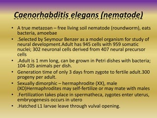
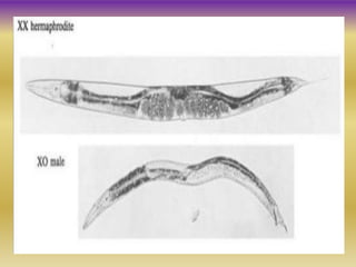
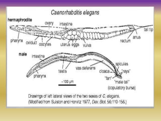
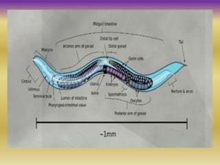
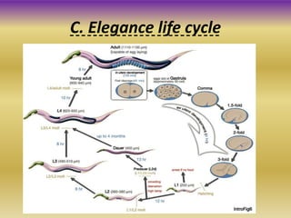
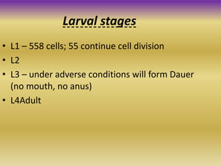
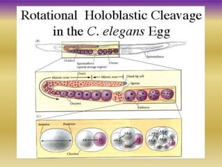
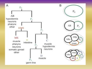
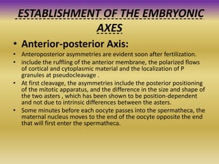
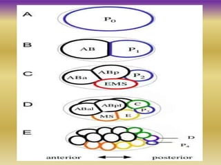
Caenorhabditis
elegans (nematode)
Synopsis
• Introduction of Caenorhabditis elegans
• Structure
• C. elegance life cycle
• Larval stages
• Early development of c. elegance
• Establishment of embryonic axis
①Anterior-posterior
②Dorso-ventral
③Left-right
• Rreference
Caenorhabditis elegans (nematode)
• A true metazoan – free living soil nematode (roundworm), eats
bacteria, amoebae
• .Selected by Seymour Benzer as a model organism for study of
neural development.Adult has 945 cells with 959 somatic
nuclei; 302 neuronal cells derived from 407 neural precursor
cells
• .Adult is 1 mm long, can be grown in Petri dishes with bacteria;
104-105 animals per dish.
• Generation time of only 3 days from zygote to fertile adult.300
progeny per adult.
• Sexually dimorphic – hermaphrodite (XX), male
(XO)Hermaphrodites may self-fertilize or may mate with males
• .Fertilization takes place in spermatheca, zygotes enter uterus,
embryogenesis occurs in utero
• .Hatched L1 larvae leave through vulval opening.
C. Elegance life cycle
Larval stages
• L1 – 558 cells; 55 continue cell division
• L2
• L3 – under adverse conditions will form Dauer
(no mouth, no anus)
• L4Adult
Early develeopment of c. elegance
• The sperm enters at the future posterior end of the embryo, opposite the
end where the oocyte pronucleus resides.
• The oocyte pronucleus, which was arrested in meiotic prophase, resumes
meiosis and both polar bodies are extruded.
• After meiosis, contractions of the anterior membrane occur and a
transient pseudocleavage furrow forms near the center of the egg. During
this time the oocyte pronucleus migrates posteriorly toward the sperm
pronucleus.
• The sperm pronucleus also migrates somewhat toward the oocyte
pronucleus, and the two meet in the posterior half of the egg. The
pronuclei break down and first cleavage divides the egg into a larger
anterior cell (AB) and a smaller posterior cell (P1). This cleavage is
unequal because of the posterior placement of the mitotic spindle.
• AB then divides with the spindle initially set up orthogonal to the
anteroposterior axis. During anaphase B, the elongating spindle skews
towards the anteroposterior axis so that one daughter, ABa, is positioned
anterior to the other, ABp.
• A few minutes later, P1 divides along the anteroposterior axis to form a
larger anterior cell (EMS) and a smaller posterior cell (P2).
Cont....
• Divisions in the AB lineage continue along axes orthogonal
to the preceding division axes, while divisions of the P1
progeny tend to place them in an anterior-posterior line,
though constraints of the eggshell skew them somewhat
off this axis.
• Gastrulation begins at the 28-cell stage; Ea and Ep are the
first cells to migrate into the center of the embryo. By the
time gastrulation ends approximately 550 cells have been
generated and most embryonic cell divisions are
complete.
• The embryo elongates inside the eggshell until it is folded
in three. During this period of elongation, muscle cells
begin to function and the embryo begins to twitch.
• At approximately 12 hours after fertilization, the L1 larva
hatches from the eggshell.
ESTABLISHMENT OF THE EMBRYONIC
AXES
• Anterior-posterior Axis:
• Anteroposterior asymmetries are evident soon after fertilization.
• include the ruffling of the anterior membrane, the polarized flows
of cortical and cytoplasmic material and the localization of P
granules at pseudocleavage .
• At first cleavage, the asymmetries include the posterior positioning
of the mitotic apparatus, and the difference in the size and shape of
the two asters , which has been shown to be position-dependent
and not due to intrinsic differences between the asters.
• Some minutes before each oocyte passes into the spermatheca, the
maternal nucleus moves to the end of the oocyte opposite the end
that will first enter the spermatheca.
• The end near the oocyte nucleus will become the anterior
end of the embryo. The polar bodies, which are a useful
mark of the place where the oocyte nucleus underwent
meiosis, are normally found at this end of the embryo
following fertilization, but occasionally can be found at the
posterior end.
• As the oocyte enters the spermatheca, a sperm enters at
the end opposite the maternal pronucleus. Experimentally
altering the position of the mitotic apparatus in early
blastomeres can generate cortical and cytoplasmic flows
similar to those of pseudocleavage, whose directions are
predicted by the position of the mitotic apparatus.
• This finding raises the possibility that the sperm asters
(analogous to the mitotic apparatus) could direct the f lows
seen during pseudocleavage, and by doing so may establish
the anteroposterior axis.
• The daughters of AB, ABa and ABp, give rise to distinct
lineages. In an experiment designed to test whether the
ABa and ABp blastomeres are initially equivalent, Priess
and Thomson (1987) switched the positions of ABa and ABp
by exerting pressure on one side of AB as it divided.
• Normal worm developed, demonstrating that ABa and ABp
are equivalent cells, in that each has the potential to
differentiate as the other normally does. The subsequent
differences in their fates must come from cell interactions.
• EMS blastomere was on the side that would have become
dorsal if unmanipulated, and a daughter of AB was on the
side that would have become ventral if unmanipulated.
The dorsoventral axis
• Left-right asymmetries are first apparent in C. elegans at the six-cell
stage with the synchronous division of ABa and ABp.
• As these cells divide, an invariant skewing of their spindles causes
the AB progeny on the left side of the embryo (ABal and ABpl) to
shift slightly anteriorly relative to the two AB progeny on the right
(ABar and ABpr).
• Wood (1991) has shown that the left-right handedness of the worm
is established via this shift. When the left side progeny of AB were
pushed posteriorly at the six-cell stage these cells executed lineages
appropriate for the right hand side.
• Similarly, the cells on the right hand side executed left hand
lineages. A worm developed but structures normally found on the
worm’s right side had developed on the left, and vice versa.
The left-right axis
• Cell and molecular biology
By Gerald karp
• Cell and molecular biology
P. K. Gupta
• http://www.wormbook.org/chapters/www_asym
celldiv/asymcelldiv.html
• http://www.devbio.biology.gatech.edu/?page_id
=41
• http://vle.du.ac.in/mod/book/view.php?id=1327
6&chapterid=29164
Reference
Thank you

C. elegans early develeopment

  • 1.
  • 2.
    Synopsis • Introduction ofCaenorhabditis elegans • Structure • C. elegance life cycle • Larval stages • Early development of c. elegance • Establishment of embryonic axis ①Anterior-posterior ②Dorso-ventral ③Left-right • Rreference
  • 3.
    Caenorhabditis elegans (nematode) •A true metazoan – free living soil nematode (roundworm), eats bacteria, amoebae • .Selected by Seymour Benzer as a model organism for study of neural development.Adult has 945 cells with 959 somatic nuclei; 302 neuronal cells derived from 407 neural precursor cells • .Adult is 1 mm long, can be grown in Petri dishes with bacteria; 104-105 animals per dish. • Generation time of only 3 days from zygote to fertile adult.300 progeny per adult. • Sexually dimorphic – hermaphrodite (XX), male (XO)Hermaphrodites may self-fertilize or may mate with males • .Fertilization takes place in spermatheca, zygotes enter uterus, embryogenesis occurs in utero • .Hatched L1 larvae leave through vulval opening.
  • 9.
  • 10.
    Larval stages • L1– 558 cells; 55 continue cell division • L2 • L3 – under adverse conditions will form Dauer (no mouth, no anus) • L4Adult
  • 11.
    Early develeopment ofc. elegance • The sperm enters at the future posterior end of the embryo, opposite the end where the oocyte pronucleus resides. • The oocyte pronucleus, which was arrested in meiotic prophase, resumes meiosis and both polar bodies are extruded. • After meiosis, contractions of the anterior membrane occur and a transient pseudocleavage furrow forms near the center of the egg. During this time the oocyte pronucleus migrates posteriorly toward the sperm pronucleus. • The sperm pronucleus also migrates somewhat toward the oocyte pronucleus, and the two meet in the posterior half of the egg. The pronuclei break down and first cleavage divides the egg into a larger anterior cell (AB) and a smaller posterior cell (P1). This cleavage is unequal because of the posterior placement of the mitotic spindle. • AB then divides with the spindle initially set up orthogonal to the anteroposterior axis. During anaphase B, the elongating spindle skews towards the anteroposterior axis so that one daughter, ABa, is positioned anterior to the other, ABp. • A few minutes later, P1 divides along the anteroposterior axis to form a larger anterior cell (EMS) and a smaller posterior cell (P2).
  • 15.
    Cont.... • Divisions inthe AB lineage continue along axes orthogonal to the preceding division axes, while divisions of the P1 progeny tend to place them in an anterior-posterior line, though constraints of the eggshell skew them somewhat off this axis. • Gastrulation begins at the 28-cell stage; Ea and Ep are the first cells to migrate into the center of the embryo. By the time gastrulation ends approximately 550 cells have been generated and most embryonic cell divisions are complete. • The embryo elongates inside the eggshell until it is folded in three. During this period of elongation, muscle cells begin to function and the embryo begins to twitch. • At approximately 12 hours after fertilization, the L1 larva hatches from the eggshell.
  • 16.
    ESTABLISHMENT OF THEEMBRYONIC AXES • Anterior-posterior Axis: • Anteroposterior asymmetries are evident soon after fertilization. • include the ruffling of the anterior membrane, the polarized flows of cortical and cytoplasmic material and the localization of P granules at pseudocleavage . • At first cleavage, the asymmetries include the posterior positioning of the mitotic apparatus, and the difference in the size and shape of the two asters , which has been shown to be position-dependent and not due to intrinsic differences between the asters. • Some minutes before each oocyte passes into the spermatheca, the maternal nucleus moves to the end of the oocyte opposite the end that will first enter the spermatheca.
  • 17.
    • The endnear the oocyte nucleus will become the anterior end of the embryo. The polar bodies, which are a useful mark of the place where the oocyte nucleus underwent meiosis, are normally found at this end of the embryo following fertilization, but occasionally can be found at the posterior end. • As the oocyte enters the spermatheca, a sperm enters at the end opposite the maternal pronucleus. Experimentally altering the position of the mitotic apparatus in early blastomeres can generate cortical and cytoplasmic flows similar to those of pseudocleavage, whose directions are predicted by the position of the mitotic apparatus. • This finding raises the possibility that the sperm asters (analogous to the mitotic apparatus) could direct the f lows seen during pseudocleavage, and by doing so may establish the anteroposterior axis.
  • 20.
    • The daughtersof AB, ABa and ABp, give rise to distinct lineages. In an experiment designed to test whether the ABa and ABp blastomeres are initially equivalent, Priess and Thomson (1987) switched the positions of ABa and ABp by exerting pressure on one side of AB as it divided. • Normal worm developed, demonstrating that ABa and ABp are equivalent cells, in that each has the potential to differentiate as the other normally does. The subsequent differences in their fates must come from cell interactions. • EMS blastomere was on the side that would have become dorsal if unmanipulated, and a daughter of AB was on the side that would have become ventral if unmanipulated. The dorsoventral axis
  • 22.
    • Left-right asymmetriesare first apparent in C. elegans at the six-cell stage with the synchronous division of ABa and ABp. • As these cells divide, an invariant skewing of their spindles causes the AB progeny on the left side of the embryo (ABal and ABpl) to shift slightly anteriorly relative to the two AB progeny on the right (ABar and ABpr). • Wood (1991) has shown that the left-right handedness of the worm is established via this shift. When the left side progeny of AB were pushed posteriorly at the six-cell stage these cells executed lineages appropriate for the right hand side. • Similarly, the cells on the right hand side executed left hand lineages. A worm developed but structures normally found on the worm’s right side had developed on the left, and vice versa. The left-right axis
  • 24.
    • Cell andmolecular biology By Gerald karp • Cell and molecular biology P. K. Gupta • http://www.wormbook.org/chapters/www_asym celldiv/asymcelldiv.html • http://www.devbio.biology.gatech.edu/?page_id =41 • http://vle.du.ac.in/mod/book/view.php?id=1327 6&chapterid=29164 Reference
  • 25.