Establishment of Body Axis in Caenorhabditis elegans
•
1 like•494 views

Establishment of the body axes in Caenorhabditis elegans (nematode).

Recommended
More Related Content
What's hot
What's hot (20)
Similar to Establishment of Body Axis in Caenorhabditis elegans
Similar to Establishment of Body Axis in Caenorhabditis elegans (20)
More from Syed Muhammad Khan
More from Syed Muhammad Khan (20)
Recently uploaded
Recently uploaded (20)
Establishment of Body Axis in Caenorhabditis elegans
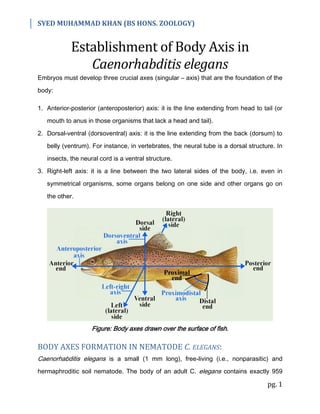
- 1. SYED MUHAMMAD KHAN (BS HONS. ZOOLOGY) pg. 1 Establishment of Body Axis in Caenorhabditis elegans Embryos must develop three crucial axes (singular – axis) that are the foundation of the body: 1. Anterior-posterior (anteroposterior) axis: it is the line extending from head to tail (or mouth to anus in those organisms that lack a head and tail). 2. Dorsal-ventral (dorsoventral) axis: it is the line extending from the back (dorsum) to belly (ventrum). For instance, in vertebrates, the neural tube is a dorsal structure. In insects, the neural cord is a ventral structure. 3. Right-left axis: it is a line between the two lateral sides of the body, i.e. even in symmetrical organisms, some organs belong on one side and other organs go on the other. Figure: Body axes drawn over the surface of fish. BODY AXES FORMATION IN NEMATODE C. ELEGANS: Caenorhabditis elegans is a small (1 mm long), free-living (i.e., nonparasitic) and hermaphroditic soil nematode. The body of an adult C. elegans contains exactly 959
- 2. SYED MUHAMMAD KHAN (BS HONS. ZOOLOGY) pg. 2 somatic cells. It also has a transparent cuticle (all of its organs are visible from the outside of its body, under a microscope). C. elegans has the rudiments (not very well developed) of nearly all the major types of bodily systems (feeding, nervous, reproductive, etc.) although there is no skeleton. 1. Anterior-Posterior Axis Formation: The egg of C. elegans is slightly elongated, and this elongated axis defines the future anterior-posterior axis of the nematode's body. The ends that will become anterior and posterior are decided by the position of the sperm pronucleus (nucleus of gametes is called pronucleus). The end of the egg from where the sperm enters becomes the posterior pole. The cell division, cell specification, and morphogenesis are coordinated by the sperm and several PAR (partitioning) proteins (i.e. PAR-2 for posterior end, and PAR-3 for anterior end, etc.). The sperm provides a protein: CYK-4, this protein activates the egg actin microfilaments to reposition PAR-3 proteins towards the anterior end. The PAR-2 proteins are positioned along the cortical (along cell membrane) cytoplasm of the posterior end. The PAR proteins localize a lot of entities including P-granules (ribonucleoprotein/RNP complexes that specify the germ cells). The P-granules are a collection of translation regulators. P-granules are randomly scattered before fertilization, but upon fertilization, they move towards the posterior end of the zygote, so that they enter only the blastomere (P1) formed from the posterior cytoplasm. At the two-cell stage, there is one blastomere that is formed from the anterior part and gives rise to the anterior structures, that cell is called AB cell, whereas the other blastomere is formed from the posterior part and gives rise to the posterior parts, that cell is called P1 cell. The AB cell retains MEX-5 protein (from the egg) in its central
- 3. SYED MUHAMMAD KHAN (BS HONS. ZOOLOGY) pg. 3 cytoplasm and PAR-3 in its cortical (near cell membrane) cytoplasm. The P1 cell inherits P-granules and in its cortical cytoplasm is PAR-2 (except the area where the two cells are connected). Hence AB and P1 cells form the basis of the anterior-posterior axis in C. elegans. Figure: The first division causes the separation of anterior and posterior determinants in AB (MEX-5 and PAR-3) and P1 cells (P-granules and PAR-2) respectively. 2. Dorsal-Ventral Axis Formation: The dorsal-ventral axis of the nematode is seen in the division of the AB cell. As the AB cell divides, it becomes longer than the eggshell is wide; this causes the cells to slide, resulting in one AB daughter cell being anterior and one being posterior (hence they are called ABa and ABp respectively). Figure: The AB cell divides to form ABa and ABp cells, these cells slide because the eggshell is not wide enough. Consequently, the ABp cell gets above EMS cell (the P1 cell divides into EMS cell and P2 cell). The ABp cell defines the future dorsal side and the EMS cell defines the future ventral side. This squeezing causes the ABp cell to take a position above the EMS cell that results from the division of the P1 blastomere (P1 blastomere divides into EMS cell and P2
- 4. SYED MUHAMMAD KHAN (BS HONS. ZOOLOGY) pg. 4 cell). The ABp cell defines the future dorsal side of the embryo, while the EMS cell (the precursor of the muscle and gut cells) marks the future ventral surface of the embryo. 3. Left-Right Axis Formation: The left-right axis is specified later, at the 12-cell stage, when the MS blastomere (from the division of the EMS cell, i.e. EMS cell divides into MS and E cells) contacts half the progeny of the ABa cell, distinguishing the right side of the body from the left side. Figure: The EMS cell divides into MS and E cells (lower side), the MS cell comes in contact with half of the progeny of ABa cell (ABal/left and ABar/right) this distinguishes the left side from the right, forming the left-right axis.