The document discusses using a U-Net convolutional neural network to automatically segment brain tumors in MRI images. It aims to eliminate the need for domain expertise by using deep learning to extract hierarchical features. The U-Net model is trained on the BRATS 2017 dataset and is able to segment tumors with 5% higher accuracy than previous methods, as measured by the Dice similarity coefficient. The system could be expanded to analyze additional MRI modalities and further improve automated tumor detection.
![International Journal of Research in Advent Technology, Vol.7, No.9, September 2019
E-ISSN: 2321-9637
Available online at www.ijrat.org
10
doi: 10.32622/ijrat.78201916
Abstract— It is very hard to detect brain tumor in the
beginning as the human brain has an extremely intricate
structure. This is a very cumbersome process for the
radiologist to detect the tumor, physically segment and
recognize the tumor in the MRI image. The machine learning
is ideal for this task as it is great at perceiving tumor locales
and there is no need of undertaking specific algorithm.
Before the advent of the deep learning applications, a
specialist in the field of medical domain used to construct the
features and it demanded the special knowledge in the
medical field. To eliminate the domain-specific knowledge;
we are using the U-Net based model to carry out the brain
tumor segmentation. The U-Net network, which is based on
deep learning, is capable of extracting the hierarchical
features present in MRI images without any manual
intervention. The idea is to use this domain-independent
algorithm to carry out the segmentation task automatically. In
this paper, we recommend using U-Net based deep
convolutional neural network to carry out brain tumor
segmentation. U-Net based Convolutional Neural Network is
expected to improve segmentation accuracy. We have used
BRATS 2017 dataset, to train the neural network and
henceforth carry out brain tumor segmentation.
Index Terms— Deep Learning, Machine Learning, Brain
Tumor, Convolutional Neural Network.
I. INTRODUCTION
Medical imaging techniques like MRI had made
tremendous progress and it had made it a reality to diagnose
the disease at earlier stages. The radiologist manually
examines these scans to interpret the presence of tumor in the
MRI. It is a very cumbersome errand to interpret such an
immense volume of scans at a stretch and daily ongoing
process. There is a huge possibility of error in this manual
process. There is a need to automate this process [3]. There
are a plethora of organizations which are making this kind of
data available for research. We are utilizing BRATS 2017
dataset to train our neural network and segment the MRI
image thereafter training. This BRATS 2017 dataset has
multimodal images like low grade and high-grade tumor MRI
scans. It is next to impossible to interpret this much volume
of information. Machine learning techniques are ideal to
mine this volume data to interpret and obtain results
automatically.
Manuscript revised on September 23, 2019 and published on October
10, 2019
Adarsh Dhiman, Dept. of Computer Engineering, DPU, Pune, India.
Prof. B.S. Satpute, Dept. of Computer Engineering, DPU, Pune, India.
The U-Net model is perfect to separate salient features
from data, which will assist the radiologist with making
better choices. U-Net model extracts various leveled data
features from MRI images to create a model that provides
insight into the data. The features extracted from the data are
automated and they provide meaningful deeper insights,
which are not even obvious to naked eye. These various
leveled features are self-learned by the U-Net based
convolutional neural network model [1].
II. LITERATURE REVIEW
A. U-Net based deep Convolutional Neural Networks
for Brain Tumor Segmentation [1]
In this paper [1], we become more acquainted with how the
U-Net based Convolutional Neural Network handles the
variable edge problem if there should be an occurrence in
MRI picture (cerebrum tumor).
The skip architecture is intended to manage variable edge
issue. This architecture has achieved promising outcomes on
MRI pictures. In this investigation, the authors have utilized
U-Net based Convolutional Neural Network as a base model.
This design has indicated great outcomes in tumor
segmentation.
B. Stacked De-noising Auto-Encoder based Deep
Learning Framework [2]
In this paper [2], deep learning is introduced to automatically
segment brain tumor and avoid the manual segmentation and
domain knowledge. This paper introduces the concept of
extracting image patches from MRI image and then provides
the gray level image patches as input to Stacked De-noising
Auto-Encoder. In order to carry out feature classification the
Stacked De-noising Auto-Encoder (SDAE), automatically
learns from the data in the MRI image. In this study, the result
is mapped on binary image and eventually the final
segmentation is achieved.
C. Deep Learning for MRI Image Analysis [3]
In this paper [3], author discussed different deep learning
neural networks like Feed-Forward Neural Networks,
Convolutional Neural Networks etc. and explained their
application in MRI Image Analysis for image segmentation
and detection of tumor.
D. Image Segmentation methods and Convolutional
Neural Network [4]
In this paper [4] author discusses, the different methods
which handles semantic division such as pixel-wise division.
Brain Tumor Segmentation in MRI Images
Adarsh Dhiman, Prof. B.S. Satpute](https://image.slidesharecdn.com/78201916-191116052427/85/Brain-Tumor-Segmentation-in-MRI-Images-1-320.jpg)
![International Journal of Research in Advent Technology, Vol.7, No.9, September 2019
E-ISSN: 2321-9637
Available online at www.ijrat.org
10
doi: 10.32622/ijrat.78201916
Abstract— It is very hard to detect brain tumor in the
beginning as the human brain has an extremely intricate
structure. This is a very cumbersome process for the
radiologist to detect the tumor, physically segment and
recognize the tumor in the MRI image. The machine learning
is ideal for this task as it is great at perceiving tumor locales
and there is no need of undertaking specific algorithm.
Before the advent of the deep learning applications, a
specialist in the field of medical domain used to construct the
features and it demanded the special knowledge in the
medical field. To eliminate the domain-specific knowledge;
we are using the U-Net based model to carry out the brain
tumor segmentation. The U-Net network, which is based on
deep learning, is capable of extracting the hierarchical
features present in MRI images without any manual
intervention. The idea is to use this domain-independent
algorithm to carry out the segmentation task automatically. In
this paper, we recommend using U-Net based deep
convolutional neural network to carry out brain tumor
segmentation. U-Net based Convolutional Neural Network is
expected to improve segmentation accuracy. We have used
BRATS 2017 dataset, to train the neural network and
henceforth carry out brain tumor segmentation.
Index Terms— Deep Learning, Machine Learning, Brain
Tumor, Convolutional Neural Network.
I. INTRODUCTION
Medical imaging techniques like MRI had made
tremendous progress and it had made it a reality to diagnose
the disease at earlier stages. The radiologist manually
examines these scans to interpret the presence of tumor in the
MRI. It is a very cumbersome errand to interpret such an
immense volume of scans at a stretch and daily ongoing
process. There is a huge possibility of error in this manual
process. There is a need to automate this process [3]. There
are a plethora of organizations which are making this kind of
data available for research. We are utilizing BRATS 2017
dataset to train our neural network and segment the MRI
image thereafter training. This BRATS 2017 dataset has
multimodal images like low grade and high-grade tumor MRI
scans. It is next to impossible to interpret this much volume
of information. Machine learning techniques are ideal to
mine this volume data to interpret and obtain results
automatically.
Manuscript revised on September 23, 2019 and published on October
10, 2019
Adarsh Dhiman, Dept. of Computer Engineering, DPU, Pune, India.
Prof. B.S. Satpute, Dept. of Computer Engineering, DPU, Pune, India.
The U-Net model is perfect to separate salient features
from data, which will assist the radiologist with making
better choices. U-Net model extracts various leveled data
features from MRI images to create a model that provides
insight into the data. The features extracted from the data are
automated and they provide meaningful deeper insights,
which are not even obvious to naked eye. These various
leveled features are self-learned by the U-Net based
convolutional neural network model [1].
II. LITERATURE REVIEW
A. U-Net based deep Convolutional Neural Networks
for Brain Tumor Segmentation [1]
In this paper [1], we become more acquainted with how the
U-Net based Convolutional Neural Network handles the
variable edge problem if there should be an occurrence in
MRI picture (cerebrum tumor).
The skip architecture is intended to manage variable edge
issue. This architecture has achieved promising outcomes on
MRI pictures. In this investigation, the authors have utilized
U-Net based Convolutional Neural Network as a base model.
This design has indicated great outcomes in tumor
segmentation.
B. Stacked De-noising Auto-Encoder based Deep
Learning Framework [2]
In this paper [2], deep learning is introduced to automatically
segment brain tumor and avoid the manual segmentation and
domain knowledge. This paper introduces the concept of
extracting image patches from MRI image and then provides
the gray level image patches as input to Stacked De-noising
Auto-Encoder. In order to carry out feature classification the
Stacked De-noising Auto-Encoder (SDAE), automatically
learns from the data in the MRI image. In this study, the result
is mapped on binary image and eventually the final
segmentation is achieved.
C. Deep Learning for MRI Image Analysis [3]
In this paper [3], author discussed different deep learning
neural networks like Feed-Forward Neural Networks,
Convolutional Neural Networks etc. and explained their
application in MRI Image Analysis for image segmentation
and detection of tumor.
D. Image Segmentation methods and Convolutional
Neural Network [4]
In this paper [4] author discusses, the different methods
which handles semantic division such as pixel-wise division.
Brain Tumor Segmentation in MRI Images
Adarsh Dhiman, Prof. B.S. Satpute](https://image.slidesharecdn.com/78201916-191116052427/75/Brain-Tumor-Segmentation-in-MRI-Images-1-2048.jpg)
![International Journal of Research in Advent Technology, Vol.7, No.9, September 2019
E-ISSN: 2321-9637
Available online at www.ijrat.org
11
doi: 10.32622/ijrat.78201916
In this case a small patch of picture is used to order the
middle pixel, and the fully convolutional designs where the
system is provided with whole picture and the output is a
semantic division volume. This study used Convolutional
Neural Network for MRI image segmentation.
E. Brain Tumor Segmentation with aid of Convolutional
Neural Network [5]
In this paper [5], author used Convolutional Neural Network
for brain tumor segmentation in medical image. In this study
the preprocessing step consists of bias correction and patch
normalization. In this study the existing low-grade glioma
(LGG) samples are rotated to produce new LGG samples.
The deep Convolutional Neural Network is built using 3X3
kernels on top of convolutional layers.
III. MACHINE LEARNING FOR BRAIN TUMOR
SEGMENTATION
The images are manually labelled by domain expert to train
the neural network. The idea is to use the fully Convolutional
Neural Network based on deep learning which will be trained
on the input data and will learn the features present in MRI
image without any manual intervention. The model generated
in due course will be used to segment the MRI image and
detect the necrotic and enhancing tumor.
A. Existing System and Disadvantages
Traditionally the features were hand crafted by the domain
specialist. This process required the domain specific
knowledge. These images were then used to train the neural
network. This process consumes a lot of time and labor
intensive. The loss function used in the current system is not
apt for the problem in hand. The following are the
disadvantages of established system:
• Error Prone
• Time Consuming
• Manual
• Requires Domain Knowledge
• Human Fatigue
B. Proposed System and Advantages
In proposed system we used deep learning techniques based
on U-Net architecture. The system performs computer aided
segmentation and detection of tumor. The success of the
proposed system comes from the following factors:
• Use of high end CPUs and GPUs
• Discovery of Hierarchical Features
• Development of Learning Algorithms
a. Overview of System Architecture Diagram
The system architecture consists of the following
components:
Fig. 1. System Architecture Diagram
b. U-Net Architecture Diagram
Fig. 2. U-Net Convolutional Neural Network Architecture
In this study the U-Net based network architecture is used,
which consists of a down-sampling path and an up-sampling
path as indicated in “Fig. 2”. The contraction path or
down-sampling consists of four blocks. In this scheme every
block has 3X3 convolutional layers and ReLU activation.
The down sampling or contraction path consists of a 2×2 Max
Pool which is added to the end of every block, to reduce
feature map. The expansion path or up-sampling path has
also four blocks. All the blocks in the expansion or
up-sampling path consists of a de-convolutional layer, a link
to the corresponding feature map in the down-sampling path
and 3X3 convolutional layers and ReLU activation.
c. Segmentation Algorithm
The algorithm used in U-Net based Convolutional Neural
Network is explained below in “Fig. 3”. This architecture
has a contraction path and an expansion path.](https://image.slidesharecdn.com/78201916-191116052427/85/Brain-Tumor-Segmentation-in-MRI-Images-2-320.jpg)
![International Journal of Research in Advent Technology, Vol.7, No.9, September 2019
E-ISSN: 2321-9637
Available online at www.ijrat.org
13
doi: 10.32622/ijrat.78201916
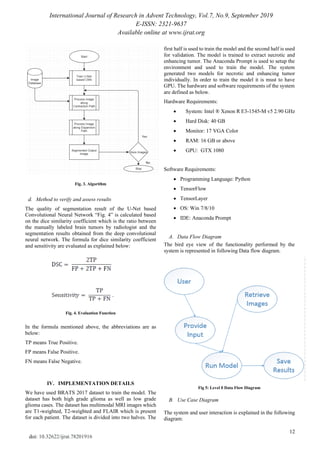
Fig 6: Use Case Diagram
V. RESULT AND DISCUSSION
In this study, we used U-Net based fully convolutional neural
network for brain MRI image segmentation. Deep learning or
deep convolutional neural networks are giving promising
results in medical image segmentation based on Dice
Similarity Coefficient as compared to other techniques.
The input to the network is a MRI image “Fig. 7”, and output
is “Fig. 8”, the segmented image.
Input Image:
Fig 7: Input MRI Image
Segmented Image:
Fig 8: Segmented MRI Image
The chart in “Fig. 9” shows the dice similarity coefficient
(ranges between 0 and 1) of the base method and the new
proposed method in case of tumor demarcation. The dice
similarity coefficient of the proposed method has improved
by 5%.
Fig 9: Dice Similarity Coefficient
This network can further be used to study other modalities
available in the MRI images to prove the validity and range
of network to segment brain MRI images.
VI. CONCLUSION AND FUTURE SCOPE
This paper prescribes to utilize Convolutional Neural
Network based deep learning methods to segment the MRI
image. The progress in image segmentation methods will
incredibly aid in automated identification of tumor. The
ongoing improvement in deep learning has revealed the
cellular features in the MRI images. Deep learning strategies
have indicated promising outcomes over various ailments
like Alzheimer, Pneumonia and so on; be that as it may, there
is still scope for development.
REFERENCES
[1] Hao Dong, Guang Yang, Fangde Liu, Yuanhan Mo, Yike Guo,
“Automatic Brain Tumor Detection and Segmentation Using U-Net
Based Fully Convolutional Networks”, arXiv:1705.03820 [cs.CV], 3
June 2017.
[2] Zhe Xiao, Ruohan Huang, Yi Ding, Tian Lan, RongFeng Dong and
Zhiguang Qin, “A Deep Learning-Based Segmentation Method for
Brain Tumor in MR Images”, presented at 2016 IEEE 6th International
Conference on Computational Advances in Bio and Medical Sciences
(ICCABS), Atlanta, GA, USA, Oct. 13-15, 2016.
[3] Shen, Guorong Wu and Heung-Il Suk, “Deep Learning in Medical
Image Analysis”, Annual Review of Biomedical Engineering, vol. 19,
pp. 221-248, June 2017.
[4] Manda SSSNMSRL Pavan, P. Jagadeesh, “Brain Tumor Segmentation
using Convolutional Neural Network in MRI Images”, International
Journal of Pure and Applied Mathematics, vol. 119 no. 17, pp.
1585-1592, 2018.
[5] Sérgio Pereira, Adriano Pinto, Victor Alves, and Carlos A. Silva,
“Brain Tumor Segmentation Using Convolutional Neural Networks in
MRI Images”, IEEE Transactions on Medical Imaging, vol. 35, no. 5,
pp. 1240-1251, May 2016.
[6] Hapsari Peni Agustin Tjahyaningtijas, “Brain Tumor Image
Segmentation in MRI Image”, 2018 IOP Conf. Ser.: Mater. Sci. Eng.
336 012012, vol. 336, conference 1.
[7] Adnan Qayyuma, Syed Muhammad Anwar, Muhammad Majid,
Muhammad Awais, Majdi Alnowamid, “Medical Image Analysis
using Convolutional Neural Networks: A Review”, Springer US, vol.
42, pp. 226, Oct. 2018.
[8] Kavitha Angamuthu Rajasekaran, Chellamuthu Chinna Gounder,
“Advanced Brain Tumor Segmentation from MRI Images”,
High-Resolution Neuroimaging - Basic Physical Principles and
Clinical Applications, ch. 6, pp. 735, 2018.
[9] Adarsh Dhiman, B.S. Satpute, “Brain Tumor Segmentation in MRI
Images”, International Journal of Engineering and Techniques, vol. 5
Issue 1, pp. 112, Jan-Feb 2019.](https://image.slidesharecdn.com/78201916-191116052427/85/Brain-Tumor-Segmentation-in-MRI-Images-4-320.jpg)
![International Journal of Research in Advent Technology, Vol.7, No.9, September 2019
E-ISSN: 2321-9637
Available online at www.ijrat.org
14
doi: 10.32622/ijrat.78201916
[10] Adarsh Dhiman and B.S. Satpute, “Brain Tumor Segmentation in MRI
Images”, Eighth Post Graduate Conference of Computer Engineering,
2018-19.
AUTHORS PROFILE
Adarsh Dhiman, M.E., Dept. of Computer Engineering,
DPU, Pune.
Prof. B.S. Satpute, Associate Professor, Dept. of
Computer Engineering, DPU, Pune.](https://image.slidesharecdn.com/78201916-191116052427/85/Brain-Tumor-Segmentation-in-MRI-Images-5-320.jpg)