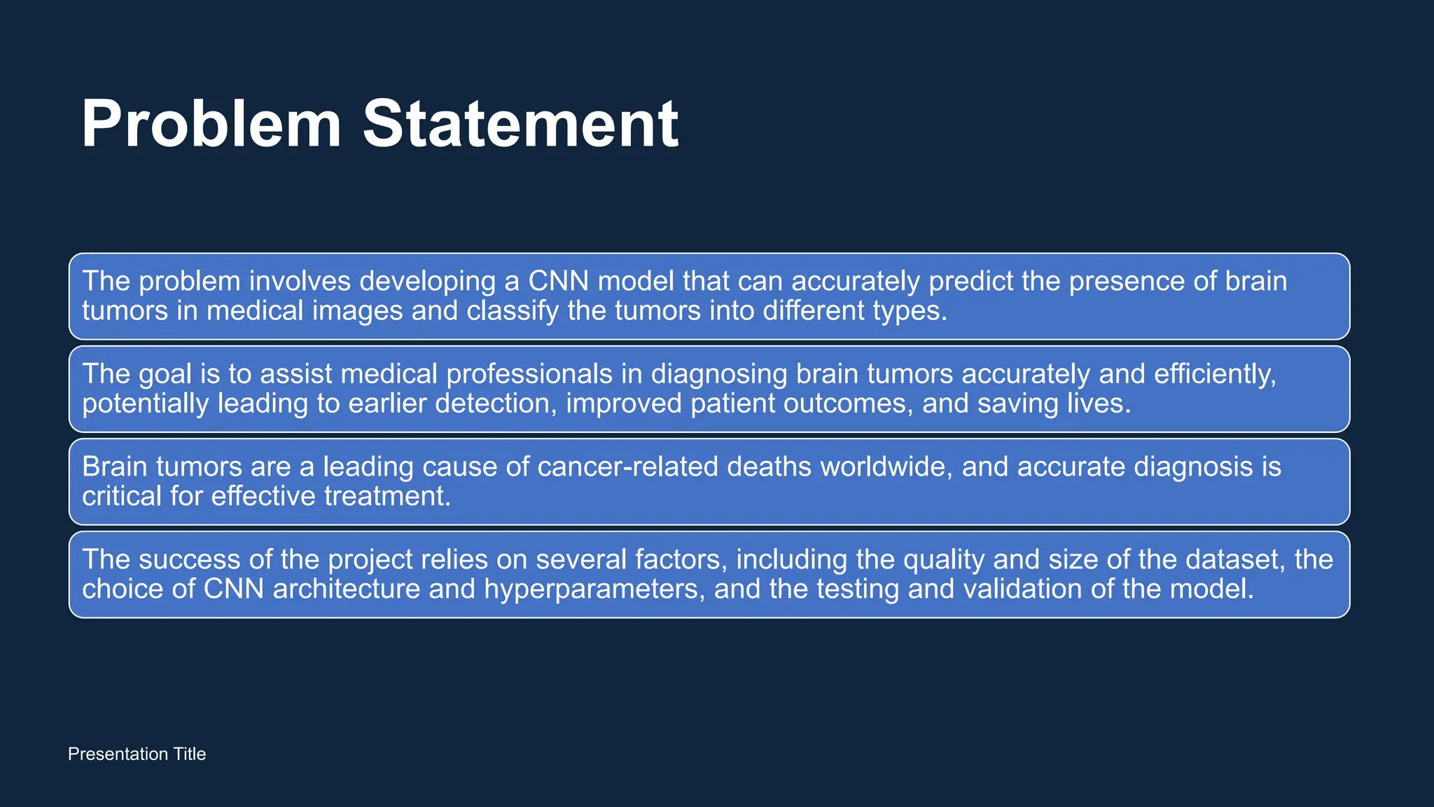
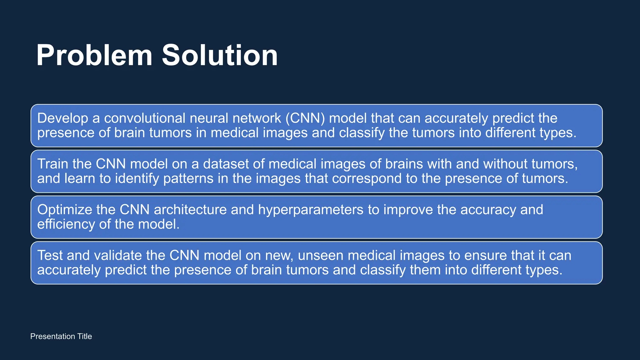
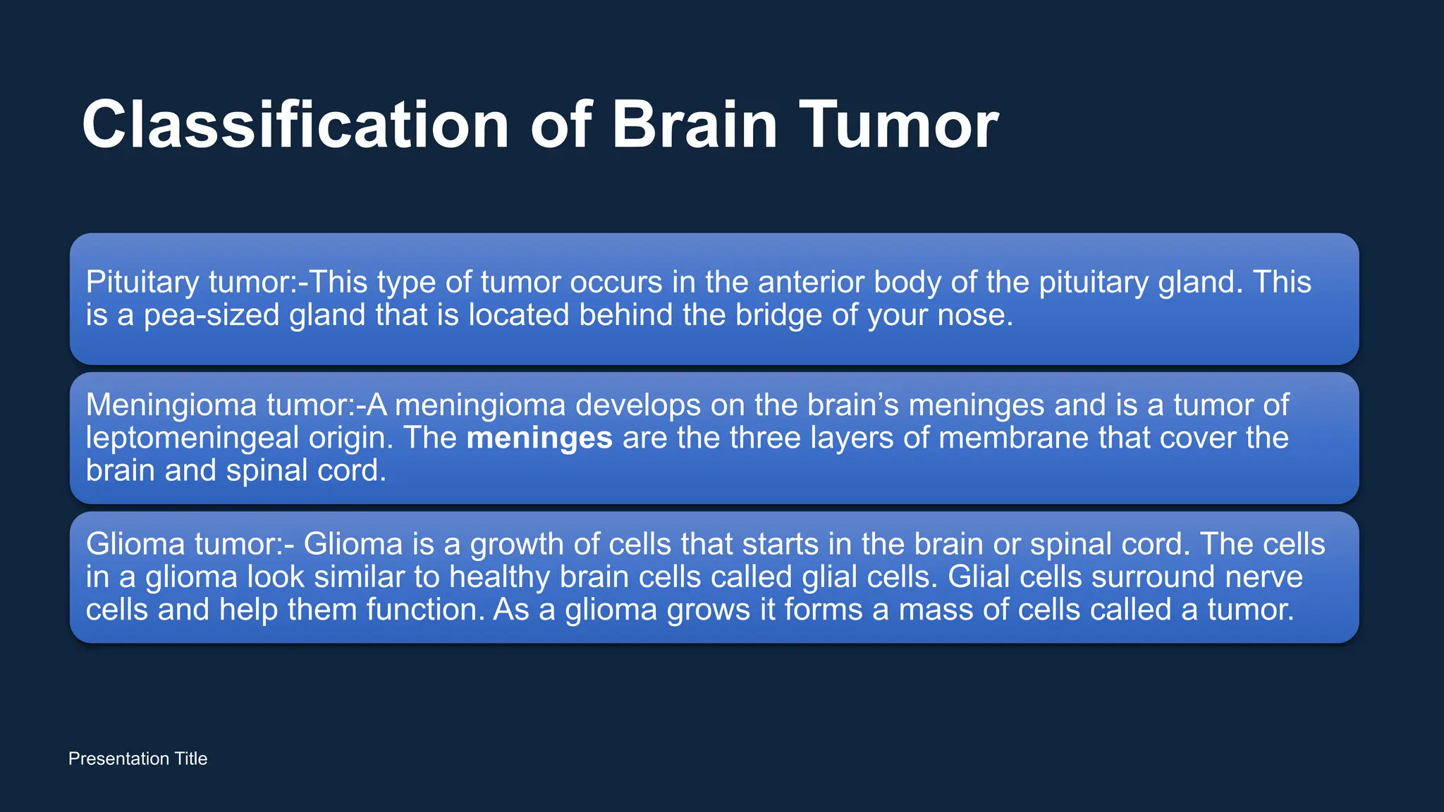
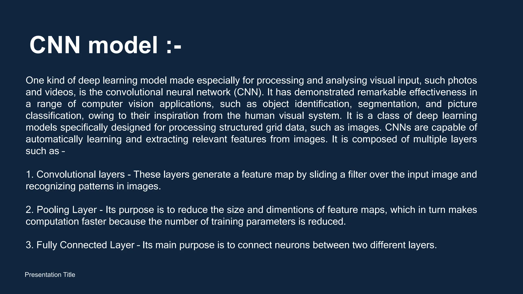
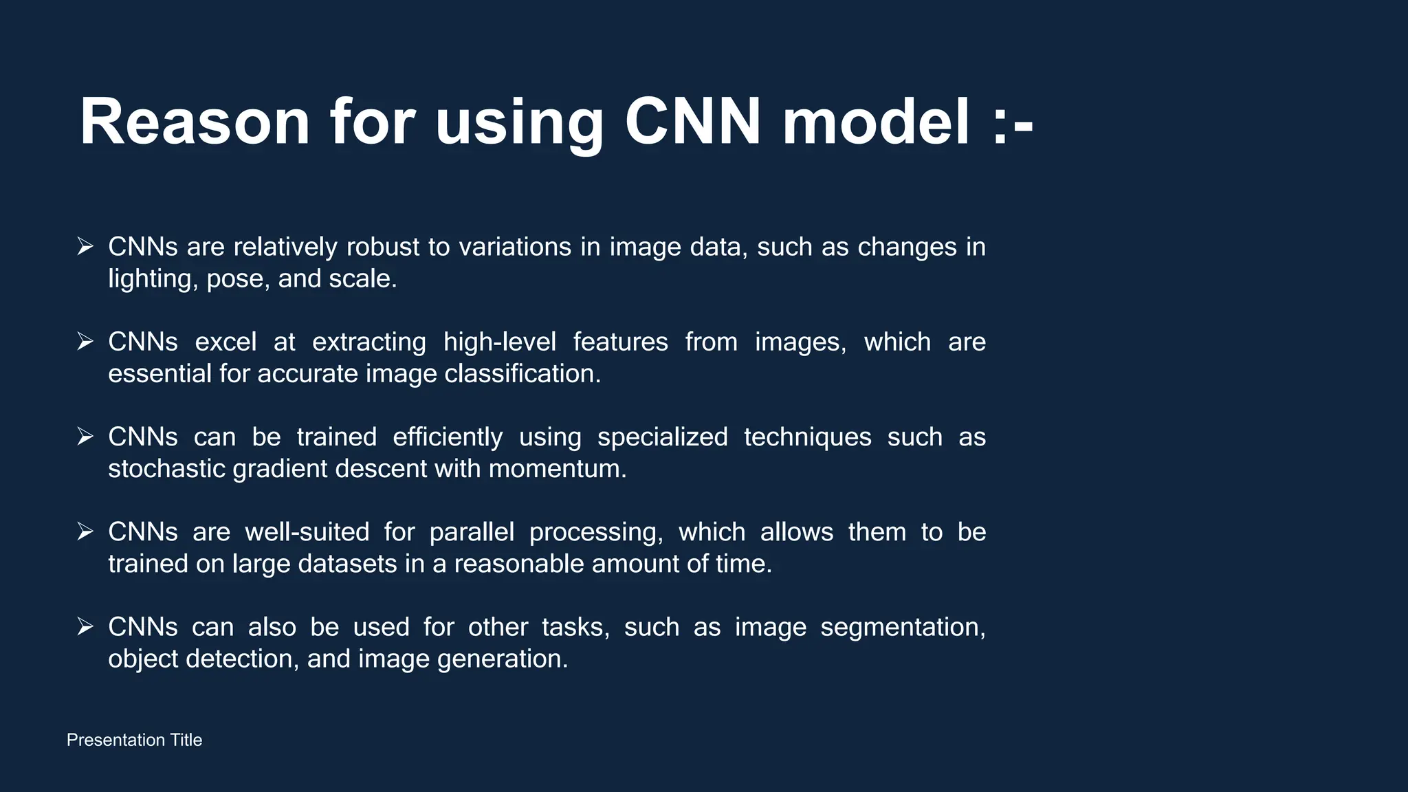
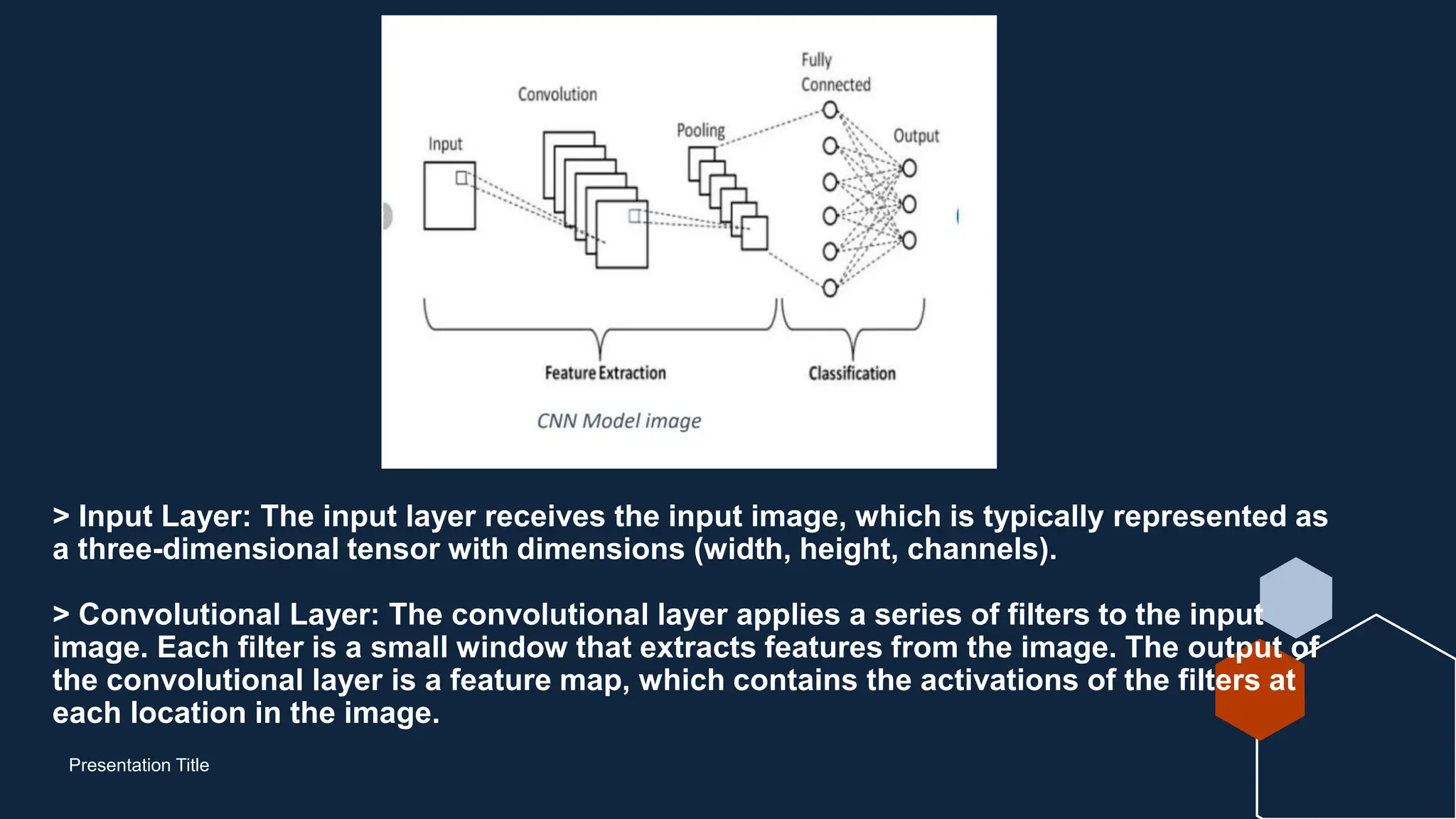
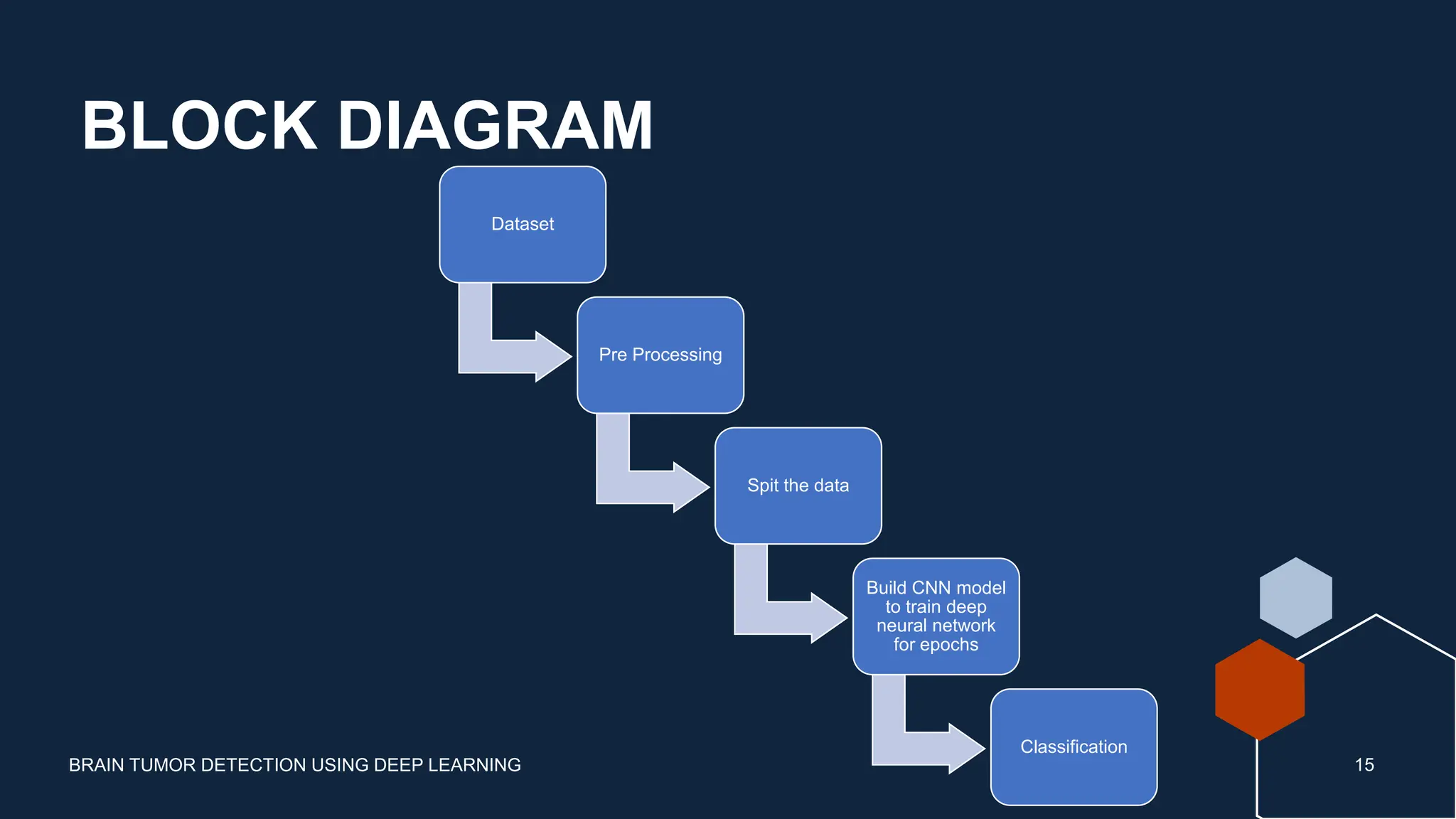
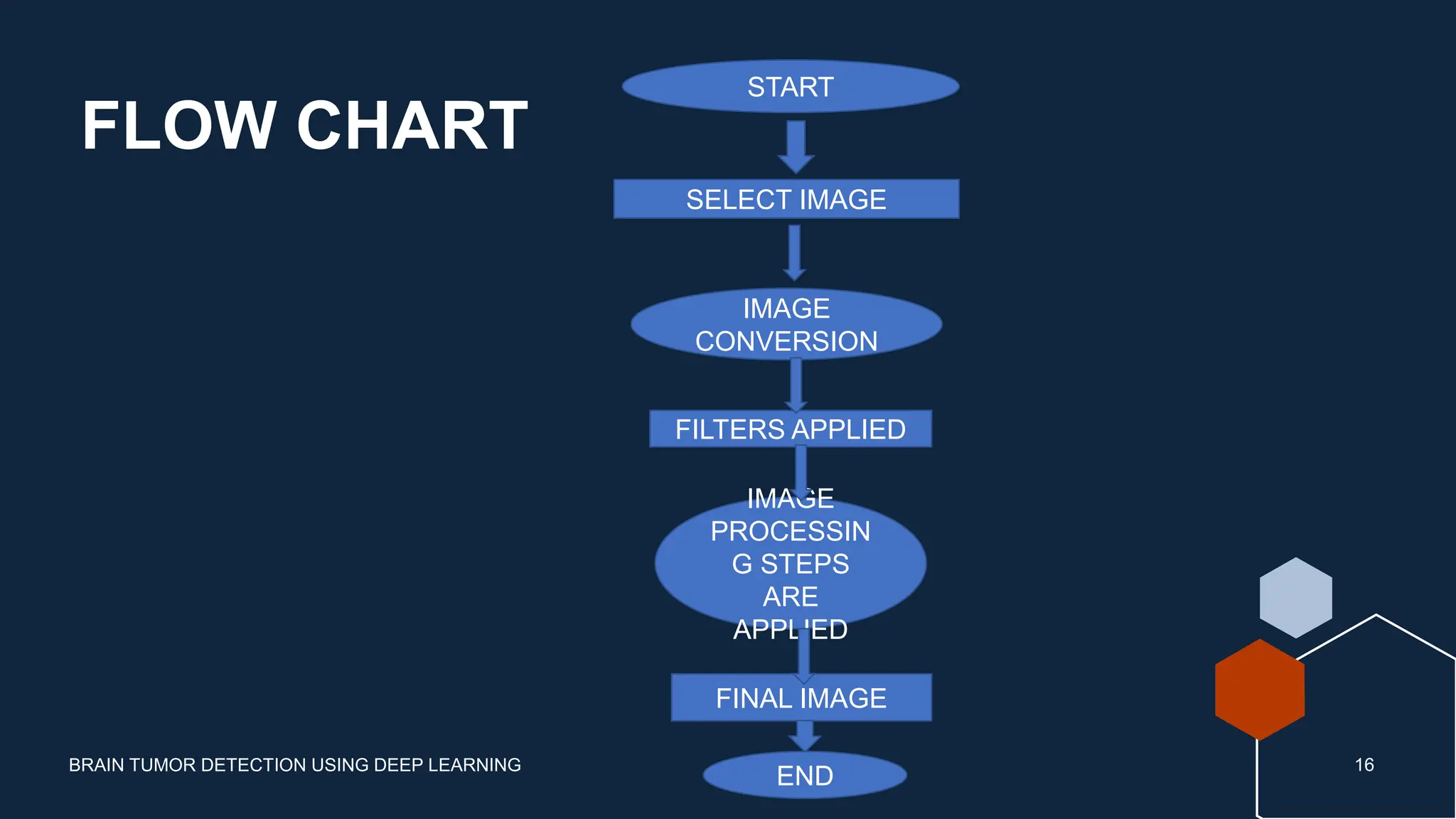
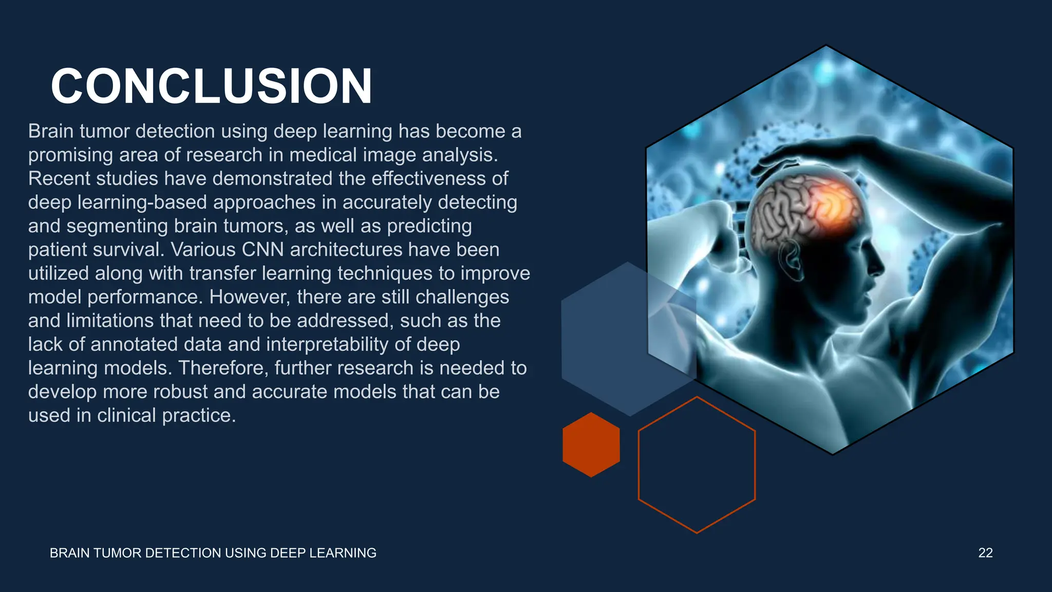
Brain Tumor
Detection Using
Deep Learning
Nitin Raj
Mohit Khajuria
Neraj Roy
BRAIN TUMOR
DETECTION USING DEEP
LEARNING
Agenda
• INTRODUTION
• PROBLEM
STATEMENT
• PROBLEM SOLUTION
• PROJECT
OUTCOMES
• LITERATURE
REVIEW
• COMPONENT
REQUIRED
• BLOCK DIAGRAM &
FLOW CHART
• WORKING OF
PROJECT
• CONCLUSION
• FUTURE SCOPE
Introduction
Brain tumors are a serious and potentially life-threatening medical condition
that requires prompt diagnosis and treatment. However, accurately detecting
brain tumors can be a challenging task for medical professionals, and
misdiagnosis can have severe consequences. In recent years, deep learning
has emerged as a powerful tool for medical image analysis, and brain tumor
detection is one area where it has shown great promise. This project focuses
on using deep learning algorithms, such as convolutional neural networks
(CNNs), to aid in the detection and diagnosis of brain tumors using medical
imaging data. By leveraging the power of deep learning, we aim to improve
the accuracy and speed of brain tumor diagnosis, ultimately leading to better
patient outcomes.
3
BRAIN TUMOR DETECTION USING DEEP LEARNING
Problem Statement
Presentation Title
The problem involves developing a CNN model that can accurately predict the presence of brain
tumors in medical images and classify the tumors into different types.
The goal is to assist medical professionals in diagnosing brain tumors accurately and efficiently,
potentially leading to earlier detection, improved patient outcomes, and saving lives.
Brain tumors are a leading cause of cancer-related deaths worldwide, and accurate diagnosis is
critical for effective treatment.
The success of the project relies on several factors, including the quality and size of the dataset, the
choice of CNN architecture and hyperparameters, and the testing and validation of the model.
Problem Solution
Presentation Title
Develop a convolutional neural network (CNN) model that can accurately predict the
presence of brain tumors in medical images and classify the tumors into different types.
Train the CNN model on a dataset of medical images of brains with and without tumors,
and learn to identify patterns in the images that correspond to the presence of tumors.
Optimize the CNN architecture and hyperparameters to improve the accuracy and
efficiency of the model.
Test and validate the CNN model on new, unseen medical images to ensure that it can
accurately predict the presence of brain tumors and classify them into different types.
Classification of Brain Tumor
Presentation Title
Pituitary tumor:-This type of tumor occurs in the anterior body of the pituitary gland. This
is a pea-sized gland that is located behind the bridge of your nose.
Meningioma tumor:-A meningioma develops on the brain’s meninges and is a tumor of
leptomeningeal origin. The meninges are the three layers of membrane that cover the
brain and spinal cord.
Glioma tumor:- Glioma is a growth of cells that starts in the brain or spinal cord. The cells
in a glioma look similar to healthy brain cells called glial cells. Glial cells surround nerve
cells and help them function. As a glioma grows it forms a mass of cells called a tumor.
CNN model :-
Presentation Title
One kind of deep learning model made especially for processing and analysing visual input, such photos
and videos, is the convolutional neural network (CNN). It has demonstrated remarkable effectiveness in
a range of computer vision applications, such as object identification, segmentation, and picture
classification, owing to their inspiration from the human visual system. It is a class of deep learning
models specifically designed for processing structured grid data, such as images. CNNs are capable of
automatically learning and extracting relevant features from images. It is composed of multiple layers
such as –
1. Convolutional layers - These layers generate a feature map by sliding a filter over the input image and
recognizing patterns in images.
2. Pooling Layer - Its purpose is to reduce the size and dimentions of feature maps, which in turn makes
computation faster because the number of training parameters is reduced.
3. Fully Connected Layer – Its main purpose is to connect neurons between two different layers.
Reason for using CNN model :-
Presentation Title
 CNNs are relatively robust to variations in image data, such as changes in
lighting, pose, and scale.
 CNNs excel at extracting high-level features from images, which are
essential for accurate image classification.
 CNNs can be trained efficiently using specialized techniques such as
stochastic gradient descent with momentum.
 CNNs are well-suited for parallel processing, which allows them to be
trained on large datasets in a reasonable amount of time.
 CNNs can also be used for other tasks, such as image segmentation,
object detection, and image generation.
> Input Layer: The input layer receives the input image, which is typically represented as
a three-dimensional tensor with dimensions (width, height, channels).
> Convolutional Layer: The convolutional layer applies a series of filters to the input
image. Each filter is a small window that extracts features from the image. The output of
the convolutional layer is a feature map, which contains the activations of the filters at
each location in the image.
Presentation Title
> Pooling Layer: The pooling layer reduces the size of the feature map by applying a pooling
operation to each region of the map. Common pooling operations include max pooling and
average pooling.
> Fully Connected Layer: The fully connected layer takes the flattened output of the pooling
layer and transforms it into a probability distribution over the possible classes.
> Output Layer: The output layer outputs the probability distribution over the possible classes.
The class with the highest probability is the detected class for the input image.
Presentation Title
Dataset used :-
Presentation Title
No Tumor Pituitary Tumor Glioma Tumor Meningioma Tumor
PROJECT OUTCOME
BRAIN TUMOR DETECTION USING DEEP LEARNING 12
The expected outcomes from a project on brain tumor detection using deep learning may vary depending
on the specific goals and objectives of the project. However, some potential outcomes include:
 Improved accuracy and efficiency of brain tumor detection: By using deep learning algorithms, the
accuracy and efficiency of brain tumor detection can be improved. This could lead to earlier detection
of tumors, better treatment planning, and improved patient outcomes.
 Development of new algorithms and methods: A project in this field may involve the development of
new deep learning algorithms and methods that can be used to improve brain tumor detection. These
algorithms could have broader applications in medical imaging and other fields.
PROJECT OUTCOME
BRAIN TUMOR DETECTION USING DEEP LEARNING 13
 Validation and evaluation of existing algorithms: A project may involve the validation and evaluation of
existing deep learning algorithms for brain tumor detection. This could help to identify areas for
improvement and provide insights into how these algorithms can be optimized for specific applications.
 Improved understanding of brain tumor characteristics: By analyzing large datasets of brain tumor
images, a project could help to improve our understanding of the characteristics and properties of
different types of brain tumors. This could lead to better treatment options and improved patient
outcomes.
 Increased collaboration between medical professionals and computer scientists: A project in this field
may involve collaboration between medical professionals and computer scientists, which could lead to
increased knowledge sharing and interdisciplinary research. This could result in the development of
new solutions and innovations that would not have been possible through traditional research methods.
NumPy module
works with the
numerical da
NumPy is a Python
library used
for working with
arrays. ta.
for data analysis
and associated
manipulation of
tabular data in
DataFrames.
Is used to Create a
graph or we can
say used for 2d
visualization
Used to do deep
learning and also
develop CNN
model
provides the facility
to establish the
interaction between
the user and the
operating system
NUMPY
COMPONENTS
REQUIRED
PANDAS
MATPLOTLIB
and SEABORN
TENSORFLOW OS
BRAIN TUMOR DETECTION USING DEEP LEARNING
BLOCK DIAGRAM
BRAIN TUMOR DETECTION USING DEEP LEARNING 15
Dataset
Pre Processing
Spit the data
Build CNN model
to train deep
neural network
for epochs
Classification
FLOW CHART
BRAIN TUMOR DETECTION USING DEEP LEARNING 16
START
SELECT IMAGE
IMAGE
CONVERSION
FILTERS APPLIED
IMAGE
PROCESSIN
G STEPS
ARE
APPLIED
FINAL IMAGE
END
WORKING OF PROJECT
BRAIN TUMOR DETECTION USING DEEP LEARNING
Data Collection: Gather a dataset of brain MRI or CT scan images, consisting of both tumor-
positive and tumor-negative cases. The dataset should be appropriately labeled.
Data Preprocessing: Prepare the data for training by performing preprocessing steps such as
resizing the images, normalizing pixel values, and splitting the dataset into training and testing sets.
Model Training: Train the CNN model using the training dataset. During training, the model learns
to recognize patterns and features indicative of brain tumors. This is done by optimizing the model's
weights and biases through an iterative process called backpropagation using a loss function and
an optimizer.
WORKING OF PROJECT
BRAIN TUMOR DETECTION USING DEEP LEARNING
Model Evaluation: Evaluate the trained model using the testing dataset to assess its performance.
Common evaluation metrics include accuracy, precision, recall. These metrics provide insights into
the model's ability to correctly classify brain tumors.
Prediction and Visualization: Apply the trained model to unseen brain images to predict the
presence of tumors. Visualize the predictions and compare them against ground truth labels. This
step helps validate the model's effectiveness and identify any potential areas for improvement.
Deployment: Once the model achieves satisfactory performance, it can be deployed as a tool for
assisting medical professionals in diagnosing brain tumors. The model can take new brain images
as input and provide predictions or probability scores indicating the likelihood of the presence of a
tumor.
Confusion Metrics
Presentation Title
Accuracy and
loss during
epochs
Presentation Title
Dataset
distribution
Presentation Title
CONCLUSION
Brain tumor detection using deep learning has become a
promising area of research in medical image analysis.
Recent studies have demonstrated the effectiveness of
deep learning-based approaches in accurately detecting
and segmenting brain tumors, as well as predicting
patient survival. Various CNN architectures have been
utilized along with transfer learning techniques to improve
model performance. However, there are still challenges
and limitations that need to be addressed, such as the
lack of annotated data and interpretability of deep
learning models. Therefore, further research is needed to
develop more robust and accurate models that can be
used in clinical practice.
22
BRAIN TUMOR DETECTION USING DEEP LEARNING
Thank you

Brain Tumor Detection Using Deep Learning ppt new made.pptx

  • 1.
    Brain Tumor Detection Using DeepLearning Nitin Raj Mohit Khajuria Neraj Roy
  • 2.
    BRAIN TUMOR DETECTION USINGDEEP LEARNING Agenda • INTRODUTION • PROBLEM STATEMENT • PROBLEM SOLUTION • PROJECT OUTCOMES • LITERATURE REVIEW • COMPONENT REQUIRED • BLOCK DIAGRAM & FLOW CHART • WORKING OF PROJECT • CONCLUSION • FUTURE SCOPE
  • 3.
    Introduction Brain tumors area serious and potentially life-threatening medical condition that requires prompt diagnosis and treatment. However, accurately detecting brain tumors can be a challenging task for medical professionals, and misdiagnosis can have severe consequences. In recent years, deep learning has emerged as a powerful tool for medical image analysis, and brain tumor detection is one area where it has shown great promise. This project focuses on using deep learning algorithms, such as convolutional neural networks (CNNs), to aid in the detection and diagnosis of brain tumors using medical imaging data. By leveraging the power of deep learning, we aim to improve the accuracy and speed of brain tumor diagnosis, ultimately leading to better patient outcomes. 3 BRAIN TUMOR DETECTION USING DEEP LEARNING
  • 4.
    Problem Statement Presentation Title Theproblem involves developing a CNN model that can accurately predict the presence of brain tumors in medical images and classify the tumors into different types. The goal is to assist medical professionals in diagnosing brain tumors accurately and efficiently, potentially leading to earlier detection, improved patient outcomes, and saving lives. Brain tumors are a leading cause of cancer-related deaths worldwide, and accurate diagnosis is critical for effective treatment. The success of the project relies on several factors, including the quality and size of the dataset, the choice of CNN architecture and hyperparameters, and the testing and validation of the model.
  • 5.
    Problem Solution Presentation Title Developa convolutional neural network (CNN) model that can accurately predict the presence of brain tumors in medical images and classify the tumors into different types. Train the CNN model on a dataset of medical images of brains with and without tumors, and learn to identify patterns in the images that correspond to the presence of tumors. Optimize the CNN architecture and hyperparameters to improve the accuracy and efficiency of the model. Test and validate the CNN model on new, unseen medical images to ensure that it can accurately predict the presence of brain tumors and classify them into different types.
  • 6.
    Classification of BrainTumor Presentation Title Pituitary tumor:-This type of tumor occurs in the anterior body of the pituitary gland. This is a pea-sized gland that is located behind the bridge of your nose. Meningioma tumor:-A meningioma develops on the brain’s meninges and is a tumor of leptomeningeal origin. The meninges are the three layers of membrane that cover the brain and spinal cord. Glioma tumor:- Glioma is a growth of cells that starts in the brain or spinal cord. The cells in a glioma look similar to healthy brain cells called glial cells. Glial cells surround nerve cells and help them function. As a glioma grows it forms a mass of cells called a tumor.
  • 7.
    CNN model :- PresentationTitle One kind of deep learning model made especially for processing and analysing visual input, such photos and videos, is the convolutional neural network (CNN). It has demonstrated remarkable effectiveness in a range of computer vision applications, such as object identification, segmentation, and picture classification, owing to their inspiration from the human visual system. It is a class of deep learning models specifically designed for processing structured grid data, such as images. CNNs are capable of automatically learning and extracting relevant features from images. It is composed of multiple layers such as – 1. Convolutional layers - These layers generate a feature map by sliding a filter over the input image and recognizing patterns in images. 2. Pooling Layer - Its purpose is to reduce the size and dimentions of feature maps, which in turn makes computation faster because the number of training parameters is reduced. 3. Fully Connected Layer – Its main purpose is to connect neurons between two different layers.
  • 8.
    Reason for usingCNN model :- Presentation Title  CNNs are relatively robust to variations in image data, such as changes in lighting, pose, and scale.  CNNs excel at extracting high-level features from images, which are essential for accurate image classification.  CNNs can be trained efficiently using specialized techniques such as stochastic gradient descent with momentum.  CNNs are well-suited for parallel processing, which allows them to be trained on large datasets in a reasonable amount of time.  CNNs can also be used for other tasks, such as image segmentation, object detection, and image generation.
  • 9.
    > Input Layer:The input layer receives the input image, which is typically represented as a three-dimensional tensor with dimensions (width, height, channels). > Convolutional Layer: The convolutional layer applies a series of filters to the input image. Each filter is a small window that extracts features from the image. The output of the convolutional layer is a feature map, which contains the activations of the filters at each location in the image. Presentation Title
  • 10.
    > Pooling Layer:The pooling layer reduces the size of the feature map by applying a pooling operation to each region of the map. Common pooling operations include max pooling and average pooling. > Fully Connected Layer: The fully connected layer takes the flattened output of the pooling layer and transforms it into a probability distribution over the possible classes. > Output Layer: The output layer outputs the probability distribution over the possible classes. The class with the highest probability is the detected class for the input image. Presentation Title
  • 11.
    Dataset used :- PresentationTitle No Tumor Pituitary Tumor Glioma Tumor Meningioma Tumor
  • 12.
    PROJECT OUTCOME BRAIN TUMORDETECTION USING DEEP LEARNING 12 The expected outcomes from a project on brain tumor detection using deep learning may vary depending on the specific goals and objectives of the project. However, some potential outcomes include:  Improved accuracy and efficiency of brain tumor detection: By using deep learning algorithms, the accuracy and efficiency of brain tumor detection can be improved. This could lead to earlier detection of tumors, better treatment planning, and improved patient outcomes.  Development of new algorithms and methods: A project in this field may involve the development of new deep learning algorithms and methods that can be used to improve brain tumor detection. These algorithms could have broader applications in medical imaging and other fields.
  • 13.
    PROJECT OUTCOME BRAIN TUMORDETECTION USING DEEP LEARNING 13  Validation and evaluation of existing algorithms: A project may involve the validation and evaluation of existing deep learning algorithms for brain tumor detection. This could help to identify areas for improvement and provide insights into how these algorithms can be optimized for specific applications.  Improved understanding of brain tumor characteristics: By analyzing large datasets of brain tumor images, a project could help to improve our understanding of the characteristics and properties of different types of brain tumors. This could lead to better treatment options and improved patient outcomes.  Increased collaboration between medical professionals and computer scientists: A project in this field may involve collaboration between medical professionals and computer scientists, which could lead to increased knowledge sharing and interdisciplinary research. This could result in the development of new solutions and innovations that would not have been possible through traditional research methods.
  • 14.
    NumPy module works withthe numerical da NumPy is a Python library used for working with arrays. ta. for data analysis and associated manipulation of tabular data in DataFrames. Is used to Create a graph or we can say used for 2d visualization Used to do deep learning and also develop CNN model provides the facility to establish the interaction between the user and the operating system NUMPY COMPONENTS REQUIRED PANDAS MATPLOTLIB and SEABORN TENSORFLOW OS BRAIN TUMOR DETECTION USING DEEP LEARNING
  • 15.
    BLOCK DIAGRAM BRAIN TUMORDETECTION USING DEEP LEARNING 15 Dataset Pre Processing Spit the data Build CNN model to train deep neural network for epochs Classification
  • 16.
    FLOW CHART BRAIN TUMORDETECTION USING DEEP LEARNING 16 START SELECT IMAGE IMAGE CONVERSION FILTERS APPLIED IMAGE PROCESSIN G STEPS ARE APPLIED FINAL IMAGE END
  • 17.
    WORKING OF PROJECT BRAINTUMOR DETECTION USING DEEP LEARNING Data Collection: Gather a dataset of brain MRI or CT scan images, consisting of both tumor- positive and tumor-negative cases. The dataset should be appropriately labeled. Data Preprocessing: Prepare the data for training by performing preprocessing steps such as resizing the images, normalizing pixel values, and splitting the dataset into training and testing sets. Model Training: Train the CNN model using the training dataset. During training, the model learns to recognize patterns and features indicative of brain tumors. This is done by optimizing the model's weights and biases through an iterative process called backpropagation using a loss function and an optimizer.
  • 18.
    WORKING OF PROJECT BRAINTUMOR DETECTION USING DEEP LEARNING Model Evaluation: Evaluate the trained model using the testing dataset to assess its performance. Common evaluation metrics include accuracy, precision, recall. These metrics provide insights into the model's ability to correctly classify brain tumors. Prediction and Visualization: Apply the trained model to unseen brain images to predict the presence of tumors. Visualize the predictions and compare them against ground truth labels. This step helps validate the model's effectiveness and identify any potential areas for improvement. Deployment: Once the model achieves satisfactory performance, it can be deployed as a tool for assisting medical professionals in diagnosing brain tumors. The model can take new brain images as input and provide predictions or probability scores indicating the likelihood of the presence of a tumor.
  • 19.
  • 20.
  • 21.
  • 22.
    CONCLUSION Brain tumor detectionusing deep learning has become a promising area of research in medical image analysis. Recent studies have demonstrated the effectiveness of deep learning-based approaches in accurately detecting and segmenting brain tumors, as well as predicting patient survival. Various CNN architectures have been utilized along with transfer learning techniques to improve model performance. However, there are still challenges and limitations that need to be addressed, such as the lack of annotated data and interpretability of deep learning models. Therefore, further research is needed to develop more robust and accurate models that can be used in clinical practice. 22 BRAIN TUMOR DETECTION USING DEEP LEARNING
  • 23.