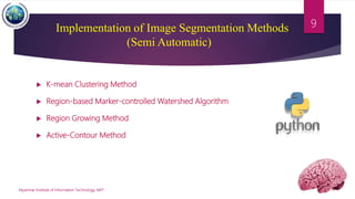
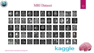
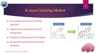
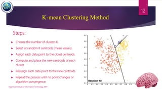
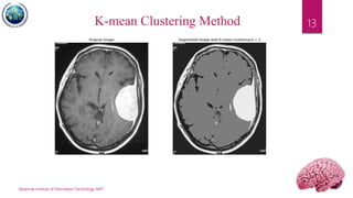
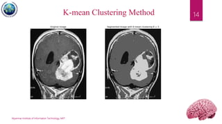
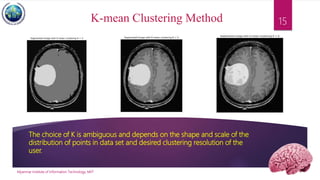
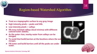
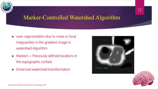
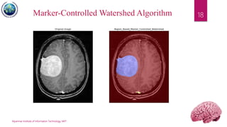
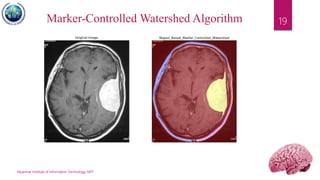
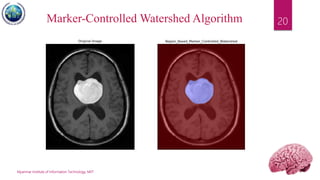
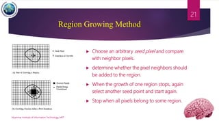
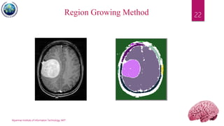
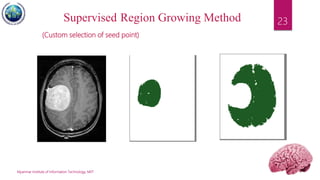
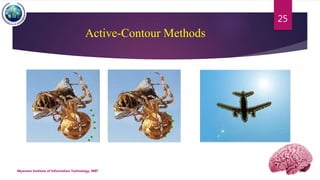
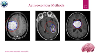
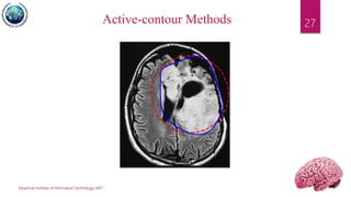
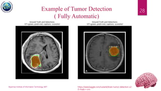
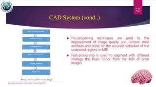
Applications of Image Segmentation
in Brain Tumor Detection
INSTRUCTOR-IN-CHARGE : DR G R SINHA
TEAM MEMBERS : 2015-MIIT-CSE-053, THEINT THIN ZAR SOE
2015-MIIT-CSE-060, ZAW HTET AUNG
2015-MIIT-ECE-003, MYAT MYINT ZU THIN
2015-MIIT-ECE-004, NILA KO
2015-MIIT-ECE-005, PHYO AUNK KYAW
1
Myanmar Institute of Information Technology, MIIT
2
Contents
Applications of Image
Segmentation in Brain
Tumor Detection
on
 What is Brain Tumor?
 What is Image Segmentation?
 Brain Tumor Segmentation Methods
 Brain Tumor Testing Machines
 Brain Tumor diagnosis using MRI
 Implementation of Image Segmentation
Methods in Python
 Computer Aided Diagnosis (CAD) System
 Artificial Intelligence in Medical Imaging
 Why do we need AI in Medical Applications?
 AI + Image Processing in Cancer Diagnosis
Brain Tumor
 What is Brain Tumor?
A brain tumor is a mass or growth of abnormal cells in your brain. We can differentiate
brain tumors in two major groups. Some brain tumors are non-cancerous (benign) and some
are cancerous (malignant).
 Benign Brain Tumor
A benign tumour is a mass of cell (tumour) that does not invade neighbouring tissue. It
is non-cancerous but it can grow slowly inside the brain.
 Malignant Brain Tumor
A malignant tumor is cancerous and rapidly spread into other parts of the brain by
sending cancerous cells into surrounding tissue.
Myanmar Institute of Information Technology, MIIT
3
Benign Vs Malignant
Myanmar Institute of Information Technology, MIIT
4
Image Segmentation
What is Image Segmentation?
 In computer vision, image segmentation is the process of
partitioning a digital image into multiple segments based
on grey level, intensity, color, texture, depth or motion.
 The goal of segmentation is to represent an image into
something that is more meaningful and easier to analyse.
Myanmar Institute of Information Technology, MIIT
5
Brain tumor segmentation plays an important role in medical image processing.
Treatment of patients with brain tumors is highly dependent on early detection of
these tumors. Early detection of brain tumors will improve the patient's life chances.
Brain Tumor Segmentation Methods
Nowadays, brain tumor segmentation methods can be organized into
different categories based on different principles. In the clinic, brain tumor
segmentation methods are usually classified into three main categories –
 Manual
 Semi-automatic
 Fully automatic segmentations based on the degree of required human
interaction.
Myanmar Institute of Information Technology, MIIT
6
How we can test these tumors in brain?
 CT Scan – where X- rays are used to build a detailed image
of our brain.
 MRI scan – where a detailed mage of our brain is produced
using a strong magnetic field.
 Electroencephalogram(EEG) – electrodes are attached to
your scalp to record our brain activity and detect any
abnormalities if it is suspected you are having epileptic fits.
 If a tumour is suspected , a biopsy may be carried out to
established the type of tumour and the most effective
treatment.
Myanmar Institute of Information Technology, MIIT
7
Brain Tumor diagnosis Using MRI
 What is MRI? ( Magnetic Resonance Image)
 providing the rich information about human soft-issue anatomy.
 In brain MRI analysis, image segmentation is commonly used for measuring
and visualizing the brain’s anatomical structures, analysing brain changes,
surgical planning and image-guided interventions.
 Tumor Segmentation from MRI is an important and time consuming manual
task performed by medical experts.
 There are different brain tumor detection and segmentation methods to detect
and segment a brain tumor from MRI images.
Myanmar Institute of Information Technology, MIIT
8
 K-mean Clustering Method
 Region-based Marker-controlled Watershed Algorithm
 Region Growing Method
 Active-Contour Method
Myanmar Institute of Information Technology, MIIT
9Implementation of Image Segmentation Methods
(Semi Automatic)
Myanmar Institute of Information Technology, MIIT
10MRI Dataset
 An unsupervised machine learning
algorithm
 Segment the interest area from the
background.
 Partition N observations into K clusters.
 Group all the pixels with some kind of
similarities.
Myanmar Institute of Information Technology, MIIT
11
K-mean Clustering Method
 Choose the number of clusters K.
 Select at random K centroids (mean values).
 Assign each data point to the closet centroids.
 Compute and place the new centroids of each
cluster
 Reassign each data point to the new centroids.
 Repeat the process until no point changes or
algorithm convergence
Myanmar Institute of Information Technology, MIIT
12
K-mean Clustering Method
Steps:
Myanmar Institute of Information Technology, MIIT
13K-mean Clustering Method
Myanmar Institute of Information Technology, MIIT
14K-mean Clustering Method
The choice of K is ambiguous and depends on the shape and scale of the
distribution of points in data set and desired clustering resolution of the
user.
Myanmar Institute of Information Technology, MIIT
15K-mean Clustering Method
Region-based Watershed Algorithm
 Treat as a topographic surface to any gray image
 high intensity pixels = peaks and hills
 Low intensity pixels = valleys
 fill every isolated valleys (local minima) with different
colored water (labels).
 As the water rises, nearby water from valleys can be
merged.
 To avoid that build barriers at the locations where the water
merges.
 Fill water and build barriers until all the peaks are under
water.
Myanmar Institute of Information Technology, MIIT
16
Marker-Controlled Watershed Algorithm
 over-segmentation due to noise or local
irregularities in the gradient image in
watershed Algorithm
 Markers = Previously defined locations in
the topographic surface
 Enhanced watershed transformation
Myanmar Institute of Information Technology, MIIT
17
Myanmar Institute of Information Technology, MIIT
18Marker-Controlled Watershed Algorithm
Myanmar Institute of Information Technology, MIIT
19Marker-Controlled Watershed Algorithm
Myanmar Institute of Information Technology, MIIT
20Marker-Controlled Watershed Algorithm
 Choose an arbitrary seed pixel and compare
with neighbor pixels.
 determine whether the pixel neighbors should
be added to the region.
 When the growth of one region stops, again
select another seed point and start again.
 Stop when all pixels belong to some region.
Myanmar Institute of Information Technology, MIIT
21
Region Growing Method
Myanmar Institute of Information Technology, MIIT
22Region Growing Method
Myanmar Institute of Information Technology, MIIT
23Supervised Region Growing Method
(Custom selection of seed point)
Active-Contour Methods
Myanmar Institute of Information Technology, MIIT
24
 Uses of energy forces and constraints for
segregation of the pixels of interest from the
image.
 Snakes: Active contour models. Kass, M.; Witkin,
A.; Terzopoulos, D. International Journal of
Computer Vision 1 (4): 321 (1988).
DOI:10.1007/BF00133570
 A snake is an energy minimizing, deformable
spline influenced by constraint and image forces
that pull it towards object contours and internal
forces that resist deformation.
Active-Contour Methods
Myanmar Institute of Information Technology, MIIT
25
Myanmar Institute of Information Technology, MIIT
26Active-contour Methods
Myanmar Institute of Information Technology, MIIT
27Active-contour Methods
Myanmar Institute of Information Technology, MIIT
28Example of Tumor Detection
( Fully Automatic)
https://www.kaggle.com/ruslankl/brain-tumor-detection-v2-
0-mask-r-cnn
Computer Aided Diagnosis ( CAD) System
 Automated system (detection) of brain tumor through MRI is basically called
Computer-Aided Diagnosis (CAD) system
 The CAD system can provide highly accurate reconstruction of the original image
i.e. the valuable outlook and accuracy of earlier brain tumor detection.
 In the initial stage, pre-processing has required after that stages post-processing
i.e. segmentation are required.
Myanmar Institute of Information Technology, MIIT
29
CAD System (cond..)
 Pre-processing techniques are used to the
improvement of image quality and remove small
artefacts and noise for the accurate detection of the
undesired regions in MRI.
 Post-processing is used to segment with different
strategy the brain tumor from the MRI of brain
images
Myanmar Institute of Information Technology, MIIT
30
Artificial Intelligence in Medical Imaging
 Magnetic Resonance Imaging (MRI) and Computed Tomography (CT) are the
most involved techniques.
 One of the most promising areas of health innovation is the application of
Artificial Intelligence (AI), primarily in medical Imaging such as image acquisition,
image segmentation, data storage, data mining and many others.
 Due to this wide range of applications, AI is expected to massively impact on the
radiologist’s daily life.
Myanmar Institute of Information Technology, MIIT
31
Why do we need AI in Medical Applications?
 Nowadays, there are new diseases that take birth daily.
 The medical field needs fast, automated, efficient and reliable techniques to
provide best to best treatments for patients
 This is possible only with AI technology. Now what is AI?
 AI is the science of simulating the most powerful functions of the human brain to
solve problems for humans and which can self-improve.
 They act as the robotic version of humans, but their working and calculations are
way faster plus accurate than that of humans.
Myanmar Institute of Information Technology, MIIT
32
Why do we need AI in Medical Applications?
 With the innovation in science and technology, healthcare has experienced
surprising advancements.
 The Artificial Intelligence-based machines can work as efficiently as the human
brain or even faster to heal people from ill health.
 The shortage of doctors, nurses, physicians, or other health workers worldwide
can be overcome by the AI machines which save hours of waiting in line by the
patients and also provide a quick diagnosis.
 The difference between AI and traditional technologies in health care is the ability
to gain information, process it and give a well-defined and reliable output for the
patients.
Myanmar Institute of Information Technology, MIIT
33
AI + Image Processing in Cancer Diagnosis
 AI applied to Image Processing plays a very vital role in healthcare.
 The super-intelligent devices created by AI with the help of machine
learning and deep learning have contributed a lot to the industry.
 Sometimes, even the most skilled and expert doctors can misdiagnose.
 That’s why intelligent machines are used for the patient’s safety and fast
recovery.
Myanmar Institute of Information Technology, MIIT
34
Myanmar Institute of Information Technology, MIIT
36
AI + Image Processing in Cancer Diagnosis
 Using laser imaging and artificial
intelligence, researchers were able to
diagnose brain tumors in under 150
seconds.
 The dark ovals are tumor cells, among
nerve fibers.
 The AI takes two and a half minutes where
as the traditional method takes more.
 In addition to speeding up the process, the new technique can also
detect some details that traditional methods may miss, like the spread of
a tumor along nerve fibers.
 AI(applied Image Processing) has been shown to be effective in the
accurate diagnosis of various medical conditions.
 AI using image processing, deep learning and neural networks,
accurately diagnosed and provided treatment decisions.
Myanmar Institute of Information Technology, MIIT
37
AI + Image Processing in Cancer Diagnosis
 The researchers used about 2.5 millions tissue samples from thousands of brain
surgery patients to train an artificial intelligence system to identify about 10 most
common types of brain tumor.
 Experiment : AI Vs Humans
 The experiment involved brain tissues from 278 patients, analyzed while the
surgery was still going on.
 Each sample was split, with half going to A.I. and half to a neuropathologist.
 Result  Humans got 93.9% correct , where as AI got 94.6% correct
Myanmar Institute of Information Technology, MIIT
38
AI + Image Processing in Cancer Diagnosis
Myanmar Institute of Information Technology, MIIT
39
AI + Image Processing in Cancer Diagnosis
 Reduce Mortality Rate
 Fast and Accurate Diagnosis
 Reduce Errors Related to Human Fatigue
 Improved Radiology
With this technology, cancer operations are safer and more effective than ever before.
Advantages of AI + Image Processing in Cancer Diagnosis
References
 https://scikit-image.org/docs/dev/auto_examples/edges/plot_active_contours.html
 https://www.thepythoncode.com/article/kmeans-for-image-segmentation-opencv-python
 https://scikit-image.org/docs/dev/user_guide/tutorial_segmentation.html
 https://github.com/Spinkoo/Region-Growing
 Video Source :
https://www.youtube.com/watch?v=QjfaCnCz6Bc&list=LLQn3P8EmnR2e8wmrZGCwm2w&in
dex=3
 https://www.healthcentral.com/slideshow/8-ways-artificial-intelligence-is-affecting-the-medical-
field
 https://www.nytimes.com/2020/01/06/health/artificial-intelligence-brain-cancer.html
Myanmar Institute of Information Technology, MIIT
40
Thank You
41
Applications of Image Segmentation in Brain Tumor Detection
Any Questions!

Application of-image-segmentation-in-brain-tumor-detection

  • 1.
    Applications of ImageSegmentation in Brain Tumor Detection INSTRUCTOR-IN-CHARGE : DR G R SINHA TEAM MEMBERS : 2015-MIIT-CSE-053, THEINT THIN ZAR SOE 2015-MIIT-CSE-060, ZAW HTET AUNG 2015-MIIT-ECE-003, MYAT MYINT ZU THIN 2015-MIIT-ECE-004, NILA KO 2015-MIIT-ECE-005, PHYO AUNK KYAW 1
  • 2.
    Myanmar Institute ofInformation Technology, MIIT 2 Contents Applications of Image Segmentation in Brain Tumor Detection on  What is Brain Tumor?  What is Image Segmentation?  Brain Tumor Segmentation Methods  Brain Tumor Testing Machines  Brain Tumor diagnosis using MRI  Implementation of Image Segmentation Methods in Python  Computer Aided Diagnosis (CAD) System  Artificial Intelligence in Medical Imaging  Why do we need AI in Medical Applications?  AI + Image Processing in Cancer Diagnosis
  • 3.
    Brain Tumor  Whatis Brain Tumor? A brain tumor is a mass or growth of abnormal cells in your brain. We can differentiate brain tumors in two major groups. Some brain tumors are non-cancerous (benign) and some are cancerous (malignant).  Benign Brain Tumor A benign tumour is a mass of cell (tumour) that does not invade neighbouring tissue. It is non-cancerous but it can grow slowly inside the brain.  Malignant Brain Tumor A malignant tumor is cancerous and rapidly spread into other parts of the brain by sending cancerous cells into surrounding tissue. Myanmar Institute of Information Technology, MIIT 3
  • 4.
    Benign Vs Malignant MyanmarInstitute of Information Technology, MIIT 4
  • 5.
    Image Segmentation What isImage Segmentation?  In computer vision, image segmentation is the process of partitioning a digital image into multiple segments based on grey level, intensity, color, texture, depth or motion.  The goal of segmentation is to represent an image into something that is more meaningful and easier to analyse. Myanmar Institute of Information Technology, MIIT 5 Brain tumor segmentation plays an important role in medical image processing. Treatment of patients with brain tumors is highly dependent on early detection of these tumors. Early detection of brain tumors will improve the patient's life chances.
  • 6.
    Brain Tumor SegmentationMethods Nowadays, brain tumor segmentation methods can be organized into different categories based on different principles. In the clinic, brain tumor segmentation methods are usually classified into three main categories –  Manual  Semi-automatic  Fully automatic segmentations based on the degree of required human interaction. Myanmar Institute of Information Technology, MIIT 6
  • 7.
    How we cantest these tumors in brain?  CT Scan – where X- rays are used to build a detailed image of our brain.  MRI scan – where a detailed mage of our brain is produced using a strong magnetic field.  Electroencephalogram(EEG) – electrodes are attached to your scalp to record our brain activity and detect any abnormalities if it is suspected you are having epileptic fits.  If a tumour is suspected , a biopsy may be carried out to established the type of tumour and the most effective treatment. Myanmar Institute of Information Technology, MIIT 7
  • 8.
    Brain Tumor diagnosisUsing MRI  What is MRI? ( Magnetic Resonance Image)  providing the rich information about human soft-issue anatomy.  In brain MRI analysis, image segmentation is commonly used for measuring and visualizing the brain’s anatomical structures, analysing brain changes, surgical planning and image-guided interventions.  Tumor Segmentation from MRI is an important and time consuming manual task performed by medical experts.  There are different brain tumor detection and segmentation methods to detect and segment a brain tumor from MRI images. Myanmar Institute of Information Technology, MIIT 8
  • 9.
     K-mean ClusteringMethod  Region-based Marker-controlled Watershed Algorithm  Region Growing Method  Active-Contour Method Myanmar Institute of Information Technology, MIIT 9Implementation of Image Segmentation Methods (Semi Automatic)
  • 10.
    Myanmar Institute ofInformation Technology, MIIT 10MRI Dataset
  • 11.
     An unsupervisedmachine learning algorithm  Segment the interest area from the background.  Partition N observations into K clusters.  Group all the pixels with some kind of similarities. Myanmar Institute of Information Technology, MIIT 11 K-mean Clustering Method
  • 12.
     Choose thenumber of clusters K.  Select at random K centroids (mean values).  Assign each data point to the closet centroids.  Compute and place the new centroids of each cluster  Reassign each data point to the new centroids.  Repeat the process until no point changes or algorithm convergence Myanmar Institute of Information Technology, MIIT 12 K-mean Clustering Method Steps:
  • 13.
    Myanmar Institute ofInformation Technology, MIIT 13K-mean Clustering Method
  • 14.
    Myanmar Institute ofInformation Technology, MIIT 14K-mean Clustering Method
  • 15.
    The choice ofK is ambiguous and depends on the shape and scale of the distribution of points in data set and desired clustering resolution of the user. Myanmar Institute of Information Technology, MIIT 15K-mean Clustering Method
  • 16.
    Region-based Watershed Algorithm Treat as a topographic surface to any gray image  high intensity pixels = peaks and hills  Low intensity pixels = valleys  fill every isolated valleys (local minima) with different colored water (labels).  As the water rises, nearby water from valleys can be merged.  To avoid that build barriers at the locations where the water merges.  Fill water and build barriers until all the peaks are under water. Myanmar Institute of Information Technology, MIIT 16
  • 17.
    Marker-Controlled Watershed Algorithm over-segmentation due to noise or local irregularities in the gradient image in watershed Algorithm  Markers = Previously defined locations in the topographic surface  Enhanced watershed transformation Myanmar Institute of Information Technology, MIIT 17
  • 18.
    Myanmar Institute ofInformation Technology, MIIT 18Marker-Controlled Watershed Algorithm
  • 19.
    Myanmar Institute ofInformation Technology, MIIT 19Marker-Controlled Watershed Algorithm
  • 20.
    Myanmar Institute ofInformation Technology, MIIT 20Marker-Controlled Watershed Algorithm
  • 21.
     Choose anarbitrary seed pixel and compare with neighbor pixels.  determine whether the pixel neighbors should be added to the region.  When the growth of one region stops, again select another seed point and start again.  Stop when all pixels belong to some region. Myanmar Institute of Information Technology, MIIT 21 Region Growing Method
  • 22.
    Myanmar Institute ofInformation Technology, MIIT 22Region Growing Method
  • 23.
    Myanmar Institute ofInformation Technology, MIIT 23Supervised Region Growing Method (Custom selection of seed point)
  • 24.
    Active-Contour Methods Myanmar Instituteof Information Technology, MIIT 24  Uses of energy forces and constraints for segregation of the pixels of interest from the image.  Snakes: Active contour models. Kass, M.; Witkin, A.; Terzopoulos, D. International Journal of Computer Vision 1 (4): 321 (1988). DOI:10.1007/BF00133570  A snake is an energy minimizing, deformable spline influenced by constraint and image forces that pull it towards object contours and internal forces that resist deformation.
  • 25.
    Active-Contour Methods Myanmar Instituteof Information Technology, MIIT 25
  • 26.
    Myanmar Institute ofInformation Technology, MIIT 26Active-contour Methods
  • 27.
    Myanmar Institute ofInformation Technology, MIIT 27Active-contour Methods
  • 28.
    Myanmar Institute ofInformation Technology, MIIT 28Example of Tumor Detection ( Fully Automatic) https://www.kaggle.com/ruslankl/brain-tumor-detection-v2- 0-mask-r-cnn
  • 29.
    Computer Aided Diagnosis( CAD) System  Automated system (detection) of brain tumor through MRI is basically called Computer-Aided Diagnosis (CAD) system  The CAD system can provide highly accurate reconstruction of the original image i.e. the valuable outlook and accuracy of earlier brain tumor detection.  In the initial stage, pre-processing has required after that stages post-processing i.e. segmentation are required. Myanmar Institute of Information Technology, MIIT 29
  • 30.
    CAD System (cond..) Pre-processing techniques are used to the improvement of image quality and remove small artefacts and noise for the accurate detection of the undesired regions in MRI.  Post-processing is used to segment with different strategy the brain tumor from the MRI of brain images Myanmar Institute of Information Technology, MIIT 30
  • 31.
    Artificial Intelligence inMedical Imaging  Magnetic Resonance Imaging (MRI) and Computed Tomography (CT) are the most involved techniques.  One of the most promising areas of health innovation is the application of Artificial Intelligence (AI), primarily in medical Imaging such as image acquisition, image segmentation, data storage, data mining and many others.  Due to this wide range of applications, AI is expected to massively impact on the radiologist’s daily life. Myanmar Institute of Information Technology, MIIT 31
  • 32.
    Why do weneed AI in Medical Applications?  Nowadays, there are new diseases that take birth daily.  The medical field needs fast, automated, efficient and reliable techniques to provide best to best treatments for patients  This is possible only with AI technology. Now what is AI?  AI is the science of simulating the most powerful functions of the human brain to solve problems for humans and which can self-improve.  They act as the robotic version of humans, but their working and calculations are way faster plus accurate than that of humans. Myanmar Institute of Information Technology, MIIT 32
  • 33.
    Why do weneed AI in Medical Applications?  With the innovation in science and technology, healthcare has experienced surprising advancements.  The Artificial Intelligence-based machines can work as efficiently as the human brain or even faster to heal people from ill health.  The shortage of doctors, nurses, physicians, or other health workers worldwide can be overcome by the AI machines which save hours of waiting in line by the patients and also provide a quick diagnosis.  The difference between AI and traditional technologies in health care is the ability to gain information, process it and give a well-defined and reliable output for the patients. Myanmar Institute of Information Technology, MIIT 33
  • 34.
    AI + ImageProcessing in Cancer Diagnosis  AI applied to Image Processing plays a very vital role in healthcare.  The super-intelligent devices created by AI with the help of machine learning and deep learning have contributed a lot to the industry.  Sometimes, even the most skilled and expert doctors can misdiagnose.  That’s why intelligent machines are used for the patient’s safety and fast recovery. Myanmar Institute of Information Technology, MIIT 34
  • 36.
    Myanmar Institute ofInformation Technology, MIIT 36 AI + Image Processing in Cancer Diagnosis  Using laser imaging and artificial intelligence, researchers were able to diagnose brain tumors in under 150 seconds.  The dark ovals are tumor cells, among nerve fibers.  The AI takes two and a half minutes where as the traditional method takes more.
  • 37.
     In additionto speeding up the process, the new technique can also detect some details that traditional methods may miss, like the spread of a tumor along nerve fibers.  AI(applied Image Processing) has been shown to be effective in the accurate diagnosis of various medical conditions.  AI using image processing, deep learning and neural networks, accurately diagnosed and provided treatment decisions. Myanmar Institute of Information Technology, MIIT 37 AI + Image Processing in Cancer Diagnosis
  • 38.
     The researchersused about 2.5 millions tissue samples from thousands of brain surgery patients to train an artificial intelligence system to identify about 10 most common types of brain tumor.  Experiment : AI Vs Humans  The experiment involved brain tissues from 278 patients, analyzed while the surgery was still going on.  Each sample was split, with half going to A.I. and half to a neuropathologist.  Result  Humans got 93.9% correct , where as AI got 94.6% correct Myanmar Institute of Information Technology, MIIT 38 AI + Image Processing in Cancer Diagnosis
  • 39.
    Myanmar Institute ofInformation Technology, MIIT 39 AI + Image Processing in Cancer Diagnosis  Reduce Mortality Rate  Fast and Accurate Diagnosis  Reduce Errors Related to Human Fatigue  Improved Radiology With this technology, cancer operations are safer and more effective than ever before. Advantages of AI + Image Processing in Cancer Diagnosis
  • 40.
    References  https://scikit-image.org/docs/dev/auto_examples/edges/plot_active_contours.html  https://www.thepythoncode.com/article/kmeans-for-image-segmentation-opencv-python https://scikit-image.org/docs/dev/user_guide/tutorial_segmentation.html  https://github.com/Spinkoo/Region-Growing  Video Source : https://www.youtube.com/watch?v=QjfaCnCz6Bc&list=LLQn3P8EmnR2e8wmrZGCwm2w&in dex=3  https://www.healthcentral.com/slideshow/8-ways-artificial-intelligence-is-affecting-the-medical- field  https://www.nytimes.com/2020/01/06/health/artificial-intelligence-brain-cancer.html Myanmar Institute of Information Technology, MIIT 40
  • 41.
    Thank You 41 Applications ofImage Segmentation in Brain Tumor Detection Any Questions!

Editor's Notes

  • #7 In the semi-automatic brain tumor methods, the user needs to input some parameters and is responsible for analyzing the visual information and providing feedback response for the software computing. For fully automatic brain tumor segmentation, the computer determines the segmentation of brain tumor without any human interaction. In general, a fully automatic segmentation algorithm combines artificial intelligence and prior knowledge.
  • #11 Kaggle allows users to find and publish data sets, explore and build models in a web-based data-science environment, work with other data scientists and machine learning engineers, and enter competitions to solve data science challenges.
  • #17 depending on the peaks (gradients) nearby, water from different valleys, obviously with different colors will start to merge. To avoid that, you build barriers in the locations where water merges. You continue the work of filling water and building barriers until all the peaks are under water. Then the barriers you created gives you the segmentation result. This is the “philosophy” behind the watershed. However, in practice, this transform produces an important over-segmentation due to noise or local irregularities in the gradient image
  • #19 A major enhancement of the watershed transformation consists in flooding the topographic surface from a previously defined set of markers. OpenCV implemented a marker-based watershed algorithm where you specify which are all valley points are to be merged and which are not. It is an interactive image segmentation. What we do is to give different labels for our object we know. Label the region which we are sure of being the foreground or object with one color (or intensity), label the region which we are sure of being background or non-object with another color and finally the region which we are not sure of anything, label it with 0. That is our marker. Then apply watershed algorithm. Then our marker will be updated with the labels we gave, and the boundaries of objects will have a value of -1.
  • #23 Current region dominates the growth process -- ambiguities around edges of adjacent regions may not be resolved correctly. Different choices of seeds may give different segmentation results. Problems can occur if the (arbitrarily chosen) seed point lies on an edge.
  • #25 In mathematics, a spline is a special function defined piecewise by polynomials. In interpolating problems, spline interpolation is often preferred to polynomial interpolation because it yields similar results, even when using low degree polynomials, while avoiding Runge's phenomenon for higher degrees.
  • #26 Current contour is the sum of external and internal energy. External energy is the minimum at object boundary. Internal energy regulates the shape of contour, controlling its curvature, shapes and regularity.
  • #27 snakes have multiple advantages: They autonomously and adaptively search for the minimum state. External image forces act upon the snake in an intuitive manner. Incorporating Gaussian smoothing in the image energy function introduces scale sensitivity. They can be used to track dynamic objects. The key drawbacks of the traditional snakes are They are sensitive to local minima states, which can be counteracted by simulated annealing techniques. Minute features are often ignored during energy minimization over the entire contour. Their accuracy depends on the convergence policy.
  • #29 CNN method IoU means Intersection over Union which is used in object detection to determine the accuracy of an object detector.